. 2015 Apr 29;35(17):6752-69.
doi: 10.1523/JNEUROSCI.2663-14.2015.

Intracellular FGF14 (iFGF14) Is Required for Spontaneous and Evoked Firing in Cerebellar Purkinje Neurons and for Motor Coordination and Balance

Affiliations

Intracellular FGF14 (iFGF14) Is Required for Spontaneous and Evoked Firing in Cerebellar Purkinje Neurons and for Motor Coordination and Balance

Marie K Bosch et al. J Neurosci. .

Abstract

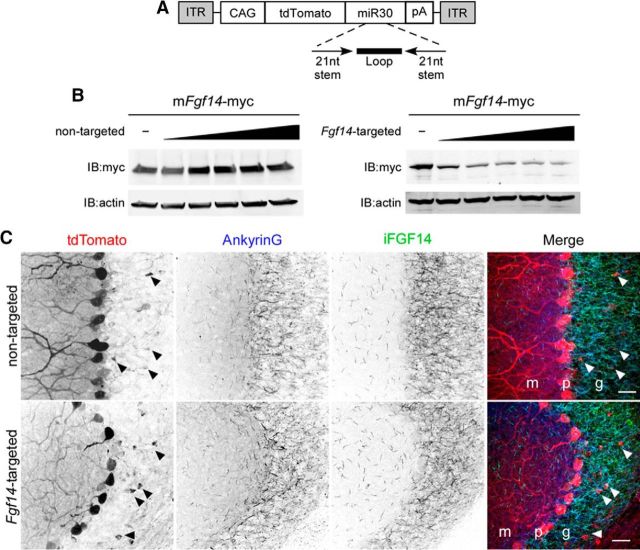
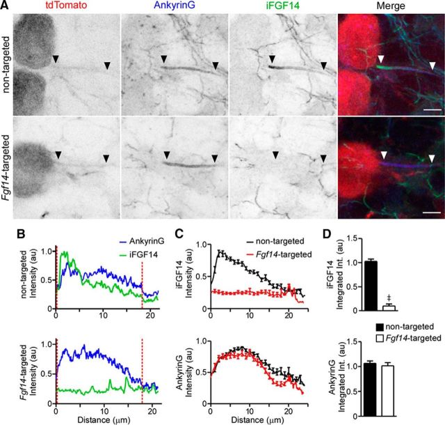
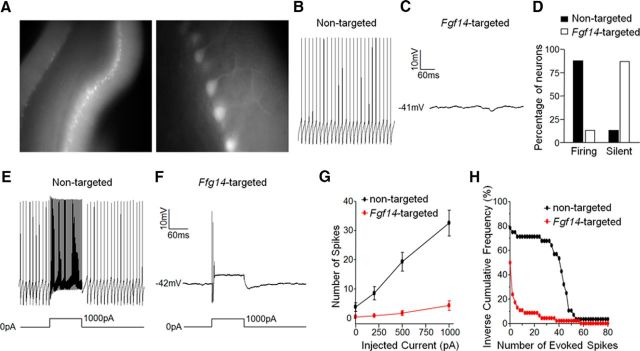
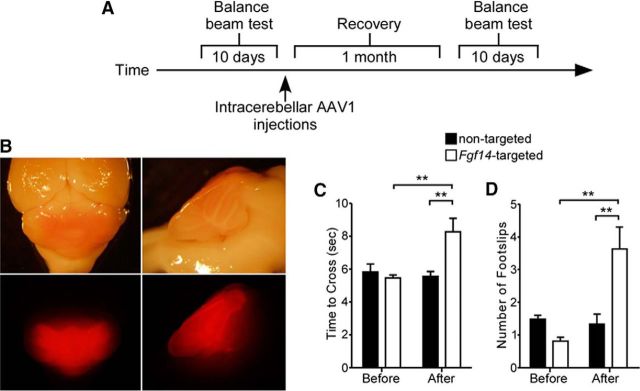
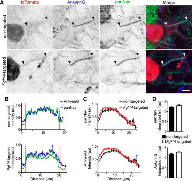
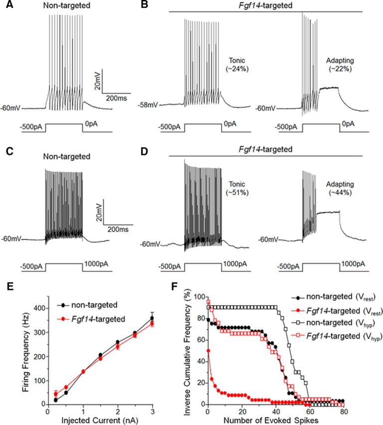
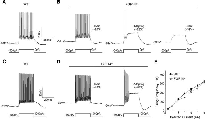
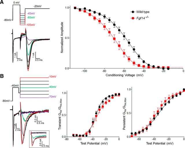
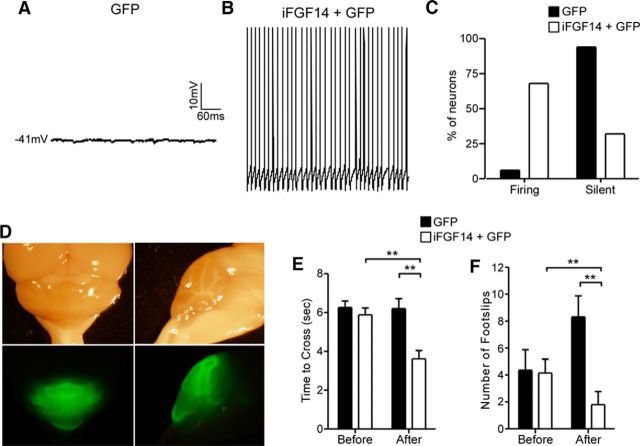
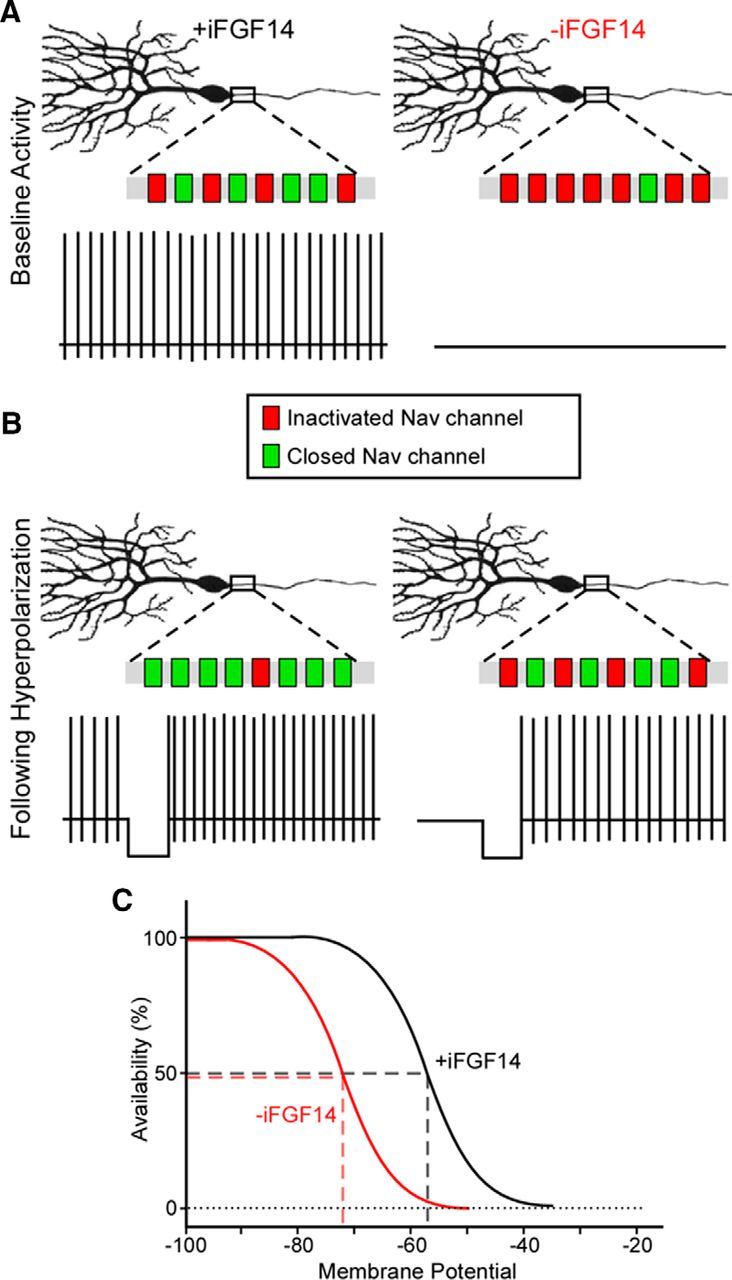
Mutations in FGF14, which encodes intracellular fibroblast growth factor 14 (iFGF14), have been linked to spinocerebellar ataxia (SCA27). In addition, mice lacking Fgf14 (Fgf14(-/-)) exhibit an ataxia phenotype resembling SCA27, accompanied by marked changes in the excitability of cerebellar granule and Purkinje neurons. It is not known, however, whether these phenotypes result from defects in neuronal development or if they reflect a physiological requirement for iFGF14 in the adult cerebellum. Here, we demonstrate that the acute and selective Fgf14-targeted short hairpin RNA (shRNA)-mediated in vivo "knock-down" of iFGF14 in adult Purkinje neurons attenuates spontaneous and evoked action potential firing without measurably affecting the expression or localization of voltage-gated Na(+) (Nav) channels at Purkinje neuron axon initial segments. The selective shRNA-mediated in vivo "knock-down" of iFGF14 in adult Purkinje neurons also impairs motor coordination and balance. Repetitive firing can be restored in Fgf14-targeted shRNA-expressing Purkinje neurons, as well as in Fgf14(-/-) Purkinje neurons, by prior membrane hyperpolarization, suggesting that the iFGF14-mediated regulation of the excitability of mature Purkinje neurons depends on membrane potential. Further experiments revealed that the loss of iFGF14 results in a marked hyperpolarizing shift in the voltage dependence of steady-state inactivation of the Nav currents in adult Purkinje neurons. We also show here that expressing iFGF14 selectively in adult Fgf14(-/-) Purkinje neurons rescues spontaneous firing and improves motor performance. Together, these results demonstrate that iFGF14 is required for spontaneous and evoked action potential firing in adult Purkinje neurons, thereby controlling the output of these cells and the regulation of motor coordination and balance.

Keywords: FGF14; channel inactivation; fibroblast growth factor homologous factor 4 (FHF4); intrinsic excitability; spinocerebellar ataxia 27; voltage-gated sodium (Nav) channels.


Figures

Figure 1.
Figure 1.
Generation and validation of Fgf14-targeted shRNA. A, Schematic representation of the AAV transfer vector with an shRNA embedded in a miRNA (miR30) context in the 3′-UTR of tdTomato. Expression is driven by the chicken β-actin (with a CMV enhancer) promoter (CAG). ITR, Inverted terminal repeat; pA, polyadenylation signal. B, Mouse Fgf14-myc was expressed in CHL cells with increasing amounts of either the nontargeted or the Fgf14-targeted shRNA plasmid. Western blot analyses using an anti-myc antibody revealed that the Fgf14-targeted, but not the nontargeted, shRNA reduced iFGF14 protein expression in a dose-dependent manner. Blots were also probed with an anti-actin antibody to verify equal sample loading. C, Wild-type mice were injected into the cerebellum with the nontargeted shRNA-targeted (top) or the Fgf14-targeted (bottom) shRNA-expressing AAV1. Parasaggital sections were cut and stained with anti-Ankyrin G (blue) and anti-FGF14 (green) antibodies. In A, Representative low-magnification images of nontargeted shRNA- and Fgf14-targeted shRNA-transduced cells are shown. Viral-transduced neurons were identified by tdTomato expression and cell type was determined by morphology and location. Purkinje neurons, for example, were readily identified by large somata and extensively branched dendritic trees extending into the molecular layer; TdTomato expression in Purkinje neurons is robust. Arrowheads indicate the few tdTomato expressing (virally transduced) granule neurons. Scale bars, 25 μm. m, Molecular layer; p, Purkinje layer; g, granule layer. Overall anti-FGF14 labeling in the granule layer is similar in sections prepared after injections of the nontargeted and Fgf14-targeted shRNA-expressing AAV1.
Figure 2.
Figure 2.
Ankyrin G expression at the axon initial segment is robust in Purkinje neurons transduced with the Fgf14-targeted shRNA. A, Representative images of nontargeted and Fgf14-targeted shRNA-transduced Purkinje neurons identified by tdTomato fluorescence (red) and stained with anti-Ankyrin G (blue) and anti-iFGF14 (green) specific antibodies. In each panel, the arrowheads indicate the AIS. Scale bars, 5 μm. B, Representative line scans of anti-Ankyrin G and anti-iFGF14 immunofluorescence intensities along the AIS of a nontargeted (top) and an Fgf14-targeted (bottom) shRNA-transduced Purkinje neuron. Vertical (red) dotted lines indicate the start and end of each AIS. C, Mean ± SEM anti-iFGF14 (top) and anti-Ankyrin G (bottom) immunofluorescence intensities along the AIS of Purkinje neurons transduced with either the nontargeted (n = 29 AIS, 2 animals) or the Fgf14-targeted (n = 26 AIS, 2 animals) shRNA. D, Mean ± SEM anti-iFGF14 (top) integrated immunofluorescence intensity was reduced significantly (≠p < 0.0001; Student's t test) in Fgf14-targeted shRNA-expressing (n = 26) compared with nontargeted shRNA-expressing (n = 29) Purkinje neurons, whereas mean ± SEM anti-ankyrin G integrated immunofluorescence intensities were not measurably affected.
Figure 3.
Figure 3.
Spontaneous and evoked firing is attenuated in Purkinje neurons transduced with the Fgf14-targeted shRNA. A, Representative fluorescence images of acute cerebellar slices prepared from a wild-type mouse injected with tdTomato-shRNA AAV1. Low-magnification image (left) shows widespread infectivity of an entire lobule of the cerebellum and the high-magnification image (right) reveals infected Purkinje neurons. B, C, Representative recordings of spontaneous activity in a nontargeted shRNA-transduced Purkinje neuron (B) and a quiescent Fgf14-targeted shRNA-transduced Purkinje neuron (C). D, Percentages of nontargeted (n = 30) and Fgf14-targeted (n = 46) shRNA-transduced Purkinje neurons that fired spontaneously or were silent. The vast majority (>85%) of the nontargeted shRNA-transduced cells were spontaneously active, whereas most (>85%) of the Fgf14-targeted shRNA-transduced Purkinje neurons were silent. E, F, Representative recordings from nontargeted (E) and Fgf14-targeted (F) shRNA-transduced Purkinje neurons before and during a prolonged (250 ms) 1000 pA depolarizing current injection. G, Mean ± SEM numbers of action potentials evoked in nontargeted (n = 28) and Fgf14-targeted (n = 46) shRNA transduced Purkinje neurons are plotted as a function of the amplitude of the injected current. H, Inverse cumulative frequency plots of spike numbers (elicited by 250 ms 1000 pA depolarizing current injections) in nontargeted (n = 28) and Fgf14-targeted (n = 46) shRNA-transduced Purkinje neurons. The frequency distribution in Fgf14-targeted shRNA-transduced neurons is shifted significantly (p < 0.0001, Kolmogorov–Smirnov test) to the left compared with nontargeted shRNA-transduced cells.
Figure 4.
Figure 4.
Motor performance and coordination are impaired in mice expressing the Fgf14-targeted shRNA in Purkinje neurons. A, Motor performance and coordination were evaluated in the balance beam test (see Materials and Methods) before and 4 weeks after intracerebellar injections of the nontargeted (n = 11) or the Fgf14-targeted (n = 9) shRNA-expressing AAV1. B, Representative bright-field (top) and fluorescence (bottom) images of the posterior part of a brain dissected from a wild-type mouse 4 weeks after 2 stereotaxic injections of tdTomato-shRNA AAV1 into the cerebellum. Left, Dorsal views; right, midsagittal views. C, D, The time to traverse a 5 mm beam (C), as well as the number of hindlimb placement errors (foot slips) during crossing (D), were evaluated before and after intracerebellar injections of the nontargeted (n = 11) or the Fgf14-targeted (n = 9) shRNA AAV1. Times to traverse the beam and number of foot slips were indistinguishable before and after injection of the nontargeted shRNA. The mean ± SEM time to cross was significantly (**p < 0.01, two-way ANOVA) longer and the mean ± SEM number of foot slips was significantly (**p < 0.01, two-way ANOVA) higher, however, in the animals injected with the Fgf14-targeted shRNA AAV1 whether compared with the same animals before injection or to the animals injected with the nontargeted shRNA AAV1.
Figure 5.
Figure 5.
Anti-Nav α-subunit immunofluorescence intensity and localization at Purkinje neuron AIS are unaffected by Fgf14-targeted shRNA. After injections of the nontargeted (top) or the Fgf14-targeted (bottom) shRNA AAV1, parasagittal sections were cut and stained with anti-Ankyrin G (blue) and anti-panNav α-subunit- (green) specific antibodies. A, Representative images of nontargeted and Fgf14-targeted shRNA-transduced Purkinje neurons, identified by tdTomato fluorescence (red); in each panel, arrowheads indicate AIS regions and scale bars are 5 μm. B, Representative line scans of anti-Ankyrin G and anti-panNav immunofluorescence intensities along the AIS of a nontargeted (top) and an Fgf14-targeted (bottom) shRNA-transduced Purkinje neuron. Vertical (red) dotted lines indicate the starts and ends of the AIS. C, Mean ± SEM immunofluorescence intensities of anti-pan Nav (top) and anti-Ankyrin G (bottom) along the AIS of Purkinje neurons transduced with either the nontargeted (n = 51 AIS, 2 animals) or the Fgf14-targeted (n = 60 AIS, 2 animals) shRNA. D, Mean ± SEM integrated immunofluorescence intensity of anti-panNav (top) and anti-Ankyrin G (bottom) staining along the AIS of Purkinje neurons transduced with the nontargeted (n = 51 AIS, 2 animals) or the Fgf14-targeted (n = 60 AIS, 2 animals) shRNA. Mean ± SEM anti-Ankyrin G and anti-Nav α-subunit labeling intensities are indistinguishable (Student's t test) in adult Purkinje neurons transduced with the nontargeted and the Fgf14-targeted shRNAs.
Figure 6.
Figure 6.
Membrane hyperpolarization rescues repetitive firing in Purkinje neurons transduced with Fgf14-targeted shRNA. AD, Representative evoked firing recorded in nontargeted (A,C) and Fgf14-targeted (B,D) shRNA-transduced Purkinje neurons in response to membrane depolarization from a hyperpolarized membrane potential. Removal of a (500 pA) hyperpolarizing current injection resulted in spontaneous firing in most (20 of 25) of the nontargeted shRNA-transduced Purkinje neurons (A) and in 19 of 41 Fgf14-targeted shRNA-transduced Purkinje neurons: 10 of these cells fired tonically and 9 adapted; the remaining (12) cells were silent. Depolarizing (1000 pA) current injections (250 ms) after membrane hyperpolarization elicited high-frequency repetitive firing in most (23 of 25) Purkinje neurons expressing the nontargeted shRNA, as well as in most (39 of 41) Purkinje neurons expressing the Fgf14-targeted shRNA (D). Mean ± SEM firing frequencies in nontargeted and Fgf14-targeted shRNA-transduced Purkinje neurons in response to (250 ms) depolarizing current injections of varying amplitudes presented after membrane hyperpolarization are plotted in E. Firing rates were indistinguishable (two-way ANOVA) in nontargeted and Fgf14-targeted shRNA-transduced Purkinje neurons. F, Inverse cumulative frequency plots of numbers of action potentials evoked by 250 ms, 1000 pA depolarizing current injections after membrane hyperpolarization (Vhyp) in nontargeted and Fgf14-targeted shRNA-transduced Purkinje neurons are plotted. Histograms of spike numbers elicited from rest (Vrest) are replotted from Figure 3H for comparison purposes. Membrane hyperpolarization significantly (p < 0.0001) shifted the frequency distributions to the right (more spikes) in both nontargeted shRNA and Fgf14-targeted shRNA-transduced Purkinje neurons. In addition, the distribution of spikes evoked from a hyperpolarized potential in Fgf14-targeted shRNA-transduced Purkinje neurons is not significantly different from the distribution of spikes evoked from rest in Purkinje neurons expressing the nontargeted shRNA.
Figure 7.
Figure 7.
Membrane hyperpolarization rescues repetitive firing in adult Fgf14−/− Purkinje neurons. AD, Representative recordings from WT and Fgf14−/− Purkinje neurons obtained using the protocols described in the legend to Figure 5. Removal of the (500 pA) hyperpolarizing current resulted in repetitive firing in most (24 of 32) of the WT neurons (A) and in 11 of the 23 Fgf14−/− neurons (B). Depolarizing (1000 pA) current injections, presented from hyperpolarized membrane potentials, resulted in repetitive firing in most (29 of 32) WT (C) and (21 of 23) Fgf14−/− (D) Purkinje neurons. E, Mean ± SEM firing frequencies in WT and Fgf14−/− Purkinje neurons in response to prolonged (250 ms) depolarizing current injections of varying amplitudes. Firing rates increased as a function of the amplitudes of the depolarizing current injections and were indistinguishable (two-way ANOVA) in WT (n = 32) and Fgf14−/− (n = 23) Purkinje neurons.
Figure 8.
Figure 8.
iFGF14 shifts the voltage dependence of steady-state inactivation of the Nav currents in cerebellar Purkinje neurons. Whole-cell voltage-clamp recordings were obtained at 33 ± 1°C from WT and Fgf14−/− Purkinje neurons in acute cerebellar slices as described in Materials and Methods. A, Representative recordings of Nav currents (left) evoked in a WT cerebellar Purkinje neuron from various conditioning voltages; the voltage-clamp paradigm is illustrated above the records and the currents are shown in the color of the corresponding voltage step. Note that the persistent currents were digitally subtracted (see Materials and Methods) and only the inactivating, transient components of the currents are shown. The transient Nav currents evoked at 0 mV were measured and normalized (in the same cell) to the current evoked from the most hyperpolarized test potential (of −110 mV). Mean ± SEM normalized transient Nav current amplitudes in WT and Fgf14−/− Purkinje neurons were then plotted (right) as a function of the conditioning membrane potential and fitted using a Boltzmann equation (see Materials and Methods). The transient Nav current is inactivated at a significantly (p < 0.001) more hyperpolarized membrane potential in Fgf14−/− (V1/2 = −71.1 ± 0.9 mV, slope = 11.3 ± 1.0; n = 10), than in WT (V1/2 = −58.1 ± 0.9 mV, slope = 10.3 ± 1.0; n = 11) Purkinje neurons. B, Representative recordings of the transient and persistent Nav currents (left) evoked in a WT Purkinje neuron at various test potentials; the voltage-clamp paradigm is illustrated above the records and the currents are shown in the color of the corresponding voltage step; the raw, unsubtracted current records used to quantify the transient and persistent compoents of the Nav currents are shown in the inset. The transient and persistent Nav conductances for the currents evoked at each test potential were determined and normalized (in the same cell) to the maximal transient and persistent Nav conductances and mean ± SEM normalized transient and persistent Nav conductances in WT and Fgf14−/− Purkinje neurons were plotted (right) as a function of the test potential. Although well described by single Boltzmann functions (see Materials and Methods), the voltage dependences of activation of both the transient and the persistent components of the Nav currents (right) are similar in WT and Fgf14−/− Purkinje neurons.
Figure 9.
Figure 9.
Viral-mediated expression of iFGF14 in adult Fgf14−/− Purkinje neurons. A, Schematic of the AAV transfer plasmid (see Materials and Methods) chicken β actin with CMV enhancer promoter (CAG). IRES, Internal ribosome entry site; ITR, inverted terminal repeat; pA, polyadenylation signal. B, C, Representative low-magnification (B) and high magnification (C) images of parasagital sections of adult Fgf14−/− mouse cerebellum injected with AAV1 expressing GFP alone (top panels) or in combination with the AAV1 expressing iFGF14 (bottom panels). Scale bars: B, 25 μm; C, 5 μm. Anti-iFGF14 and anti-Ankyrin G immunostaining are shown in red and blue, respectively. Transduced Purkinje neurons were identified by GFP fluorescence.
Figure 10.
Figure 10.
Acute expression of iFGF14 in Fgf14−/− Purkinje neurons rescues spontaneous firing and improves motor coordination in Fgf14−/− mice. Representative recordings of a quiescent GFP-transduced Fgf14−/− Purkinje neuron (A) and of spontaneous activity in an Fgf14−/− Purkinje neuron cotransduced with the iFGF14 and GFP viruses (B). C, Percentages of GFP (n = 33) and GFP + iFGF14 (n = 57) transduced Purkinje neurons that fired spontaneously or were silent. The vast majority (>93%) of the GFP-transduced Fgf14−/− Purkinje neurons were silent, whereas most (>68%) of the Fgf14−/− Purkinje neurons transduced with iFGF14 were spontaneously active. D, Representative bright-field (top) and fluorescence (bottom) images of the posterior part of a brain dissected from a wild-type mouse 4 weeks after 2 stereotaxic injections of GFP virus into the cerebellum. Left, Dorsal views; right, midsagittal views. The mean ± SEM times to traverse a 12 mm beam (E) and numbers of hindlimb foot slips (F) in Fgf14−/− mice (n = 8) before and after intracerebellar injections of the GFP virus are not significantly different. In the Fgf14−/− mice injected with both the iFGF14 and GFP viruses (n = 9), however, the mean ± SEM time to cross the beam (E) was significantly (**p < 0.001) faster and the mean ± SEM number of foot slips (F) was significantly (**p < 0.01) lower after the injections compared with the same animals before the injections. The measured values are also significantly (**p < 0.01) different from the Fgf14−/− mice injected with the GFP virus.
Figure 11.
Figure 11.
Schematic illustration of the modulatory effect of iFGF14 on Nav channel availability and repetitive firing in Purkinje neurons. A, Nav channel availability at rest is high in WT Purkinje neurons, which express iFGF14, and low in iFGF14 deficient Purkinje neurons. In addition, WT Purkinje neurons are spontaneously active, whereas most iFGF14-deficient Purkinje neurons are quiescent. B, Hyperpolarization of the membrane potential increases Nav channel availability in both WT and iFGF14-deficient Purkinje neurons and, importantly, “rescues” repetitive firing in the iFGF14-deficient cells. The total numbers of Nav channels are unchanged; only the numbers of closed channels that are available to open are affected by the membrane hyperpolarization. In addition, the availability of Nav channels is similar in iFGF14-deficient Purkinje neurons at hyperpolarized membrane potentials and in control Purkinje neurons at rest. C, In the presence of iFGF14, Nav channel availability in Purkinje neurons is shifted markedly to the right (depolarized).

Similar articles

Cited by

References

    1. Apps R, Garwicz M. Anatomical and physiological foundations of cerebellar information processing. Nat Rev Neurosci. 2005;6:297–311. doi: 10.1038/nrn1646. - DOI - PubMed
    1. Bosch MK, Nerbonne JM, Ornitz DM. Dual transgene expression in murine cerebellar Purkinje neurons by viral transduction in vitro. PLoS One. 2014;9:e104062. doi: 10.1371/journal.pone.0104062. - DOI - PMC - PubMed
    1. Brusse E, de Koning I, Maat-Kievit A, Oostra BA, Heutink P, van Swieten JC. Spinocerebellar ataxia associated with a mutation in the fibroblast growth factor 14 gene (SCA27): A new phenotype. Mov Disord. 2006;21:396–401. doi: 10.1002/mds.20708. - DOI - PubMed
    1. Buttermore ED, Piochon C, Wallace ML, Philpot BD, Hansel C, Bhat MA. Pinceau organization in the cerebellum requires distinct functions of neurofascin in Purkinje and basket neurons during postnatal development. J Neurosci. 2012;32:4724–4742. doi: 10.1523/JNEUROSCI.5602-11.2012. - DOI - PMC - PubMed
    1. Carter RJ, Morton J, Dunnett SB. Motor coordination and balance in rodents. Curr Protoc Neurosci. 2001 doi: 10.1002/0471142301.ns0812s15. Chapter 8:Unit 8.12. - DOI - PubMed

Publication types

MeSH terms

LinkOut - more resources