. 2015 Jul 10;290(28):17041-54.
doi: 10.1074/jbc.M114.610204. Epub 2015 Apr 29.

Inside-out Regulation of Ectodomain Cleavage of Cluster-of-Differentiation-44 (CD44) and of Neuregulin-1 Requires Substrate Dimerization

Affiliations

Inside-out Regulation of Ectodomain Cleavage of Cluster-of-Differentiation-44 (CD44) and of Neuregulin-1 Requires Substrate Dimerization

Monika Hartmann et al. J Biol Chem. .

Abstract

Ectodomain shedding of transmembrane precursor proteins generates numerous life-essential molecules, such as epidermal growth factor receptor ligands. This cleavage not only releases the regulatory growth factor, but it is also the required first step for the subsequent processing by γ-secretase and the release of gene regulatory intracellular fragments. Signaling within the cell modifies the cytoplasmic tails of substrates, a step important in starting the specific and regulated cleavage of a large number of studied substrates. Ectodomain cleavage occurs, however, on the outside of the plasma membrane and is carried out by membrane-bound metalloproteases. How the intracellular domain modification communicates with the ectodomain of the substrate to allow for cleavage to occur is unknown. Here, we show that homodimerization of a cluster-of-differentiation-44 or of pro-neuregulin-1 monomers represents an essential pre-condition for their regulated ectodomain cleavage. Both substrates are associated with their respective metalloproteases under both basal or cleavage-stimulated conditions. These interactions only turn productive by specific intracellular signal-induced intracellular domain modifications of the substrates, which in turn regulate metalloprotease access to the substrates' ectodomain and cleavage. We propose that substrate intracellular domain modification induces a relative rotation or other positional change of the dimerization partners that allow metalloprotease cleavage in the extracellular space. Our findings fill an important gap in understanding substrate-specific inside-out signal transfer along cleaved transmembrane proteins and suggest that substrate dimerization (homo- or possibly heterodimerization) might represent a general principle in ectodomain shedding.

Keywords: ADAM; ADAM10; ADAM17; actin; adhesion molecule; angiotensin; ezrin; metalloprotease; neuregulin.


Figures

FIGURE 1.
FIGURE 1.
TPA-induced and ADAM10-dependent processing of CD44 depends on the CD44ICD. A and B, ADAM10 is a major protease acting on CD44. A, wild type (Wt) MEFs or MEFs with the disruption of either the Adam10 or Adam17 or both genes (A10−/−/A17−/−) were transfected with Myc-tagged CD44 (C-terminal c-Myc) wild type, the noncleavable mutant CD44-KR-Mt, or with an empty vector (V). The cells were grown at low cell density, and CD44 cleavage was stimulated by treatment with 100 ng/ml TPA for 30 min. DAPT (5 μm) was added to the cells to prevent degradation of the CD44ΔE cleavage product by γ-secretase. Control cells were treated with DMSO alone (solvent for TPA and DAPT). Subsequently, CD44 full-length (CD44fl) and the membrane-bound C-terminal cleavage product CD44ΔE were detected by c-Myc antibody. CD44fl forms a double band, most likely because of differential glycosylation. Induced CD44 cleavage occurs only in the Adam17 null MEFs but not in the cells with disruption of the Adam10 gene. B, RPM-MC cells transfected with doubly tagged CD44 (N-terminal FLAG and C-terminal c-Myc) were grown at low cell density. A, TPA treatment. Expression of ADAM10 (A10) or ADAM17 (A17) was down-regulated by siRNA (to 3.8 and 1.6% as calculated from the blot by ImageJ). Nontargeting siRNA (C) was used as a control. The released ectodomain was precipitated from culture supernatant by TCA prior to SDS-PAGE. Cleaved ectodomain (solCD44E), CD44fl, and CD44ΔE were detected by FLAG and c-Myc antibodies, respectively. The efficiency of siRNA knockdowns was monitored by detection of ADAM10 and ADAM17 proteins as indicated (seen are the pro- (P) and mature (M) forms). Only ADAM10 knockdown significantly reduced basal and induced release of solCD44E and CD44ΔE. B′, histogram shows mean values of relative level of solCD44E ± S.D. from three independent experiments. ns, p = 0.031081; ****, p < 0.0001; ns, not significant. C, RPM-MC cells transfected with doubly tagged CD44 (N-terminal FLAG, C-terminal c-Myc) were grown at low cell density. For inhibition of translation the cells were pre-incubated with 50 μg/ml of cycloheximide (CHX). CD44 cleavage was stimulated by treatment with 100 ng/ml TPA for 4 h. WB, Western blot.
FIGURE 2.
FIGURE 2.
Substrates CD44 and NRG1 are associated with their ADAM in the absence of cleavage induction. A, association of ADAM10 and CD44 determined by BiFC was analyzed in NIH3T3 cells and RPM-MC cells under inhibition of ADAM activity. The coding regions in expression constructs of bovine ADAM10 (A10) and human CD44 proteins (Wt or KR-Mt) were fused to nonfluorescent N-terminal (V1) and C-terminal (V2) fragments of the Venus protein, respectively. Similarly modified adiponectin receptor (ARV1 versus ARV2) served as positive control (known to interact), and adiponectin receptor versus ADAM10 served as negative control. A metalloprotease inhibitor was added (10 μm batimastat) to prevent cleavage. For microscopy, the cells were fixed with 4% PFA and stained with Texas Red® wheat germ agglutinin prior to permeabilization (see “Experimental Procedures”). Scale bar, 10 μm. Photographs show representative results obtained in NIH3T3 cells. CD44 WT or KR-Mt are associated with ADAM10, with or without TPA stimulation. A′, histogram shows mean % values of BiFC-positive cells among all living transfected single cells ± S.D., as assessed by flow cytometry in RPM-MC cells. p ≥ 0.05 (ns). Flow cytometry revealed around 10% of CD44V2/A10V1 BiFC-positive cells in TPA-stimulated RPM-MC cells. The experiments were repeated four times in NIH3T3 cells and three times in RPM-MC cells. B, CD44-ADAM10 association shown by co-IP in RIPA buffer after protein cross-linking. Equal amounts of endogenous ADAM10 co-immunoprecipitated with transfected FLAG-tagged CD44 WT or KR-Mt, independent of TPA stimulation. Cells and treatments were as in Fig. 1B. B′, histogram shows quantitation from three independent experiments. Cleavage was inhibited by batimastat. p ≥ 0.05 (ns). C, ADAM10-CD44 co-IP. Equal amounts of FLAG-tagged CD44 WT or KR-Mt were co-immunoprecipitated with endogenous ADAM10 in TPA-treated RPM-MC cells. Cleavage was inhibited by GM-6001. C′, histogram shows quantification of three independent experiments. p ≥ 0.05 (ns). D, ADAM10-CD44 co-IP. Equal amounts of endogenous CD44 were co-immunoprecipitated with endogenous ADAM10 in resting and TPA-treated NIH3T3 cells. Cleavage was inhibited by GM-6001. D′, histogram shows quantification of three independent experiments. p ≥ 0.05 (ns). E, association of NRG1 and ADAM17 under inhibition of ADAM activity shown by co-IP (lower panel); whole cell lysate is shown in the upper panel for comparison. NRG1 and ADAM17 interact with or without TPA or AngII treatment. In contrast, NRG1 cleavage regulatory PKCδ is co-precipitated only after TPA or AngII stimulation. Cleavage was inhibited by GM-6001. The relative values of binding of PKCδ are shown within the blot. E′, histograms show quantification from three independent experiments. p ≥ 0.05 (ns). ns, not significant; WB, Western blot; V, vector; IP, immunoprecipitation; WGA, wheat germ agglutinin.
FIGURE 3.
FIGURE 3.
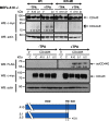
Modification of the ICD of ADAM10 is not required for cleavage regulation. CD44Wt or KR-Mt was co-transfected into ADAM10-deficient MEF cells with either ADAM10 WT (A10) or one of two ADAM10 ICD deletion mutants (Δ1 and Δ2, as indicated in the schematic). ADAM10 WT and both ADAM10 mutants rescued TPA-induced cleavage of CD44 WT as indicated by release of CD44ΔE (upper panel) or solCD44E (lower panel). CD44 KR-Mt remained uncleavable under either condition (upper panel). The experiments were repeated three times for each mutant. WB, Western blot; V, empty vector; C, control.
FIGURE 4.
FIGURE 4.
CD44 and NRG1 form homodimers. A and B, CD44 and NRG1 dimerization. CD44WT (A) and NRG1 (B) homodimerization in living cells was determined by BiFC (NIH3T3 and RPM-MC cells) under inhibition of ADAM activity. Plasmids encoding human CD44 proteins or mouse NRG1 were fused to nonfluorescent N-terminal and C-terminal fragments of the Venus protein. Adiponectin receptor monomers fused to subunits of Venus protein (ARV1 and ARV2) served as positive control, and ARV2 and ADAM10V1 as negative controls (see also Fig. 2A). To stabilize the interaction between CD44 and ADAM10, a metalloprotease inhibitor was added (10 μm batimastat). Histogram in A′/B′ shows percentage of BiFC-positive cells among all living transfected single cells ± S.D., as assessed by flow cytometry in RPM-MC cells. p ≥ 0.05. Around 75% of WtV1/WtV2 and around 68% of NRGV1/NRGV2 cells were BiFC-positive. The experiments were repeated four times in NIH3T3 cells and three times in RPM-MC cells. C, NRG1 dimer co-precipitation. Equal amounts of differentially labeled NRG1 molecules (C-terminal GFP or Myc tags) were expressed in NIH3T3 cells. Precipitation by antibodies to GFP co-precipitated Myc-tagged NRG1, indicating homodimerization. The left panel shows co-precipitation from lysates of cells that have not been treated with TPA, and the right panel shows similar co-precipitation after TPA stimulus. The experiments were repeated three times. ns, not significant; WB, Western blot; IP, immunoprecipitation; WGA, wheat germ agglutinin.
FIGURE 5.
FIGURE 5.
CD44 dimerization promotes ectodomain cleavage. A, CD44 dimerization is stabilized by disulfide bonds. RPM-MC cells were transfected with either CD44 WT, CD44 cysteine mutants (mutations as indicated in the box), or the constitutively cleaved mutant S291A. The constructs were tagged as in Fig. 1. By the use of a nonreducing gel, cysteine bridge-stabilized dimers are visualized. Cleavage was determined by detection of released ectodomains in the culture supernatant (solCD44E) and of the membrane-bound C-terminal cleavage products, which remained dimerized due to the two cysteines still present in the product (CD44ΔE dimer). Cysteine mutations decreased, whereas CD44 S291A increased basal and TPA induced dimerization and cleavage as compared with CD44 WT. The experiment was repeated three times. B, CD44 dimers are preferentially cleaved by ADAM10. Dimers, but not monomers, disappear upon induced cleavage by ADAM10. Cells, treatments, and cleavage detection were as in Fig. 1B. Where indicated, expression of ADAM10 (A10) or ADAM17 (A17) was down-regulated by siRNA. Nontargeting siRNA C was used as a control. ADAM protein knockdown (not shown) was monitored as in Fig. 1B. Reduced levels of dimers and concomitant increase of cleavage product are detected in cells expressing ADAM10 (control C and A17 lanes). Cleavage is nevertheless increased by treatment with TPA. The experiment was repeated three times. WB, Western blot; V, empty vector; C, control.
FIGURE 6.
FIGURE 6.
Disruption of CD44 dimers reduces ectodomain cleavage. A, excess of soluble ectodomain prevents homodimerization and ectodomain cleavage. Soluble CD44E was co-expressed (visible as a 70-kDa band, upper panel). Cells, treatments, and cleavage detection were as in Fig. 1B. A′, histogram shows mean values of relative level of solCD44E ± S.D. and CD44fl dimer ± S.D. from three independent experiments. **, p = 0.003930; ****, p < 0.0001. B, antibody complexes attached to CD44 prevent dimerization and ectodomain cleavage. Treating cells with 20 μg/ml primary mouse CD44 ectodomain-specific 5G8 antibody alone increased dimerization and cleavage. Adding 20 μg/ml secondary polyclonal goat anti-mouse antibody to the primary antibody before incubation inhibited dimerization and cleavage, suggesting sterical hindrance of cleavage by the bound primary-secondary antibody complex. Dimers were detected on nonreducing SDS-polyacrylamide gels. B′, histogram shows mean values of relative level of solCD44E ± S.D. and CD44fl dimer ± S.D. from three independent experiments; solCD44E “−” (no antibody) versus1st” (primary antibody), p = 0.002470; CD44fldimer “−“ versus1st”, p = 0.006427. WB, Western blot; V, empty vector.
FIGURE 7.
FIGURE 7.
Enforced dimerization enhances TPA-induced ectodomain cleavage. A and B, RPM-MC cells were transfected with HA-tagged CD44 WT containing a single (A) or a double (B) copy of the FK506 dimerization domain of FKBP added to its ICD. Dimerization was induced by the artificial FKBP ligand AP20187 (100 nm) 16 h prior to TPA. Control cells were treated with ethanol or DMSO alone (ethanol was solvent for AP20187). FKPB increased dimerization (CD44-FKBP dimer, nonreducing gel, upper panel), but TPA stimulation was still required for cleavage (CD44-FKBPΔE, second panel from bottom). A′/B′, histogram shows mean values of relative level of CD44-FKBPΔE or CD44–2×FKBPΔE ± S.D. from three independent experiments; ****, p < .0001; ***, p = 0.000113. WB, Western blot.
FIGURE 8.
FIGURE 8.
Co-regulation of dimer formation and ectodomain cleavage. A, noncleavable CD44 mutants do not dimerize. RPM-MC cells were transfected with plasmids encoding doubly tagged wild type CD44 WT or KR-Mt or CD44 with mutations of serine 291 mimicking dephosphorylation or phosphorylation (S291A and S291D, respectively). Treatments were as in Fig. 1B. Dimers were detected on nonreducing SDS-polyacrylamide gels. The noncleavable mutants KR-Mt and S291D did not dimerize. Spontaneously cleaved CD44 S291A showed increased basal and induced dimerization. B and C, constitutively cleaved CD44 lacking its ICD predominantly occurs as a dimer. RPM-MC cells were transected with plasmid encoding CD44 with deletion of the entire cytoplasmic domain and treated as in Fig. 1B. An additional mutation of cysteine 286 to alanine was introduced into CD44ΔICD, and cleavage and dimerization of this mutant were also analyzed. Mutation of cysteine 286 reduced dimer formation but did not affect cleavage. Dimers were detected on nonreducing SDS-polyacrylamide gels. D, tumor suppressor merlin reduces dimerization of CD44. Plasmids encoding CD44 WT were co-transfected with plasmids encoding constitutively active merlin (S518A) or inactive merlin (S518D). RPM-MC cells were grown at low density, which maintains endogenous merlin inactive. Treatments and cleavage detection were as in Fig. 1B. Cleavage inhibitory constitutively active merlin (S518A) inhibited dimerization. The experiments in A–D were repeated three times. WB, Western blot; V, vector.
FIGURE 9.
FIGURE 9.
Schematic representation of cleavage regulation through dimerization. CD44 monomers and dimers co-exist on the cell surface. CD44 dimers are stabilized by putative cysteine bridges in the ICD and by putative ectodomain interactions. Our data suggest that ectodomain cleavage regulation depends on ICD modification and interaction with either ERM proteins or merlin on CD44 dimers. At high cell density merlin is dephosphorylated and active and bound to the phosphorylated CD44 ICD. Under low cell density or after TPA stimulation (PKC activator), Ser-291 is dephosphorylated, and phosphorylated/activated ERM proteins displace merlin. This releases a restrictive ICD conformation in the dimer and leads to a positional structural change of the dimerization partners that enable ectodomain accessibility to the ADAM protease. CD44 without its ICD is missing the restrictive ICD conformation and is spontaneously cleaved. A link of ERM proteins to the actin cytoskeleton is possibly important in this regulation. How can CD44S291 be dephosphorylated in response to TPA-dependent activation of PKC? PP1/2 serine phosphatase can indeed be activated by PKC and is regulated by endogenous PKC-activated inhibitors (8, 41, 46).

Similar articles

Cited by

References

    1. van der Vorst E. P., Keijbeck A. A., de Winther M. P., Donners M. M. (2012) A disintegrin and metalloproteases: molecular scissors in angiogenesis, inflammation and atherosclerosis. Atherosclerosis 224, 302–308 - PubMed
    1. Klein T., Bischoff R. (2011) Active metalloproteases of the a disintegrin and metalloprotease (ADAM) family: biological function and structure. J. Proteome Res. 10, 17–33 - PubMed
    1. Hartmann M., Herrlich A., Herrlich P. (2013) Who decides when to cleave an ectodomain? Trends Biochem. Sci. 38, 111–120 - PubMed
    1. Dang M., Armbruster N., Miller M. A., Cermeno E., Hartmann M., Bell G. W., Root D. E., Lauffenburger D. A., Lodish H. F., Herrlich A. (2013) Regulated ADAM17-dependent EGF family ligand release by substrate-selecting signaling pathways. Proc. Natl. Acad. Sci. U.S.A. 110, 9776–9781 - PMC - PubMed
    1. Dang M., Dubbin K., D'Aiello A., Hartmann M., Lodish H., Herrlich A. (2011) Epidermal growth factor (EGF) ligand release by substrate-specific a disintegrin and metalloproteases (ADAMs) involves different protein kinase C (PKC) isoenzymes depending on the stimulus. J. Biol. Chem. 286, 17704–17713 - PMC - PubMed

Publication types

MeSH terms

LinkOut - more resources