. 2015 Jun 12;290(24):15197-209.
doi: 10.1074/jbc.M114.628164. Epub 2015 Apr 28.

Gastrin-stimulated Gα13 Activation of Rgnef Protein (ArhGEF28) in DLD-1 Colon Carcinoma Cells

Affiliations

Gastrin-stimulated Gα13 Activation of Rgnef Protein (ArhGEF28) in DLD-1 Colon Carcinoma Cells

Miriam Masià-Balagué et al. J Biol Chem. .

Abstract

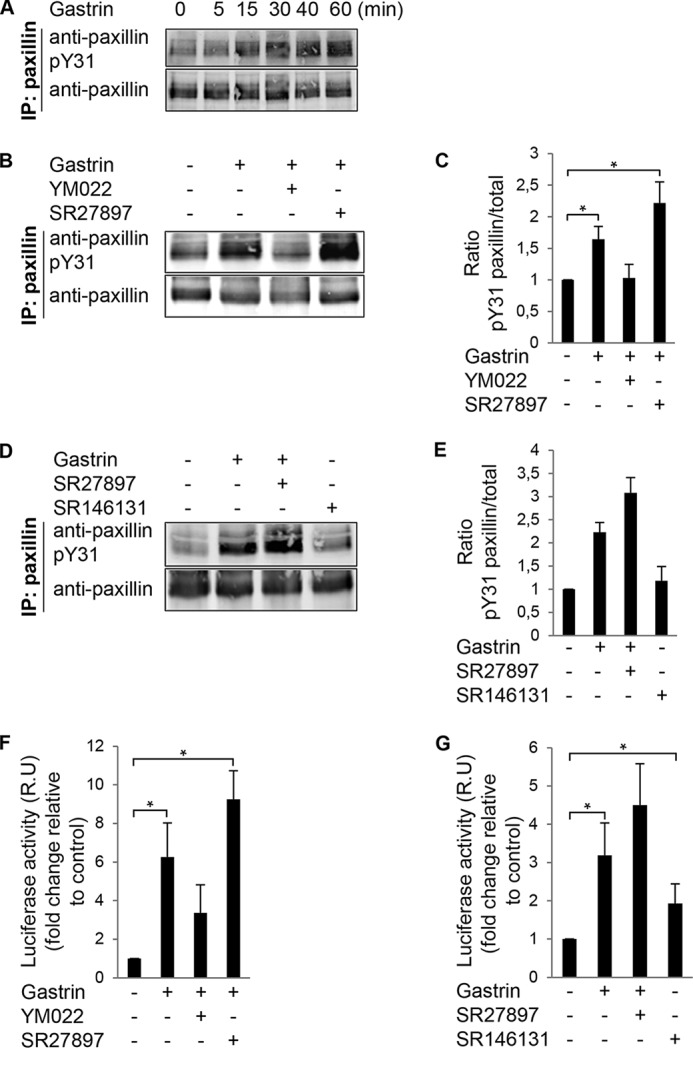
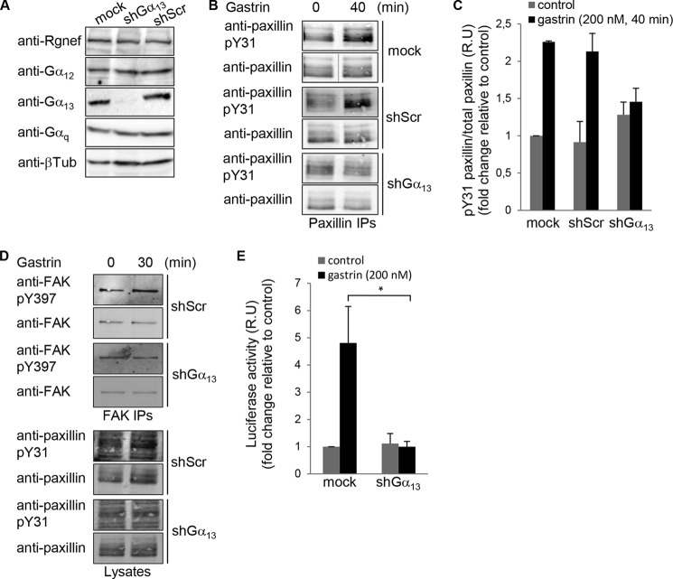
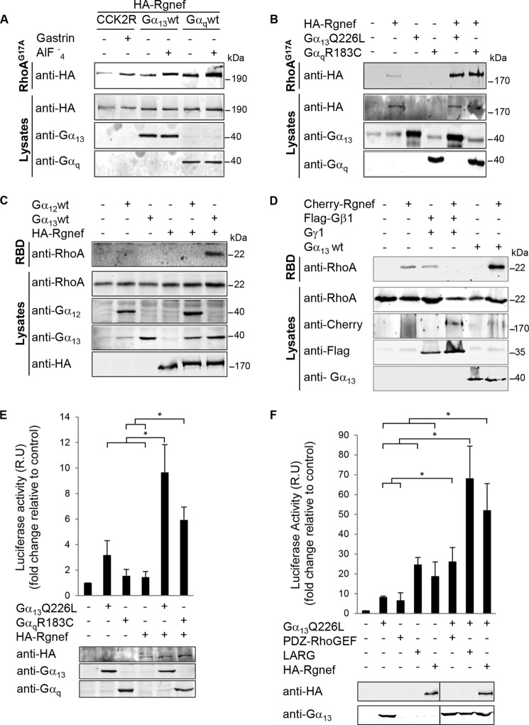
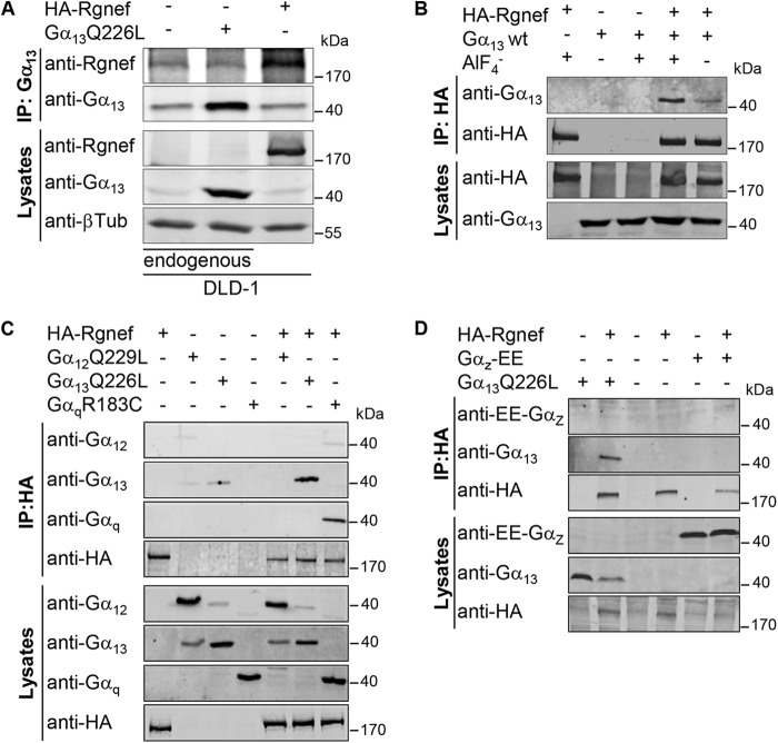
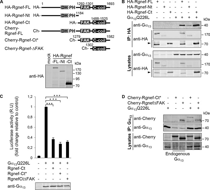
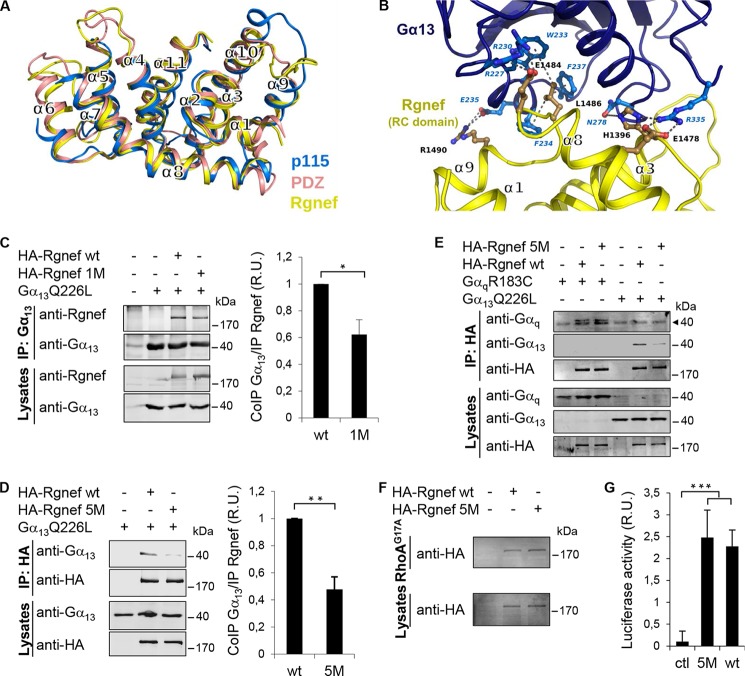
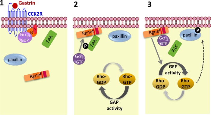
The guanine nucleotide exchange factor Rgnef (also known as ArhGEF28 or p190RhoGEF) promotes colon carcinoma cell motility and tumor progression via interaction with focal adhesion kinase (FAK). Mechanisms of Rgnef activation downstream of integrin or G protein-coupled receptors remain undefined. In the absence of a recognized G protein signaling homology domain in Rgnef, no proximal linkage to G proteins was known. Utilizing multiple methods, we have identified Rgnef as a new effector for Gα13 downstream of gastrin and the type 2 cholecystokinin receptor. In DLD-1 colon carcinoma cells depleted of Gα13, gastrin-induced FAK Tyr(P)-397 and paxillin Tyr(P)-31 phosphorylation were reduced. RhoA GTP binding and promoter activity were increased by Rgnef in combination with active Gα13. Rgnef co-immunoprecipitated with activated Gα13Q226L but not Gα12Q229L. The Rgnef C-terminal (CT, 1279-1582) region was sufficient for co-immunoprecipitation, and Rgnef-CT exogenous expression prevented Gα13-stimulated SRE activity. A domain at the C terminus of the protein close to the FAK binding domain is necessary to bind to Gα13. Point mutations of Rgnef-CT residues disrupt association with active Gα13 but not Gαq. These results show that Rgnef functions as an effector of Gα13 signaling and that this linkage may mediate FAK activation in DLD-1 colon carcinoma cells.

Keywords: G protein-coupled receptor (GPCR); Gastrin; cell signaling; guanine nucleotide exchange factor (GEF); heterotrimeric G protein; regulator of G protein signaling.


Figures

FIGURE 1.
FIGURE 1.
Gastrin stimulation of paxillin tyrosine phosphorylation and SRE activation in DLD-1 cells. A, gastrin (200 nm) was added to DLD-1 cells for the indicated times. Protein lysates were analyzed by paxillin IP and phosphospecific paxillin (pY31) or total paxillin immunoblotting. B, DLD-1 cells were pretreated with 2 μm SR27987 (CCK1R antagonist) or 2 μm YM022 (CCK2R antagonist) for 20 min. Gastrin (200 nm) was added, and lysates were prepared for paxillin IP after 40 min as in A. C, LI-COR image quantification of Tyr(P)-31 paxillin to total paxillin ratio from experiments in B. Values were set to 1 and are means ± S.E. fold increase of three experiments conducted in triplicate (*, p < 0.05, two-tailed t test). D, DLD-1 cells were pretreated with 2 μm SR27987 (CCK1R antagonist). Cells were pretreated with gastrin (200 nm) or SR146131 (CCK1R agonist) for 40 min before anti-paxillin IP as in A. E, image quantification of Tyr(P)-31 paxillin to total paxillin ratio from experiments in D. Values were set to 1. Data are the mean ± S.E. fold increase for three independent experiments. F, DLD-1 cells were transfected with pSRE.L and Renilla luciferase (RLuc) vectors. Cells were serum-starved and then pretreated with 2 μm SR27987 (CCK1R antagonist) or 2 μm YM022 (CCK2R antagonist) for 20 min. Gastrin (200 nm) or vehicle was added for 5 h, and lysates were prepared for luciferase detection. G, DLD-1 cells were transfected as above and were pretreated with 2 μm SR27987 (CCK1R antagonist). Gastrin (200 nm) or SR146131 (CCK1R agonist) was added as indicated for 5 h, and lysates were prepared for luciferase detection. F and G, values (mean ± S.E. of three independent experiments in triplicate) were normalized based on expression of RLuc and expressed (relative units, R.U.) as fold induction over serum-starved conditions (*, p < 0.05, two-tailed t test).
FIGURE 2.
FIGURE 2.
13 is necessary for gastrin-stimulated paxillin tyrosine phosphorylation and SRE activation. A, lysates of Mock, Gα13, or Scr shRNA-expressing DLD-1 cells were immunoblotted with antibodies to Rgnef, Gα12, Gα13, Gαq, and β-tubulin. B, representative paxillin IPs from lysates of Mock, Scr-, and Gα13 shRNA-expressing DLD-1 cells pretreated with gastrin (200 nm, 40 min). Phosphospecific paxillin Tyr(P)-31 (pY31) was followed by detection of total paxillin levels. C, image quantification from paxillin IPs shown in B. Control was set to 1, and values are means ± S.E. for fold induction of three independent experiments. D, representative FAK IPs from lysates of Scr- and Gα13 shRNA-expressing DLD-1 cells that stimulated with gastrin (200 nm, 30 min). Phosphospecific FAK-Tyr(P)-397 (pY397) was followed by detection of total FAK levels by immunoblotting. Phosphospecific paxillin Tyr(P)-31 was followed by detection of total paxillin levels by immunoblotting on the corresponding lysates. E, DLD-1 cells transduced with anti-Gα13 or Scr shRNA were transfected with pSRE.L and RLuc vectors. After 24 h, cells were serum-starved and then treated with vehicle or gastrin (200 nm) for 6 h, and lysates were prepared for luciferase detection. Values were set to 1, and data are means ± S.E. fold induction (relative units, R.U.) from three independent experiments (*, p < 0.05, two-tailed t test).
FIGURE 3.
FIGURE 3.
Gastrin, Gα13, and Gαq promote Rgnef and RhoA activation. A and B, affinity binding to a nucleotide-free mutant of RhoA (G17A) was used to evaluate Rgnef activation. HEK293 cells were transfected with expression vectors for HA-Rgnef, CCK2R, Gα13wt, or Gαqwt (A) or HA-Rgnef and Gα13Q226L or GαqR183C (B) as indicated. Cells were either left unstimulated or stimulated with 100 nm gastrin or 10 μm AlF4 for 40 min. Active Rgnef bound to GST-RhoAG17A beads was visualized by anti-HA immunoblotting (top blots), and protein expression in total cell lysates was evaluated by anti-HA, -Gα13, and -Gαq immunoblotting. Experiments were repeated three times with similar results. C and D, active RhoA was measured by the GST-Rhotekin RBD pulldown assay. HEK293 cells were transfected with expression vectors for HA-Rgnef, Gα13wt, or Gαqwt (C) or mCherry-Rgnef, FLAG-Gβ1, Gγ1, or Gα13wt (D) as indicated. Active RhoA in the pulldown was visualized by anti-RhoA immunoblotting (top blots), and protein expression in total cell lysates was evaluated by anti-RhoA, -mCherry, -FLAG, and -Gα13 immunoblotting. Experiments were repeated three times with similar results. E and F, SRE reporter transcriptional activity was measured in HEK293 cells transfected with expression vectors for HA-Rgnef, Gα13Q226L, and GαqR183C (E) or expression vectors for Gα13QL, PDZ-RhoGEF, LARG, or Rgnef (F) as indicated. Cells were serum-starved overnight, and luciferase activity was measured. Control empty vector-transfected cell values were set to 1. Data are means ± S.E. fold induction (relative units, R.U.) from three independent experiments (*, p < 0.05; two-tailed t test). Anti-HA (Rgnef), -Gα13, and -Gαq immunoblotting was used to verify transfected construct expression.
FIGURE 4.
FIGURE 4.
Rgnef association with Gα13 and Gαq proteins. A, DLD-1 cells were transfected with expression vectors for Gα13Q226L and HA-Rgnef as indicated. IP analyses with anti-Gα13 antibodies were used to detect complexes of endogenous and exogenous Gα13 with Rgnef. Protein expression in total cell lysates was evaluated by anti-HA (Rgnef), -Gα13, and β-tubulin immunoblotting. B, HEK293 cells transfected with Gα13 and HA-Rgnef in the presence or absence of 10 μm AlF4. IP analyses with anti-HA antibodies were used to detect complexes with Gα13 with anti-HA and -Gα13 immunoblotting. Protein expression levels were evaluated immunoblotting of total lysates. C and D, HEK293 cells were transfected with expression vectors encoding HA-Rgnef, Gα12Q229L, Gα13Q226L, or GαqR183C (C) or HA-Rgnef, Gα13Q226L, or Gαz-EE (D) as indicated. C, IP analyses with anti-HA antibodies were used to detect complexes with Gα12, Gα13, and Gαq by anti-HA and -Gα12, -Gα13, and -Gαq immunoblotting. D, IP analyses with anti-HA antibodies were used to detect complexes with Gα13 and Gαz by anti-HA, -Gα13, and -EE tag immunoblotting. Protein expression levels were evaluated by immunoblotting of total cell lysates. A–D, experiments were repeated at least three times with similar results.
FIGURE 5.
FIGURE 5.
Rgnef C-terminal domain association with activated Gα13Q226L. A, schematic of full-length and truncated Rgnef constructs as HA- or mCherry- fusion proteins. Lower panel, expression of HA-tagged full-length, Nt(1–1184), and Ct(1185–1693) Rgnef constructs in HEK293 cells by anti-HA immunoblotting. B, HEK293 cells were transfected with HA-tagged vectors encoding for Rgnef full-length, Rgnef-Nt(1–1184), Rgnef-Ct(1185–1693), and/or Gα13Q226L. Cell lysates were immunoprecipitated as described before. Data are representative of at least three independent experiments. Arrow points to Rgned-Ct. C, Rgnef-Ct domain constructs inhibit Gα13Q226L-mediated SRF activation. HEK293 cells were transfected with pSRE.L and pRL-TK, together with either empty vector, vector encoding Gα13QL, and/or Rgnef -Ct(1185–1693), Rgnef-Ct*(1279–1582), and Rgnef ΔFAK(1302–1582). After 24 h of transfection, cells were serum-starved overnight, and then SRF activities of cell lysates were measured using the Dual-Luciferase assay kit (Promega). Anti-Gα13 immunoblotting shows equal expression in cell lysates. Data are means ± S.E. of four independent experiments, each conducted in triplicate (*, p < 0.05; **, p < 0.005; two-tailed t test). D, HEK293 cells expressing Gα13 in presence or absence of mCherry-Rgnef-FL, mCherry-Rgnef-Ct*, and mCherry-RgnefΔFAK were immunoprecipitated using anti-Gα13 antibody and subjected to Western blot analysis. Cells lysates were analyzed in parallel. Full-length Rgnef, Rgnef-Ct, and ΔFAK were detected by anti-Rgnef, and Gα13 was detected using anti-Gα13 immunoblotting. Data are representative of four independent experiments.
FIGURE 6.
FIGURE 6.
Sequence alignment between the RH domains of RH-RhoGEF protein family and Rgnef. Amino acids are colored based on conservation according to the Clustal scheme. Gold boxes over the alignment indicate the localization of helices based on the crystal structures. Numbers indicate the start and end of the RH-like domain in the respective sequence as follows: p115(h):Q92888, Lsc(m):Q61210, Lsc(r)Q9Z1I6, PDZ(h):O15085, PDZ(r):Q9ES67, LARG(h):Q9NZN5, LARG(m):Q8R4H2, Rgnef(h): Q8N1W1, Rgnef(m):P97433, Rgnef(r):P0C6P5. Five conserved Rgnef human (h) residues subjected to alanine mutagenesis are boxed. m, mouse.
FIGURE 7.
FIGURE 7.
Models of putative Rgnef RH-like domain and the complex with Gα13. A, structural superposition of the RH domains of human PDZ-RhoGEF (Protein Data Bank code 3CX8; salmon), p115 RhoGEF (Protein Data Bank code 3AB3; blue) and our computer model of the domain of Rgnef (yellow). B, computer model of the domain of Rgnef (yellow) in complex with Gα13 (blue). Putative residues of charged or hydrophobic side chain interactions are shown in blue. Residues mutated in the Rgnef-5 M are shown in black (corresponding to human His-1396, Glu-1478, Glu-1484, Leu-1486, and Arg-1490). C and D, HEK293 cells were transfected with vectors encoding for HA-Rgnef (full length), HA-Rgnef 1M (C), or HA-Rgnef 5M (D) with or without Gα13Q226L. HA-tagged Rgnef constructs were co-immunoprecipitated with antibodies to Gα13 and detected by Li-COR immunoblotting. Graphical results show immunoblotting band intensities from three independent experiments expressed as fold induction with respect to immunoprecipitated Rgnef wild type and normalized by the total expression level of Rgnef. Values are means ± S.D. (*, p < 0.05; **, p < 0.005; two-tailed t test). E, HEK293 cells expressing HA-Rgnef or HA-Rgnef5M with or without GαqR183C or Gα13Q226L were co-immunoprecipitated with HA tag antibodies and detected by Li-COR immunoblotting. The arrowhead points to an unspecific band detected with the anti-Gq monoclonal antibody. F, HEK293 cells expressing HA-Rgnef or HA-Rgnef 5M were incubated with RhoAG17A-GST beads and visualized by anti-HA tag immunoblotting. G, HEK293 cells were transfected with pSRE.L and pRL-TK, together with either empty vector (Ctl), vector encoding Rgnef or Rgnef5M and SRF activities were measured. Data are means ± S.E. of three independent experiments, each conducted in duplicate (***, p < 0.001).
FIGURE 8.
FIGURE 8.
Model of Gα13-dependent activation of Rgnef downstream CCK2R. 1, gastrin stimulation of CCK2 will lead to the activation of Gα13 (GTP-bound form) and dissociation from Gγβ. 2,13 protein activation will help to recruit Rgnef to the plasma membrane where it can bind and facilitate FAK-Tyr-397 phosphorylation. 3, at later points, Gα13 can facilitate activation of the Rgnef-GEF activity leading to the activation of RhoA and signaling cascade that enhances paxillin tyrosine phosphorylation and SRE promoter activity.

Similar articles

Cited by

References

    1. Jaffe A. B., Hall A. (2005) Rho GTPases: biochemistry and biology. Annu. Rev. Cell Dev. Biol. 21, 247–269 - PubMed
    1. Sahai E., Marshall C. J. (2002) RHO-GTPases and cancer. Nat. Rev. Cancer 2, 133–142 - PubMed
    1. Leve F., Morgado-Díaz J. A. (2012) Rho GTPase signaling in the development of colorectal cancer. J. Cell. Biochem. 113, 2549–2559 - PubMed
    1. van Horck F. P., Ahmadian M. R., Haeusler L. C., Moolenaar W. H., Kranenburg O. (2001) Characterization of p190RhoGEF, a RhoA-specific guanine nucleotide exchange factor that interacts with microtubules. J. Biol. Chem. 276, 4948–4956 - PubMed
    1. Lim Y., Lim S.-T., Tomar A., Gardel M., Bernard-Trifilo J. A., Chen X. L., Uryu S. A., Canete-Soler R., Zhai J., Lin H., Schlaepfer W. W., Nalbant P., Bokoch G., Ilic D., Waterman-Storer C., Schlaepfer D. D. (2008) PyK2 and FAK connections to p190Rho guanine nucleotide exchange factor regulate RhoA activity, focal adhesion formation, and cell motility. J. Cell Biol. 180, 187–203 - PMC - PubMed

Publication types

MeSH terms

Substances

LinkOut - more resources