. 2015 May 5;11(5):704-14.
doi: 10.1016/j.celrep.2015.03.058. Epub 2015 Apr 23.

Neddylation promotes ubiquitylation and release of Ku from DNA-damage sites

Affiliations

Neddylation promotes ubiquitylation and release of Ku from DNA-damage sites

Jessica S Brown et al. Cell Rep. .

Abstract

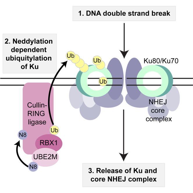
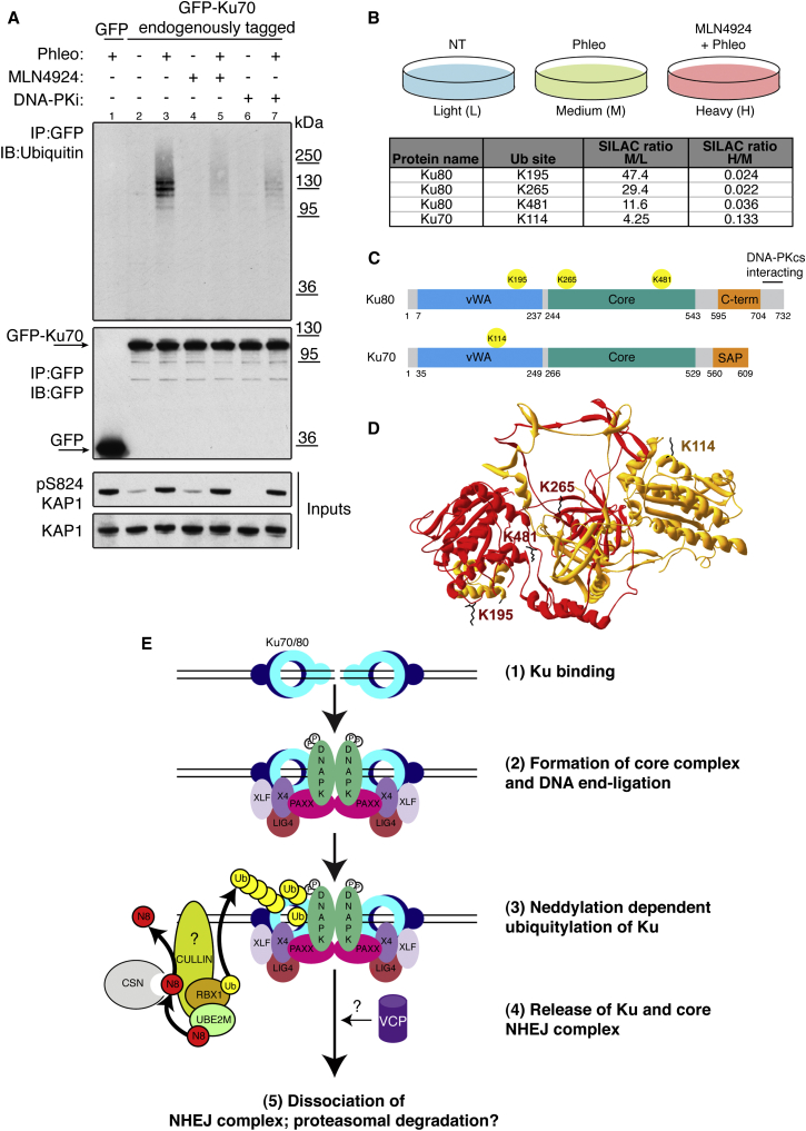
The activities of many DNA-repair proteins are controlled through reversible covalent modification by ubiquitin and ubiquitin-like molecules. Nonhomologous end-joining (NHEJ) is the predominant DNA double-strand break (DSB) repair pathway in mammalian cells and is initiated by DSB ends being recognized by the Ku70/Ku80 (Ku) heterodimer. By using MLN4924, an anti-cancer drug in clinical trials that specifically inhibits conjugation of the ubiquitin-like protein, NEDD8, to target proteins, we demonstrate that NEDD8 accumulation at DNA-damage sites is a highly dynamic process. In addition, we show that depleting cells of the NEDD8 E2-conjugating enzyme, UBE2M, yields ionizing radiation hypersensitivity and reduced cell survival following NHEJ. Finally, we demonstrate that neddylation promotes Ku ubiquitylation after DNA damage and release of Ku and Ku-associated proteins from damage sites following repair. These studies provide insights into how the NHEJ core complex dissociates from repair sites and highlight its importance for cell survival following DSB induction.


Figures

None
Graphical abstract
Figure 1
Figure 1
NEDD8 and the Neddylation Machinery Accumulate at Sites of DNA Breaks and Promote Cell Survival after NHEJ (A) Representation of major neddylation pathway components. NEDD8 (N8) is conjugated in an ATP-dependent cascade involving an E1 (NAE1-UBA3), E2 (UBE2M or F), and E3 (RBX1 or 2) to Cullin substrates (Sub). Neddylation is reversed by the CSN complex. MLN4924 inhibits UBA3. Figure adapted from Brown and Jackson (2015). (B) MLN4924 blocks NEDD8, but not ubiquitin recruitment to DNA-damage sites. U2OS-GFP-NEDD8 cells were pre-treated for 1 hr with DMSO or 3 μM MLN4924 and laser microirradiated. Cells were fixed after 20 min and visualized by immunofluorescence as indicated. Graph shows average intensity of GFP-NEDD8 at the laser line from three experiments ±SD. White bar represents 10 μM. Asterisks indicate statistically significant difference to control (p < 0.05; ∗∗p < 0.01; ∗∗∗p < 0.001; ∗∗∗∗p ≤ 0.0001). (C) GFP-UBE2F and GFP-UBE2M are recruited to DNA-damage sites. U2OS cells stably expressing GFP-UBE2F or GFP-UBE2M were laser microirradiated, fixed, and visualized as in (B). Graph shows average percentage of γH2AX positive cells with detectable GFP-UBE2M or GFP-UBE2F recruitment from five independent experiments ±SD. White bar represents 10 μM. (D) GFP-CSN5 recruitment to DNA-damage sites is blocked by MLN4924. U2OS cells stably expressing GFP-CSN5 were treated as in (B). Images were acquired by live cell imaging. Laser tracks are indicated by dashed white lines. White bar represents 10 μM (E) UBE2M depletion causes hypersensitivity to IR. Clonogenic U2OS cell survivals were performed after transfection with indicated siRNAs and doses of IR. Each point represents an average of at least three independent experiments (except UBE2M-2 which was repeated twice). Error bars correspond to SDs, and asterisks are as in (B). (F) UBE2M depletion causes an NHEJ defect. Random plasmid integration assay was performed in U2OS cells transfected with indicated siRNAs. Error bars correspond to SD of at least three independent experiments (asterisks as in B). See also Figure S1.
Figure 2
Figure 2
MLN4924 Inhibits Ku Removal from DNA Repair Sites (A and B) MLN4924 causes Ku80 foci persistence after IR. U2OS cells were pre-treated with DMSO or 3 μM MLN4924 for 1 hr and then subjected to 10 Gy IR. Samples were pre-extracted with CSK+RNase A and visualized by immunofluorescence. (A) shows representative images, and (B) shows quantification. Dotted lines indicate nuclear peripheries. Error bars correspond to SDs of at least three independent experiments (asterisks as in Figure 1B). White bar represents 10 μM. (C) U2OS-A171T UBA3 are resistant to MLN4924. U2OS cells stably expressing WT UBA3 or A171T UBA3 were treated with DMSO or 3 μM MLN4924 for 1 hr and analyzed by immunoblotting with indicated antibodies. Endogenous UBA3 was depleted with a siRNA to the 3′ UTR (Figure S2B). Neddylated conjugates are detected with a NEDD8-specific antibody. (D) MLN4924 effects on Ku80 foci retention are through UBA3 inhibition. U2OS cells stably expressing WT UBA3 or A171T UBA3 were processed as in (A), and results were quantified as in (B). (E) MLN4924 does not affect γH2AX recovery after IR. Quantification of total γH2AX intensity per nucleus in cells treated with 10 Gy IR then harvested at indicated times. Samples were prepared as in (A). Pre-treatment for 1 hr with 3 μM DNA-PK inhibitor (DNA-PKi) used as a positive control. Statistical analysis as in (B). AU, arbitrary units. See also Figure S2.
Figure 3
Figure 3
Proteomics Identifies Neddylation-Dependent Ku Interactors (A) MLN4924 causes retention of NHEJ factors on the chromatin. U2OS cells were pretreated with DMSO or 3 μM MLN4924 for 1 hr and then treated with 500 μM phleomycin (Phleo) for 1 hr. Cells were left to recover in the presence of MLN4924 or DMSO following phleomycin removal and then collected at the indicated times. Cells were pre-extracted with CSK buffer + RNase A prior to lysis (chromatin; left) or lysed as whole cell extracts (right) and immunoblotted with indicated antibodies. Black arrow indicates XRCC4. (B) RPE-1 cells stably expressing GFP or Ku70 endogenously tagged with GFP were labeled with light, medium, or heavy isotopes and treated as indicated. Cell lysates were subjected to GFP retrieval. Enriched proteins were resolved by SDS-PAGE and proteolysed in gel with trypsin, and peptides were analyzed by LC-MS/MS. The scatterplot shows the logarithmized SILAC ratio of GFP-KU70/GFP control and GFP-KU70 + MLN4924/GFP-KU70. The known Ku interactors and CUL4A (ratio 2.23) are labeled in black font and open blue circles. In pink are interactions enhanced upon MLN4924. In green are interactions decreased upon MLN4924 (see also Table S1). (C) Experiment repeated as in (B) without isotope labeling of cells. Following GFP IP, cell lysates were immunoblotted with indicated antibodies. Note that CUL1 (Postow and Funabiki, 2013) and CUL4B were not detected in Ku immunoprecipitates. See also Figure S3.
Figure 4
Figure 4
MLN4924 Blocks Ku Ubiquitylation after DNA Damage (A) In vivo ubiquitylation assay. RPE-1 cells expressing Ku70 endogenously tagged with GFP (lanes 2–7) or RPE-1 cells stably expressing GFP (lane 1) were pre-treated with DMSO, 3 μM MLN4924, or 3 μM DNA-PK inhibitor for 1 hr prior to treatment with 500 μM phleomycin (Phleo) for 1 hr as indicated. Cell lysates were immunoprecipitated (IP) with GFP-specific antibody and immunoblotted (IB) with indicated antibodies. GFP-Ku70 IP was done under stringent conditions (see Experimental Procedures). Black arrows indicate GFP-Ku70 and GFP. Phosphorylated Ser824 of KAP1 is used as a DNA-damage marker. (B) Identification of Ku ubiquitylation sites by quantitative LC-MS/MS. RPE-1 cells stably expressing Ku70 endogenously tagged with GFP were labeled with light, medium, or heavy SILAC isotopes and treated as indicated. Ku70 was enriched with GFP-Trap agarose under stringent washing conditions. Enriched proteins were resolved by SDS-PAGE and proteolysed in gel with trypsin. Peptides were extracted from gel and analyzed by LC-MS/MS. SILAC ratio M/L >2 represents induction upon DNA damage; SILAC ratio H/M < 0.5 represents inhibition by MLN4924 (see also Table S2). (C) Schematic representation of Ku70 and Ku80 domains with neddylation-dependent ubiquitylation sites identified in (B). vWA, von Willebrand factor A; C-term, C terminus; SAP, SAF-A/B, Acinus, and PIAS. (D) Positions of DNA-damage-induced neddylation-dependent ubiquitylation sites in the context of the structure of the Ku heterodimer (ID:1JEQ). Ku70 and Ku80 are in orange and red, respectively, and the ubiquitylated side chains are in black. (E) Model. (1, 2) Ku and the NHEJ complex are recruited to sites of DSBs. (3) Neddylation-dependent ubiquitylation of Ku following completion of DNA repair. (4) Ku and NHEJ factors are released from sites of DNA damage. VCP might target ubiquitylated Ku to proteasome for degradation. N8, NEDD8; Ub, ubiquitin; P, phosphorylation (see text for details). See also Figure S4.

Similar articles

Cited by

References

    1. Britton S., Coates J., Jackson S.P. A new method for high-resolution imaging of Ku foci to decipher mechanisms of DNA double-strand break repair. J. Cell Biol. 2013;202:579–595. - PMC - PubMed
    1. Brown J.S., Jackson S.P. Ubiquitylation, neddylation and the DNA damage response. Open Biol. 2015;5 - PMC - PubMed
    1. Brownell J.E., Sintchak M.D., Gavin J.M., Liao H., Bruzzese F.J., Bump N.J., Soucy T.A., Milhollen M.A., Yang X., Burkhardt A.L. Substrate-assisted inhibition of ubiquitin-like protein-activating enzymes: the NEDD8 E1 inhibitor MLN4924 forms a NEDD8-AMP mimetic in situ. Mol. Cell. 2010;37:102–111. - PubMed
    1. Ciccia A., Elledge S.J. The DNA damage response: making it safe to play with knives. Mol. Cell. 2010;40:179–204. - PMC - PubMed
    1. Cope G.A., Suh G.S., Aravind L., Schwarz S.E., Zipursky S.L., Koonin E.V., Deshaies R.J. Role of predicted metalloprotease motif of Jab1/Csn5 in cleavage of Nedd8 from Cul1. Science. 2002;298:608–611. - PubMed

Publication types

MeSH terms