Targeting the PD-1/PD-L1 Immune Evasion Axis With DNA Aptamers as a Novel Therapeutic Strategy for the Treatment of Disseminated Cancers
- PMID: 25919090
- PMCID: PMC4417124
- DOI: 10.1038/mtna.2015.11
Targeting the PD-1/PD-L1 Immune Evasion Axis With DNA Aptamers as a Novel Therapeutic Strategy for the Treatment of Disseminated Cancers
Abstract
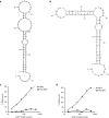
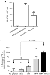
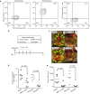
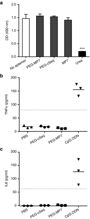
Blocking the immunoinhibitory PD-1:PD-L1 pathway using monoclonal antibodies has led to dramatic clinical responses by reversing tumor immune evasion and provoking robust and durable antitumor responses. Anti-PD-1 antibodies have now been approved for the treatment of melanoma, and are being clinically tested in a number of other tumor types as both a monotherapy and as part of combination regimens. Here, we report the development of DNA aptamers as synthetic, nonimmunogenic antibody mimics, which bind specifically to the murine extracellular domain of PD-1 and block the PD-1:PD-L1 interaction. One such aptamer, MP7, functionally inhibits the PD-L1-mediated suppression of IL-2 secretion in primary T-cells. A PEGylated form of MP7 retains the ability to block the PD-1:PD-L1 interaction, and significantly suppresses the growth of PD-L1+ colon carcinoma cells in vivo with a potency equivalent to an antagonistic anti-PD-1 antibody. Importantly, the anti-PD-1 DNA aptamer treatment was not associated with off-target TLR-9-related immune responses. Due to the inherent advantages of aptamers including their lack of immunogenicity, low cost, long shelf life, and ease of synthesis, PD-1 antagonistic aptamers may represent an attractive alternative over antibody-based anti PD-1 therapeutics.
Figures


Similar articles
-
Anti-PD-L1 DNA aptamer antagonizes the interaction of PD-1/PD-L1 with antitumor effect.J Mater Chem B. 2021 Jan 28;9(3):746-756. doi: 10.1039/d0tb01668c. J Mater Chem B. 2021. PMID: 33319876
-
A Novel PD-L1-targeting Antagonistic DNA Aptamer With Antitumor Effects.Mol Ther Nucleic Acids. 2016 Dec 13;5(12):e397. doi: 10.1038/mtna.2016.102. Mol Ther Nucleic Acids. 2016. PMID: 27959341
-
Development and Evaluation of Novel Aptamers Specific for Human PD1 Using Hybrid Systematic Evolution of Ligands by Exponential Enrichment Approach.Immunol Invest. 2020 Jul;49(5):535-554. doi: 10.1080/08820139.2020.1744639. Epub 2020 May 19. Immunol Invest. 2020. PMID: 32429721
-
The Next Immune-Checkpoint Inhibitors: PD-1/PD-L1 Blockade in Melanoma.Clin Ther. 2015 Apr 1;37(4):764-82. doi: 10.1016/j.clinthera.2015.02.018. Epub 2015 Mar 29. Clin Ther. 2015. PMID: 25823918 Free PMC article. Review.
-
PD-1/PD-L1 Blockade Therapy in Advanced Non-Small-Cell Lung Cancer: Current Status and Future Directions.Oncologist. 2019 Feb;24(Suppl 1):S31-S41. doi: 10.1634/theoncologist.2019-IO-S1-s05. Oncologist. 2019. PMID: 30819829 Free PMC article. Review.
Cited by
-
The Potential of Aptamer-Mediated Liquid Biopsy for Early Detection of Cancer.Int J Mol Sci. 2021 May 25;22(11):5601. doi: 10.3390/ijms22115601. Int J Mol Sci. 2021. PMID: 34070509 Free PMC article. Review.
-
Aptamers Chemistry: Chemical Modifications and Conjugation Strategies.Molecules. 2019 Dec 18;25(1):3. doi: 10.3390/molecules25010003. Molecules. 2019. PMID: 31861277 Free PMC article. Review.
-
Development of a DNA aptamer targeting IDO1 with anti-tumor effects.iScience. 2023 Jul 13;26(8):107367. doi: 10.1016/j.isci.2023.107367. eCollection 2023 Aug 18. iScience. 2023. PMID: 37520707 Free PMC article.
-
Applications of Cancer Cell-Specific Aptamers in Targeted Delivery of Anticancer Therapeutic Agents.Molecules. 2018 Apr 4;23(4):830. doi: 10.3390/molecules23040830. Molecules. 2018. PMID: 29617327 Free PMC article. Review.
-
Advances in Oligonucleotide Aptamers for NSCLC Targeting.Int J Mol Sci. 2020 Aug 23;21(17):6075. doi: 10.3390/ijms21176075. Int J Mol Sci. 2020. PMID: 32842557 Free PMC article. Review.
References
LinkOut - more resources
Full Text Sources
Other Literature Sources
Research Materials

