

Transcriptional activation by the thyroid hormone receptor through ligand-dependent receptor recruitment and chromatin remodelling
- PMID: 25916672
- PMCID: PMC6309829
- DOI: 10.1038/ncomms8048
Transcriptional activation by the thyroid hormone receptor through ligand-dependent receptor recruitment and chromatin remodelling
Abstract
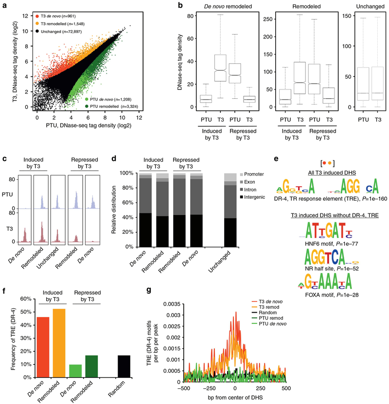
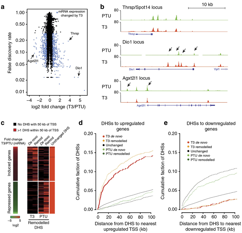
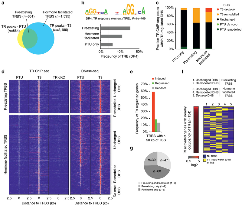
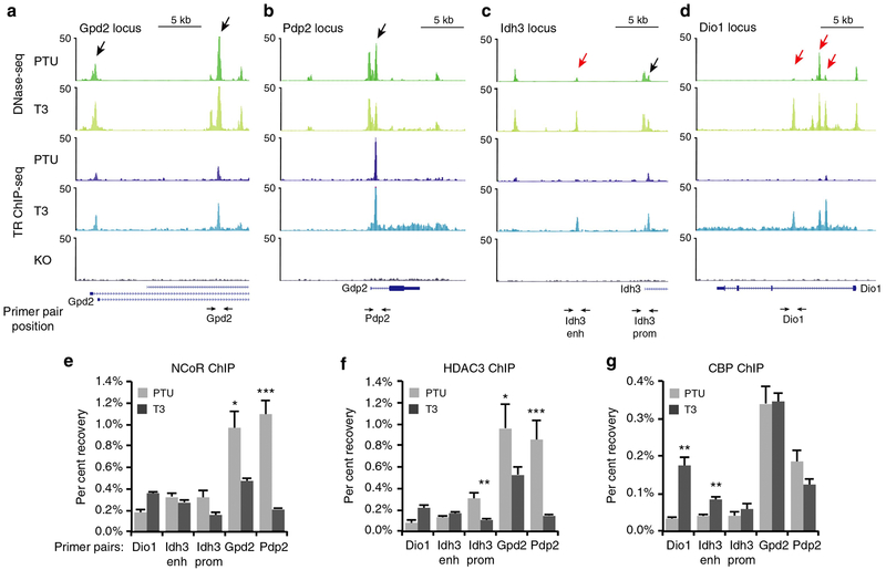
A bimodal switch model is widely used to describe transcriptional regulation by the thyroid hormone receptor (TR). In this model, the unliganded TR forms stable, chromatin-bound complexes with transcriptional co-repressors to repress transcription. Binding of hormone dissociates co-repressors and facilitates recruitment of co-activators to activate transcription. Here we show that in addition to hormone-independent TR occupancy, ChIP-seq against endogenous TR in mouse liver tissue demonstrates considerable hormone-induced TR recruitment to chromatin associated with chromatin remodelling and activated gene transcription. Genome-wide footprinting analysis using DNase-seq provides little evidence for TR footprints both in the absence and presence of hormone, suggesting that unliganded TR engagement with repressive complexes on chromatin is, similar to activating receptor complexes, a highly dynamic process. This dynamic and ligand-dependent interaction with chromatin is likely shared by all steroid hormone receptors regardless of their capacity to repress transcription in the absence of ligand.
Conflict of interest statement
Figures
Similar articles
-
Determinants of chromatin disruption and transcriptional regulation instigated by the thyroid hormone receptor: hormone-regulated chromatin disruption is not sufficient for transcriptional activation.EMBO J. 1997 Jun 2;16(11):3158-71. doi: 10.1093/emboj/16.11.3158. EMBO J. 1997. PMID: 9214633 Free PMC article.
-
Role of co-activators and co-repressors in the mechanism of steroid/thyroid receptor action.Recent Prog Horm Res. 1997;52:141-64; discussion 164-5. Recent Prog Horm Res. 1997. PMID: 9238851 Review.
-
Transcriptional activation by thyroid hormone receptor-beta involves chromatin remodeling, histone acetylation, and synergistic stimulation by p300 and steroid receptor coactivators.Mol Endocrinol. 2003 May;17(5):908-22. doi: 10.1210/me.2002-0308. Epub 2003 Feb 13. Mol Endocrinol. 2003. PMID: 12586842
-
Role of co-regulators in metabolic and transcriptional actions of thyroid hormone.J Mol Endocrinol. 2016 Apr;56(3):73-97. doi: 10.1530/JME-15-0246. Epub 2015 Dec 16. J Mol Endocrinol. 2016. PMID: 26673411 Review.
-
p300 stimulates transcription instigated by ligand-bound thyroid hormone receptor at a step subsequent to chromatin disruption.EMBO J. 1999 Oct 15;18(20):5634-52. doi: 10.1093/emboj/18.20.5634. EMBO J. 1999. PMID: 10523307 Free PMC article.
Cited by
-
Mislocalization of Cancer-associated Thyroid Hormone Receptor Mutants.Nucl Receptor Res. 2020;2020:https://web.archive.org/web/20210227193123/https://www.kenzpub.com/journals/nurr/inpress/2020/101453/. Nucl Receptor Res. 2020. PMID: 35280700 Free PMC article.
-
Ligand dependent gene regulation by transient ERα clustered enhancers.PLoS Genet. 2020 Jan 6;16(1):e1008516. doi: 10.1371/journal.pgen.1008516. eCollection 2020 Jan. PLoS Genet. 2020. PMID: 31905229 Free PMC article.
-
Thyroid hormone dependent transcriptional programming by TRβ requires SWI/SNF chromatin remodelers.Nucleic Acids Res. 2022 Feb 22;50(3):1382-1395. doi: 10.1093/nar/gkab1287. Nucleic Acids Res. 2022. PMID: 35037038 Free PMC article.
-
Multiple mechanisms regulate H3 acetylation of enhancers in response to thyroid hormone.PLoS Genet. 2020 May 26;16(5):e1008770. doi: 10.1371/journal.pgen.1008770. eCollection 2020 May. PLoS Genet. 2020. PMID: 32453730 Free PMC article.
-
Thyroid Hormone-Regulated Expression of Period2 Promotes Liver Urate Production.Front Cell Dev Biol. 2021 Apr 1;9:636802. doi: 10.3389/fcell.2021.636802. eCollection 2021. Front Cell Dev Biol. 2021. PMID: 33869182 Free PMC article.
References
-
- Baumann CT, Maruvada P, Hager GL & Yen PM Nuclear cytoplasmic shuttling by thyroid hormone receptors. multiple protein interactions are required for nuclear retention. J. Biol. Chem 276, 11237–11245 (2001). - PubMed
-
- Wong J, Shi YB & Wolffe AP A role for nucleosome assembly in both silencing and activation of the Xenopus TR beta A gene by the thyroid hormone receptor. Genes Dev 9, 2696–2711 (1995). - PubMed
Publication types
MeSH terms
Substances
Associated data
- Actions
- SRA/SRP055020
Grants and funding
LinkOut - more resources
Full Text Sources
Other Literature Sources
Molecular Biology Databases
