. 2015 May 6;86(3):726-39.
doi: 10.1016/j.neuron.2015.03.049. Epub 2015 Apr 23.

A Cdh1-APC/FMRP Ubiquitin Signaling Link Drives mGluR-Dependent Synaptic Plasticity in the Mammalian Brain

Affiliations

A Cdh1-APC/FMRP Ubiquitin Signaling Link Drives mGluR-Dependent Synaptic Plasticity in the Mammalian Brain

Ju Huang et al. Neuron. .

Abstract

Deregulation of synaptic plasticity may contribute to the pathogenesis of developmental cognitive disorders. In particular, exaggerated mGluR-dependent LTD is featured in fragile X syndrome, but the mechanisms that regulate mGluR-LTD remain incompletely understood. We report that conditional knockout of Cdh1, the key regulatory subunit of the ubiquitin ligase Cdh1-anaphase-promoting complex (Cdh1-APC), profoundly impairs mGluR-LTD in the hippocampus. Mechanistically, we find that Cdh1-APC operates in the cytoplasm to drive mGluR-LTD. We also identify the fragile X syndrome protein FMRP as a substrate of Cdh1-APC. Endogenous Cdh1-APC forms a complex with endogenous FMRP, and knockout of Cdh1 impairs mGluR-induced ubiquitination and degradation of FMRP in the hippocampus. Knockout of FMRP suppresses, and expression of an FMRP mutant protein that fails to interact with Cdh1 phenocopies, the Cdh1 knockout phenotype of impaired mGluR-LTD. These findings define Cdh1-APC and FMRP as components of a novel ubiquitin signaling pathway that regulates mGluR-LTD in the brain.


Figures

Figure 1
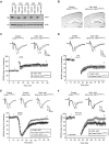
Figure 1. Hippocampal mGluR-LTD is impaired in conditional Cdh1 knockout mice
(A) Lysates of hippocampus from control or conditional Cdh1 knockout mice were immunoblotted with the Cdh1 and Actin antibodies, the latter to serve as loading control. (B) Coronal brain sections from P22 control or conditional Cdh1 knockout mice were subjected to Nissl staining. Scale bars represent 500 μm. (C) One train of high frequency stimulation (100 pulses at 100 Hz, 1 s) induced hippocampal early-LTP in both control and conditional Cdh1 knockout mice (P=0.94; control: 160.4 ± 9.3% of baseline, n=8 slices from 4 animals; Cdh1 cKO: 161.3 ± 9.6% of baseline, n=8 slices from 4 animals). In this and all subsequent electrophysiology figures, evoked synaptic responses over the course of the experiment were normalized to the average of baseline responses. The percent LTP or LTD was quantified from the average of the responses 50–60 minutes after induction. Representative recording traces from control or conditional Cdh1 knockout mice were taken at times indicated by the numbers 1 and 2. Trace 1 represents baseline responses and trace 2 represents responses one hour after LTP or LTD induction. (D) Low frequency stimulation (900 pulses at 1 Hz, 15 min) induced hippocampal NMDAR-LTD in both control and conditional Cdh1 knockout mice (P=0.98; control: 65.4 ± 8.0% of baseline, n=8 slices from 5 animals; Cdh1 cKO: 65.6 ± 6.9% of baseline, n=10 slices from 6 animals). (E) DHPG (50 μM, 10 min) induced significantly reduced hippocampal mGluR-LTD in conditional Cdh1 knockout mice as compared to the Cdh1loxP/loxP control mice (P<0.01) or Emx-Cre control mice (P<0.001) (Cdh1 cKO: 92.0 ± 2.4% of baseline, n= 13 slices from 7 animals; Cdh1loxP/loxP_ control: 74.5 ± 4.5% of baseline, n=8 slices from 5 animals; Emx-Cre_control: 71.6 ± 3.5% of baseline, n=10 slices from 5 animals). (F) Paired-pulse low frequency stimulation (900 pairs at 1 Hz with 50 ms paired-pulse interval, 15 min), in the presence of the NMDAR antagonist DL-AP5 (100 μM), induced significantly reduced hippocampal mGluR-LTD in conditional Cdh1 knockout mice as compared to control mice (P<0.01; Cdh1 cKO: 99.5 ± 10.0% of baseline, n=9 slices from 5 animals; Control: 65.2 ± 9.2% of baseline, n=10 slices from 6 animals). Supplemental Figure 1 and Supplemental Figure 2 are related to Figure 1. See also Figure S1 and S2.
Figure 2
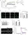
Figure 2. Cdh1-APC drives mGluR-LTD
(A) E15 mouse embryos were electroporated with the Emi1 expression plasmid or its control vector together with an mCherry expression plasmid followed by whole-cell patch clamp analyses in transfected mCherry-positive CA1 hippocampal neurons in three-week old mice. DHPG-induced mGluR-LTD was significantly reduced in CA1 neurons upon expression of Emi1 compared to control vector-transfected neurons (P<0.01; pCAG-Emi1: 96.2±8.2% of baseline, n=14 from 6 animals; pCAG vector: 57.9±8.7% of baseline, n=11 from 6 animals). (B) Immunofluorescence of surface AMPAR subunit, GluR2, in primary hippocampal neurons 14 days after plating from P0 control or conditional Cdh1 knockout mice. Neurons were treated with or without DHPG (50 μM, 10 min). 20 min after DHPG washout, neurons were fixed and subjected to immunofluorescence analyses under nonpermeabilizing condition with the antibody recognizing the ectodomain of GluR2. DHPG induced internalization of surface GluR2 in control but not conditional Cdh1 knockout hippocampal neurons. Scale bars represent 20 μm in the top panel and 5 μm in the bottom panel. (C) Quantification of surface GluR2 after DHPG treatment reveals significantly reduced surface GluR2 in control but not in conditional Cdh1 knockout hippocampal neurons (P< 0.05; Control: 55.2±14.2% of baseline, n=54 neurons from 4 animals; Cdh1 cKO: 94.6±5.7% of baseline; n=48 neurons from 4 animals). (D) Lysates of acute hippocampal slices from control or conditional Cdh1 knockout mice were treated with or without DHPG (50 μM for 10 min) and immunobloted with the ERK1/2 and phospho-ERK1/2 (Thr202/Tyr204) antibodies. (E) DHPG induction of phospho-ERK1/2 immunoreactivity is indistinguishable between control and conditional Cdh1 knockout mice (P=0.56; Control: 159.5±13.4% of baseline, n=3 animals; Cdh1 cKO: 150.4±5.3% of baseline, n=3 animals). (F) Calcium imaging on primary hippocampal neurons 14 days after plating from P0 control or conditional Cdh1 knockout mice. Neurons were loaded with Oregon Green 488 BAPTA-1AM (OGB-1) to measure the intracellular calcium. DHPG (50 μM) led to increased levels of intracellular calcium in both control and conditional Cdh1 knockout hippocampal neurons. (G) Quantification of the relative fluorescence ΔF/F from calcium imaging analyses as in (F). DHPG-induced increase in intracellular calcium was indistinguishable between control and conditional Cdh1 knockout hippocampal neurons (P=0.64; Control: 161.67±12.8% of baseline, n=36 neurons from 3 animals; Cdh1 cKO: 153.8±10.5% of baseline, n=33 neurons from 3 animals). Supplemental Figure 3 is related to Figure 2. See also Figure S3.
Figure 3
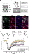
Figure 3. Cdh1-APC operates in the cytoplasm rather than the nucleus to regulate mGluR-LTD
(A) Cytosolic (Cyto) and nuclear (Nuc) fractionations were prepared from hippocampus of P15 wild type mice and immunoblotted with the Cdh1, Cdc27, α-tubulin and PARP antibodies. PARP and α-tubulin served as the nuclear and cytoplasmic marker, respectively. Equal volume of cytosolic and nuclear fractionation was loaded in western blot. (B) Schematic diagram of in utero electroporation to hippocampus at E15 embryos and whole-cell patch clamp recording of mCherry-positive CA1 neurons in three-week old conditional Cdh1 knockout mice. (C) E15 mouse embryos electroporated with a plasmid expressing GFP-NES-Cdh1 (a–d) or GFP-NLS-Cdh1 (e–h) together with a mCherry expressing plasmid were allowed to develop until P20. Brain sections were subjected to immunofluorescence analyses with the GFP and DsRed antibodies and the DNA dye bisbenzimide (Hoechst 33258). GFP-NES-Cdh1 and GFP-NLS-Cdh1 appeared to be predominantly in the cytoplasm and nucleus, respectively. Areas inside the white box (a–h) are enlarged in a’–h’. Scale bars represent 100 μm. Notably, although GFP-NLS-Cdh1 displayed modest expression in the soma in addition to robust expression in the nucleus, GFP-NES-Cdh1 was restricted to the cytoplasm and excluded from the nucleus in CA1 neurons. (D) The Cdh1 knockout-induced mGluR-LTD deficit was effectively reversed by expression of cytoplasmic Cdh1 (GFP-NES-Cdh1) (P<0.01), but not by nuclear Cdh1 (GFP-NLS-Cdh1) (P>0.05), compared with expression of pCAG empty vector in the Cdh1 knockout mice. mGluR-LTD in CA1 hippocampal neurons expressing GFP-NES-Cdh1 in the background of Cdh1 knockout is comparable in magnitude to neurons from control mice. (Control: 65.0 ± 6.8% of baseline, n=12 from 7 animals; Cdh1 CKO: 91. 6 ± 7.0% of baseline, n=12 from 6 animals; GFP-NES-Cdh1-expressing neurons in Cdh1 knockout background: 64. 9 ± 4.5% of baseline, n=11 from 7 animals; GFP-NLS-Cdh1-expressing neurons in Cdh1 knockout background: 96.3 ± 4.4% of baseline, n=12 from 6 animals; Neurons transfected with pCAG empty vector in Cdh1 knockout background: 99.8 ± 6.1% of baseline, n=12 from 5 animals). Supplemental Figure 4 is related to Figure 3. See also Figure S4.
Figure 4
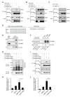
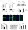
Figure 4. Cdh1-APC interacts with FMRP
(A) Lysates of 293T cells expressing Flag-Cdh1, HA-FMRP-wt and HA-FMRP-Dbm were immunoprecipitated with the Flag agarose beads followed by immunoblotting with the HA and Flag antibodies. Input was also immunoblotted with the HA and Flag antibodies. Wild type FMRP, but not D-box mutant of FMRP, formed a complex with Cdh1. (B) Lysates of 293T cells expressing Flag-Cdh1, HA-FMRP-wt and HA-FMRP-Dbm were immunoprecipitated with the HA agarose beads followed by immunoblotting with the HA and Flag antibodies. Input was also immunoblotted with the HA and Flag antibodies. Cdh1 formed a complex with wild type FMRP but not D-box mutant of FMRP. (C) Lysates of 293T cells expressing Myc-Cdc20 and HA-FMRP-wt were immunoprecipitated with the Myc antibody followed by immunoblotting with the HA and Myc antibodies. Cdc20 failed to form a complex with FMRP. (D) Conserved sequences of FMRP D-box motif from mouse, rat and human are listed. The amino acid sequence change of D-box mutation (Dbm) in FMRP is illustrated. (E) Lysates of hippocampus from wild type mice were immunoprecipitated with the Cdc27 antibody or IgG control followed by immunoblotting with the Cdh1, FMRP and Cdc27 antibodies. Endogenous Cdc27 formed a complex with endogenous Cdh1 and endogenous FMRP. (F) Lysates of hippocampus from control or conditional Cdh1 knockout mice were immunoprecipitated with the FMRP antibody or IgG control followed by immunoblotting with the Cdh1 and FMRP antibodies. Endogenous FMRP formed a complex with endogenous Cdh1 in control but not conditional Cdh1 knockout mice. (G) Lysates of N2A cells transfected with HA-FMRP-wt, HA-FMRP-S499A or HA-FMRP-S499D together with Myc-his-Ub and pretreated with MG132 were immunopreicipitated with the HA agarose beads followed by immunoblotting with the Myc and HA antibodies. FMRP S499A was ubiquitinated at a higher level than FMRP S499D. (H) Quantification of analyses as in (G). The FMRP S499A mutant is significantly more ubiquitinated than the FMRP S499D mutant protein (P<0.05). (I) Lysates from N2A cells transfected with HA-FMRP-wt, HA-FMRP-S499A or HA-FMRP-S499D, together with Flag-Cdh1, were immunopreicipitated with the Flag agarose beads followed by immunoblotting with the HA and Flag antibodies. FMRP S499A interacted with Cdh1 more effectively than FMRP S499D. (J) Quantification of analyses in (I). The FMRP S499A mutant associates with Cdh1 significantly more effectively than the FMRP S499D mutant protein (P<0.01).
Figure 5
Figure 5. Cdh1-APC triggers the ubiquitination and degradation of FMRP in the hippocampus
(A) Lysates of acute hippocampal slices from control or conditional Cdh1 knockout mice pretreated with MG132 (20 μM for 1 h) and then treated with DHPG (50 μM for 10 min) were immunoprecipitated with the FMRP antibody followed by immunoblotting with the ubiquitin (FK2), FMRP and Cdh1 antibodies. Endogenous level of ubiquitinated FMRP was substantially reduced in hippocampal slices from conditional Cdh1 knockout mice as compared to control mice. (B) Lysates of microdissected CA1 region from control or conditional Cdh1 knockout mice were analyzed as in (A). (C) Lysates of acute hippocampal slices from control or conditional Cdh1 knockout mice, pretreated with anisomycin (25 μM for 1 h), incubated with or without DHPG (50 μM for 10 min), and collected immediately after or 20 min after DHPG washout, were immunoblotted with FMRP and Actin antibodies. DHPG10′/0′ represents 0 min after DHPG (10 min) treatment; DHPG10′/20′ represents 20 min after DHPG (10 min) treatment. DHPG induced the downregulation of endogenous FMRP from control but not conditional Cdh1 knockout mice. (D) Quantification of DHPG-induced downregulation of FMRP protein levels as in (C). FMRP was normalized to Actin level in each sample. Endogenous. FMRP was significantly reduced 20 min after DHPG treatment in control mice but not in conditional Cdh1 knockout mice (P<0.05; Control: 71.7 ± 8.1% of baseline, n=3 animals; Cdh1 cKO: 126.5 ± 19.2% of baseline, n=3 animals). (E) Immunofluorescence analyses of acute hippocampal slices from control or conditional Cdh1 knockout mice, pretreated with anisomycin (25 μM for 1 h), incubated with or without DHPG (50 μM for 10 min) and collected 20 min after drug washout. Sections cut from acute slices were subjected to immunofluorescence analyses with the FMRP antibody (Ab17722) and the Hoechst 33258. DHPG triggered the downregulation of endogenous FMRP in the CA1 region from control but not conditional Cdh1 knockout mice. Scale bars represent 100 μm. (F) Quantification of DHPG-induced downregulation of FMRP fluorescent intensities at CA1 region as in (E). Endogenous FMRP was significantly decreased in response to DHPG treatment in control mice but not in conditional Cdh1 knockout mice (P<0.05; Control: 61.7 ± 8.4 % of baseline, n=3 animals; Cdh1 cKO: 112.2 ± 13.0% of baseline, n=3 animals). (G) Lysates of hippocampus from control or conditional Cdh1 knockout littermates were immunoblotted with the FMRP, Cdh1 and Actin antibodies. The basal levels of FMRP were increased in the hippocampus upon conditional Cdh1 knockout at two and five months of age. (H) Quantification of basal levels of FMRP as in (G). FMRP level was normalized to Actin level in each sample and then normalized to the level of control littermate (one-month old Cdh1 cKO: 117.3±4.9% of control littermates, n=3 pairs of animals; two-month old Cdh1 cKO: 146.9±12.4% of control littermates, n=3 pairs of animals; five-month old Cdh1 cKO: 166.9±19.9% of control littermates, n=4 pairs of animals). Supplemental Figure 5 is related to Figure 5. See also Figure S5.
Figure 6
Figure 6. FMRP operates downstream of Cdh1-APC in the regulation of mGluR-dependent LTD
(A) Measurement of DHPG-induced hippocampal mGluR-LTD in control (Cdh1loxP/loxP), conditional Cdh1 knockout (Emx-Cre;Cdh1loxP/loxP), FMRP knockout (FMR1−/y;Cdh1loxP/loxP), Cdh1 and FMRP double knockout (FMR1−/y;Emx-Cre;Cdh1loxP/loxP) mice, respectively. DHPG-induced mGluR-LTD in Cdh1 and FMRP double knockout mice was significantly increased as compared to Cdh1 knockout mice (P<0.001), but indistinguishable in magnitude as compared to FMRP knockout mice. (Control: 76.0 ± 3.4% of baseline, n=12 slices from 6 animals; Cdh1 cKO: 90.9 ± 1.6% of baseline, n=13 slices from 7 animals; FMRP KO: 68.0 ± 4.2% of baseline, n=12 slices from 7 animals; Cdh1 and FMRP dKO: 68.5 ± 2.9% of baseline, n=17 slices from 8 animals). (B) LTD level shown in (A) was quantified as percent reduction from baseline responses (average of the last ten minutes of recording). mGluR-LTD in Cdh1 and FMRP double knockout mice was significantly increased as compared to Cdh1 knockout mice (P<0.001), and indistinguishable in magnitude as compared to FMRP knockout mice (Cdh1 and FMRP dKO: 31.5 ± 2.9% of LTD, n=17 slices from 8 animals; Cdh1 cKO: 9.1 ± 1.6% of LTD, n=13 slices from 7 animals; FMRP KO: 32.0 ± 4.2% of LTD, n=12 slices from 7 animals; Control: 24.0 ± 3.4% of LTD, n=12 slices from 6 animals). (C) Lysates of 293T cells expressing wild type FMRP (HA-FMRP-wt) or the D-box mutant FMRP (HA-FMRP-Dbm) together with Myc-his-Ubiquitin and Flag-Cdh1 were immunoprecipitated with the HA agarose beads followed by immunoblotting with the Myc and HA antibodies. The D-box mutation inhibits the ubiquitination of FMRP. (D) E15 mouse embryos were electroporated with wild type FMRP (HA-FMRP-wt), D-box mutant FMRP (HA-FMRP-Dbm) or control vector (pCAG) together with an mCherry expression plasmid followed by whole-cell patch clamp analyses in transfected mCherry-positive CA1 hippocampal neurons of three-week old mice. DHPG-induced mGluR-LTD was significantly reduced in CA1 neurons upon expression of the FMRP D-box mutant protein when compared to wild type FMRP (P<0.05) or the control vector (P<0.05) (FMRP-Dbm: 102.3±11.4% of baseline, n=11 from 7 animals; FMRP-wt: 72.6±6.5% of baseline, n=10 from 6 animals; pCAG vector: 62.9±6.0% of baseline, n=8 from 4 animals). Supplemental Figure 6 is related to Figure 6. See also Figure S6.

Similar articles

Cited by

References

    1. Auerbach BD, Osterweil EK, Bear MF. Mutations causing syndromic autism define an axis of synaptic pathophysiology. Nature. 2011;480:63–68. - PMC - PubMed
    1. Bassell GJ, Warren ST. Fragile X syndrome: loss of local mRNA regulation alters synaptic development and function. Neuron. 2008;60:201–214. - PMC - PubMed
    1. Bateup HS, Takasaki KT, Saulnier JL, Denefrio CL, Sabatini BL. Loss of Tsc1 in vivo impairs hippocampal mGluR-LTD and increases excitatory synaptic function. The Journal of neuroscience. 2011;31:8862–8869. - PMC - PubMed
    1. Bear MF, Huber KM, Warren ST. The mGluR theory of fragile X mental retardation. Trends in neurosciences. 2004;27:370–377. - PubMed
    1. Bhakar AL, Dolen G, Bear MF. The pathophysiology of fragile X (and what it teaches us about synapses) Annual review of neuroscience. 2012;35:417–443. - PMC - PubMed

Publication types

MeSH terms