. 2015 Apr 21;11(4):e1005150.
doi: 10.1371/journal.pgen.1005150. eCollection 2015 Apr.

DNA damage response and spindle assembly checkpoint function throughout the cell cycle to ensure genomic integrity

Affiliations

DNA damage response and spindle assembly checkpoint function throughout the cell cycle to ensure genomic integrity

Katherine S Lawrence et al. PLoS Genet. .

Abstract

Errors in replication or segregation lead to DNA damage, mutations, and aneuploidies. Consequently, cells monitor these events and delay progression through the cell cycle so repair precedes division. The DNA damage response (DDR), which monitors DNA integrity, and the spindle assembly checkpoint (SAC), which responds to defects in spindle attachment/tension during metaphase of mitosis and meiosis, are critical for preventing genome instability. Here we show that the DDR and SAC function together throughout the cell cycle to ensure genome integrity in C. elegans germ cells. Metaphase defects result in enrichment of SAC and DDR components to chromatin, and both SAC and DDR are required for metaphase delays. During persistent metaphase arrest following establishment of bi-oriented chromosomes, stability of the metaphase plate is compromised in the absence of DDR kinases ATR or CHK1 or SAC components, MAD1/MAD2, suggesting SAC functions in metaphase beyond its interactions with APC activator CDC20. In response to DNA damage, MAD2 and the histone variant CENPA become enriched at the nuclear periphery in a DDR-dependent manner. Further, depletion of either MAD1 or CENPA results in loss of peripherally associated damaged DNA. In contrast to a SAC-insensitive CDC20 mutant, germ cells deficient for SAC or CENPA cannot efficiently repair DNA damage, suggesting that SAC mediates DNA repair through CENPA interactions with the nuclear periphery. We also show that replication perturbations result in relocalization of MAD1/MAD2 in human cells, suggesting that the role of SAC in DNA repair is conserved.


Conflict of interest statement

The authors have declared that no competing interests exist.

Figures

Fig 1
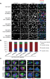
Fig 1. Both DDR and SAC components are responsive to metaphase perturbation.
(A) Cartoon of C. elegans germ line (B) Wild-type, mat-2(ts), and zyg-1(ts) germ lines stained with MAD-1, MAD-2, CENPA or P-CHK-1(Ser344) (red), α-tubulin (green), and counterstained with DAPI (blue) following growth at 25°. Scale bars = 5μM. (C) Quantification of H3S10P-positive nuclei per germ line in wild-type and zyg-1(ts) worms treated with atr, chk-1, mad-1 or control (L4440) RNAi at 25° (n ≥ 20). zyg-1(ts) mean = 9.0 ±0.5 SEM vs. WT = 5.0 ±0.4, zyg-1(ts); mad-1(RNAi) = 4.4 ±0.3, zyg-1(ts); atl-1(RNAi) = 6.2 ±0.4, zyg-1(ts); chk-1(RNAi) = 9.1±0.5 p = 0.88; ***p<0.0001. (D) Quantification of H3S10P positive nuclei per germ line in mat-2(ts) worms grown at 25° treated with control, mad-1, mad-2, mad-3, bub-3, atr, or chk-1 RNAi (n≥48). H3S10P counts between mat-2(ts) and RNAi depletions were not significant except for mad-2(RNAi), which had more H3S10P than control RNAi, p = 0.02, indicating efficient arrest.
Fig 2
Fig 2. Both DDR and SAC depletion lead to aberrant spindles and DNA morphology during metaphase arrest.
(A) mat-2(ts) germ lines treated with either control, atr, chk-1, mad-1, mad-3 or bub-3(RNAi) at 25° and stained with H3S10P (red), α-tubulin (green) and DAPI (blue). Arrows point to nuclei with aberrant DNA morphology and multiple or singular tubulin arrays. Scale bar 5μM. (B) Percentage of tubulin arrays in proliferative zones of the above genotypes at 25° (n≥10 germ lines). Percent of 2-tubulin-arrays is significantly different between mat-2(ts);control(RNAi) and mat-2(ts);atr(RNAi), mat-2(ts);chk-1(RNAi), mat-2(ts);mad-1(RNAi), all p<0.0001 (Fishers exact test). (C) mat-2(ts);chk-1(RNAi), mat-2(ts);mad-1(RNAi), or mat-2(ts);control(RNAi) metaphase nuclei stained with CENPA or SPD-2 (red), α-tubulin (green) and DAPI (blue) at 25°. The frequency of different classes is indicated. Scale bar 2μM.
Fig 3
Fig 3. MAD-2 is enriched at the nuclear periphery after DNA damage in an ATR- dependent manner.
(A) Wild-type, mad-1(gk2), chk-1(RNAi), atr(tm853), and atr(tm853);mad-1(RNAi) germ lines stained with RAD-51 (green) and counterstained with DAPI (magenta). Numbers to right indicate mean RAD-51 foci per germ line ±SEM (n = 10). (B) MAD-2 (green) staining in wild-type or atr(tm853) germ lines in the absence of damage or after HU or IR treatment counterstained with DAPI (magenta). Arrow indicates cell with nuclear MAD-2 staining. (C) Germ lines of mad-3(ok1580) and bub-3(RNAi) worms after HU stained with MAD-2 (red) and NPC (green). (D) MAD-2 localization after IR is ATR and ATM dependent. Although some MAD-2 (red) can still localize to the nuclear periphery (NPC, green) in atm-1(gk186) after 30 gy of IR, MAD-2 localization is abolished in the atm-1(gk186); atr(tm853) double mutant. Scale bars = 10μm.
Fig 4
Fig 4. SAC components function in part by delaying metaphase in the presence of DNA damage.
(A) Percent of nuclei smaller than 3.5μM, the average diameter of nuclei in untreated germ lines, after release from HU in wild-type, fzy-1(av15), mad-3(ok1580), mad-1(gk2), and mad-2(RNAi) germ lines: wild type = 30.0±1.7%, fzy-1(av15) = 40.2±2.0%; mad-3 = 52.0±1.9%; mad-1 = 54.3±2.9%; mad-2 = 52.6±1.7% (n≥24). (B) Percent of nuclei that are smaller than 3.5μM that have at least 1 RAD-51 focus in wild-type, fzy-1(av15), mad-3(ok1580), mad-1(gk2), and mad-2(RNAi) germ lines: wild type = 0.8±0.6%; mad-3 = 23.0±3.1%; mad-1 = 34.7±3.5%; mad-2 = 24.9±2.6% (n≥17). (C) Percent of nuclei that have at least 1 RAD-51 focus either—HU or 24hrs recovery in wild-type, fzy-1(av15), mad-3(ok1580), mad-1(gk2),and mad-2(RNAi) germ lines: wild type:-HU = 0.4±0.1% vs. +HU 6.9±1.7%, Δ6.5%; fzy-1(av15):-HU = 1.7±0.3% vs. +HU = 8.4±1.8%, Δ6.7%; mad-3:-HU = 3.4±0.5% vs. +HU = 19.7±3.1%, Δ16.3%; mad-1:HU = 5.3±0.5% vs. + HU = 29.5±2.3%, Δ24.2%; mad-2:—HU = 2.5±0.5% vs +HU±3.7%±23.4, Δ20.9%; (n≥ 15). (D) Percent progeny inviability—HU and after HU exposure in wild type, fzy-1(av15), mad-3(ok1580), and mad-1(gk2); wild type:-HU = 0.4±0.1 vs. +HU = 3.1±0.6%, Δ2.7%; fzy-1(av15):-HU = 2.1±1.3% vs. +HU = 8.3/-1.4%, Δ6.2%; mad-3:HU = 1.2±0.6% vs. +HU = 10.6±3.1%, Δ9.4%, p = 0.005; mad-1:—HU = 21.1±4.3% vs. +HU = 60.2±2.6% Δ39.1% (n≥10). We did not include mad-2(RNAi) due to high levels of sterility and low brood sizes. ***p<0.0001 (two-way ANOVA). Error bars indicate SEM.
Fig 5
Fig 5. Both DDR and SAC components are required for efficient DNA damage repair and progeny viability after HU.
(A) Percent of nuclei that contain at least 1 RAD-51 focus—HU or 16 hours after 5mM HU recovery in wild-type, fzy-1(av15), mad-1(gk2), mad-2(RNAi), mad-3(ok1580), bub-3(RNAi), and atr(tm853) worms. Difference between—HU and +HU is statistically different in all genotypes except wild type and fzy-1(av15), p< 0.02 (two-way ANOVA)(n≥13). (B) Percent progeny inviability—HU and after 5mM HU exposure in WT and mad-3(ok1580); n≥13. *p<0.05, **p<0.001, ***p<0.0001 (two-way ANOVA). Error bars represent SEM. (C) Proliferative zone nucleus of WT worm stained for RAD-51 (white), NPC (red), MAD-2 (green) and DAPI (blue) in the presence of HU. Image represents one slice of a z-stack taken on SIM. Scale bar = 5μm.
Fig 6
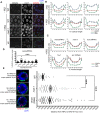
Fig 6. CENPA is regulated by both DDR and SAC components and is required for recruitment of RAD-51 to the nuclear periphery and efficient DNA damage repair.
(A) Proliferative zones of wild-type, mad-1(gk2) and atr(tm853) worms treated with HU and stained for CENPA (red), Mab414 (NPC) (green) and DAPI (blue). (B) Quantification of the average fluorescence intensity of CENPA, MAb414, and DAPI across the nucleus length binned in 10% increments-HU for wild type, mad-1(gk2)and atr(tm853), and +HU for the above genotypes (n≥30). (C) Quantification of the average fluorescence intensity of CENPA and Mab414 across the nucleus length binned in 10% increments for mad-2(RNAi), mad-3(ok1580), bub-3(RNAi), knl-1(RNAi) and fzy-1(av15) in the presence of HU (n≥30). (D) Percent of nuclei that contain at least 1 RAD-51 focus—HU or 0, 2, 4, 6, or 16 hours after release from 5mM HU with cenpa(RNAi); n≥10. Scale bars 10μm. (E) SIM images of a single nucleus from wild-type, mad-1(gk2) and cenpa(RNAi) worms treated with HU. Next to each image is the average number of RAD-51 foci observed in each genotype, as well as the average area of each RAD-51 focus (n = 4 germ lines). Images represent a projection of 3 z sections. Graph indicates the distance in nanometers between NPC and RAD-51 foci in wild-type, mad-1(gk2) and cenpa(RNAi) worms after HU. Scale bar 2 μm. Error bars indicate SEM. ***p<0.0001
Fig 7
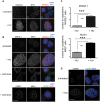
Fig 7. MAD1 and MAD2L1 are enriched in the nucleus in U2OS cells after HU exposure.
(A) Untreated or HU treated U2OS cells stained with MAD2L1 (red) and Mab414 (NPC)(green) with DAPI (blue). (B) First panels show U2OS cells stained with MAD2L1 (red) or MAD1 (green) and counterstained with DAPI (blue) in untreated cells, with HU or with colchicine. Second panels show U2OS cells treated with colchicine and stained with CREST (green), MAD1 (red) and DAPI (blue). CREST recognizes CENP-A, -B, and—C. (C) Graph of the average ratio of nucleoplasmic MAD2L1 or MAD1 fluorescence to cytoplasmic signal in the presence and absence of HU; ***p<0.0001; n≥50; Error bars indicate SEM. (D) Untreated and HU treated U2OS cells stained with CENPA (green) and counterstained with DAPI (blue). Scale bars 10 μm.
Fig 8
Fig 8. Model for DDR and SAC interactions throughout the cell cycle.
During metaphase disruptions (left), ATR (green), P-CHK-1(Ser344) (orange), MAD-1 (yellow), MAD-2 (purple), and CENPA (blue) localize to chromatin and are required for metaphase delay and stable arrest. In response to DNA damage (right), RAD-51 (red), MAD-1, MAD-2, and CENPA localize to the nuclear periphery (NPC, light green). The localization of MAD-2 is dependent on ATR and MAD-1. Additionally, the localization of CENPA is dependent on ATR, MAD-1 and MAD-2. All of these components are required for efficient DNA repair.

Similar articles

Cited by

References

    1. Ciccia A, Elledge SJ (2010) The DNA damage response: making it safe to play with knives. Mol Cell 40: 179–204. 10.1016/j.molcel.2010.09.019 - DOI - PMC - PubMed
    1. Lampert F, Westermann S (2011) A blueprint for kinetochores—new insights into the molecular mechanics of cell division. Nat Rev Mol Cell Biol 12: 407–412. 10.1038/nrm3133 - DOI - PubMed
    1. Foley EA, Kapoor TM (2013) Microtubule attachment and spindle assembly checkpoint signalling at the kinetochore. Nat Rev Mol Cell Biol 14: 25–37. 10.1038/nrm3494 - DOI - PMC - PubMed
    1. Smith J, Tho LM, Xu N, Gillespie DA (2010) The ATM-Chk2 and ATR-Chk1 pathways in DNA damage signaling and cancer. Adv Cancer Res 108: 73–112. 10.1016/B978-0-12-380888-2.00003-0 - DOI - PubMed
    1. Thompson SL, Bakhoum SF, Compton DA (2010) Mechanisms of chromosomal instability. Curr Biol 20: R285–295. 10.1016/j.cub.2010.01.034 - DOI - PMC - PubMed

Publication types