

Pneumococcal Adhesins PavB and PspC Are Important for the Interplay with Human Thrombospondin-1
- PMID: 25897078
- PMCID: PMC4505522
- DOI: 10.1074/jbc.M114.623876
Pneumococcal Adhesins PavB and PspC Are Important for the Interplay with Human Thrombospondin-1
Abstract
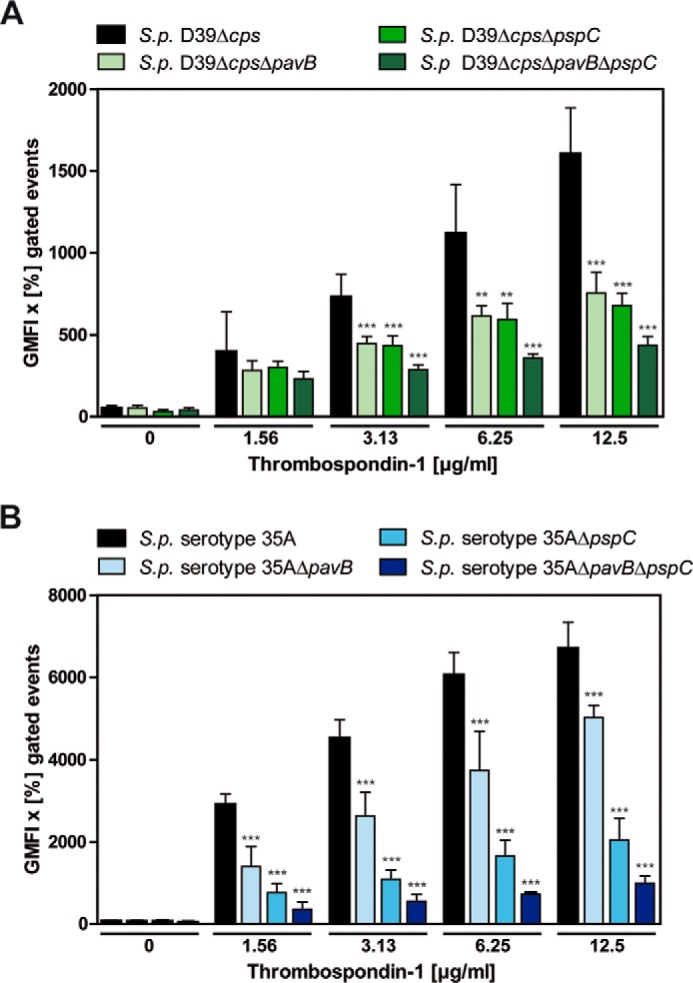
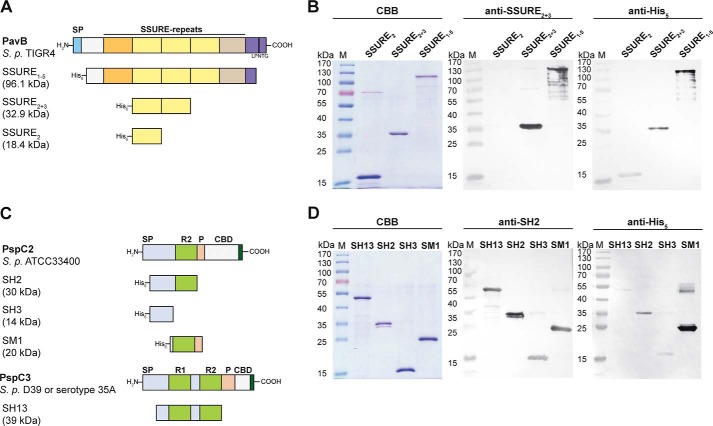
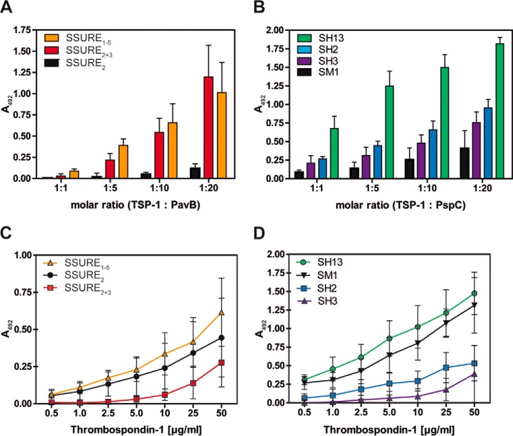
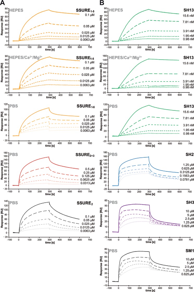
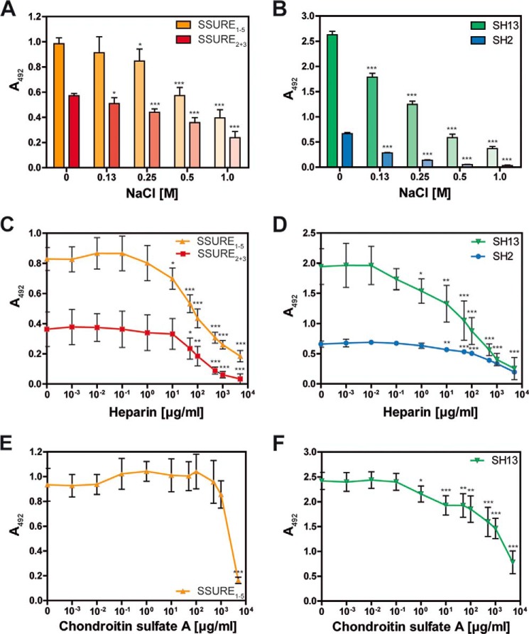
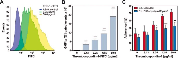
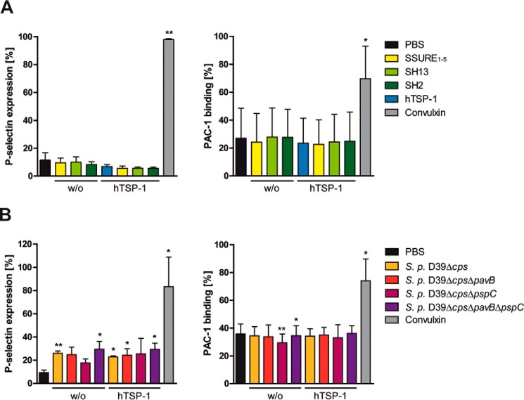
The human matricellular glycoprotein thrombospondin-1 (hTSP-1) is released by activated platelets and mediates adhesion of Gram-positive bacteria to various host cells. In staphylococci, the adhesins extracellular adherence protein (Eap) and autolysin (Atl), both surface-exposed proteins containing repeating structures, were shown to be involved in the acquisition of hTSP-1 to the bacterial surface. The interaction partner(s) on the pneumococcal surface was hitherto unknown. Here, we demonstrate for the first time that pneumococcal adherence and virulence factor B (PavB) and pneumococcal surface protein C (PspC) are key players for the interaction of Streptococcus pneumoniae with matricellular hTSP-1. PavB and PspC are pneumococcal surface-exposed adhesins and virulence factors exhibiting repetitive sequences in their core structure. Heterologously expressed fragments of PavB and PspC containing repetitive structures exhibit hTSP-1 binding activity as shown by ELISA and surface plasmon resonance studies. Binding of hTSP-1 is charge-dependent and inhibited by heparin. Importantly, the deficiency in PavB and PspC reduces the recruitment of soluble hTSP-1 by pneumococci and decreases hTSP-1-mediated pneumococcal adherence to human epithelial cells. Platelet activation assays suggested that PavB and PspC are not involved in the activation of purified human platelets by pneumococci. In conclusion, this study indicates a pivotal role of PavB and PspC for pneumococcal recruitment of soluble hTSP-1 to the bacterial surface and binding of pneumococci to host cell-bound hTSP-1 during adhesion.
Keywords: MSCRAMMs; adhesin; bacterial pathogenesis; extracellular matrix protein; platelet; pneumococci; thrombospondin.
© 2015 by The American Society for Biochemistry and Molecular Biology, Inc.
Figures
Similar articles
-
Serotype 3 pneumococci sequester platelet-derived human thrombospondin-1 via the adhesin and immune evasion protein Hic.J Biol Chem. 2017 Apr 7;292(14):5770-5783. doi: 10.1074/jbc.M116.760504. Epub 2017 Feb 16. J Biol Chem. 2017. PMID: 28209711 Free PMC article.
-
PavB is a surface-exposed adhesin of Streptococcus pneumoniae contributing to nasopharyngeal colonization and airways infections.Mol Microbiol. 2010 Jul 1;77(1):22-43. doi: 10.1111/j.1365-2958.2010.07189.x. Epub 2010 Apr 27. Mol Microbiol. 2010. PMID: 20444103
-
Repeating structures of the major staphylococcal autolysin are essential for the interaction with human thrombospondin 1 and vitronectin.J Biol Chem. 2014 Feb 14;289(7):4070-82. doi: 10.1074/jbc.M113.521229. Epub 2013 Dec 26. J Biol Chem. 2014. PMID: 24371140 Free PMC article.
-
Pneumococcal microbial surface components recognizing adhesive matrix molecules targeting of the extracellular matrix.Mol Microbiol. 2010 Jul 1;77(1):1-5. doi: 10.1111/j.1365-2958.2010.07190.x. Epub 2010 Apr 27. Mol Microbiol. 2010. PMID: 20444102 Free PMC article. Review.
-
Cellular interactions by LPxTG-anchored pneumococcal adhesins and their streptococcal homologues.Cell Microbiol. 2011 Feb;13(2):186-97. doi: 10.1111/j.1462-5822.2010.01560.x. Epub 2010 Dec 28. Cell Microbiol. 2011. PMID: 21199258 Review.
Cited by
-
Modular Architecture and Unique Teichoic Acid Recognition Features of Choline-Binding Protein L (CbpL) Contributing to Pneumococcal Pathogenesis.Sci Rep. 2016 Dec 5;6:38094. doi: 10.1038/srep38094. Sci Rep. 2016. PMID: 27917891 Free PMC article.
-
Platelets, Bacterial Adhesins and the Pneumococcus.Cells. 2022 Mar 25;11(7):1121. doi: 10.3390/cells11071121. Cells. 2022. PMID: 35406684 Free PMC article. Review.
-
Syndecan-1 Promotes Streptococcus pneumoniae Corneal Infection by Facilitating the Assembly of Adhesive Fibronectin Fibrils.mBio. 2020 Dec 8;11(6):e01907-20. doi: 10.1128/mBio.01907-20. mBio. 2020. PMID: 33293379 Free PMC article.
-
Contribution of Human Thrombospondin-1 to the Pathogenesis of Gram-Positive Bacteria.J Innate Immun. 2019;11(4):303-315. doi: 10.1159/000496033. Epub 2019 Feb 27. J Innate Immun. 2019. PMID: 30814475 Free PMC article. Review.
-
Role of Streptococcus pneumoniae Proteins in Evasion of Complement-Mediated Immunity.Front Microbiol. 2017 Feb 20;8:224. doi: 10.3389/fmicb.2017.00224. eCollection 2017. Front Microbiol. 2017. PMID: 28265264 Free PMC article. Review.
References
-
- Garenne M., Ronsmans C., Campbell H. (1992) The magnitude of mortality from acute respiratory infections in children under 5 years in developing countries. World Health Stat. Q. 45, 180–191 - PubMed
-
- Bogaert D., De Groot R., Hermans P. W. (2004) Streptococcus pneumoniae colonisation: the key to pneumococcal disease. Lancet Infect. Dis. 4, 144–154 - PubMed
-
- Gamez G., Hammerschmidt S. (2012) Combat pneumococcal infections: adhesins as candidates for protein-based vaccine development. Curr. Drug Targets 13, 323–337 - PubMed
-
- Gray B. M., Converse G. M., 3rd, Dillon H. C., Jr. (1980) Epidemiologic studies of Streptococcus pneumoniae in infants: acquisition, carriage, and infection during the first 24 months of life. J. Infect. Dis. 142, 923–933 - PubMed
Publication types
MeSH terms
Substances
LinkOut - more resources
Full Text Sources
Medical
Molecular Biology Databases
