

Identification of epithelial ovarian tumor-specific aptamers
- PMID: 25894736
- PMCID: PMC4440997
- DOI: 10.1089/nat.2014.0522
Identification of epithelial ovarian tumor-specific aptamers
Abstract
Ovarian cancer is often diagnosed in late stages with few treatment options and poor long-term prognosis. New clinical tools for early detection of ovarian malignancies will significantly help reduce mortality and improve current long-term survival rates. The objective of this work was to identify ovarian tumor-specific single-stranded DNA aptamers that bind to malignant ovarian tumor cells and internalize with high affinity and specificity. Aptamers can identify unique tumor biomarkers, can aid in early detection and diagnosis of neoplastic disorders, and can be functionalized by conjugation to small molecules. To identify aptamers from random single-stranded DNA pools (60 bases long), we used whole Cell-SELEX (systematic evolution of ligands by exponential enrichment) to enrich and isolate tumor-specific aptamers that bind to tumor-specific receptors in their native state on the cell surface. Next-Generation sequencing identified seven novel aptamers and detailed analyses of three are described. Aptamers bound to, and were internalized by, target Caov-3 cell populations, but not nontarget nonmalignant ovarian epithelial HOSE 6-3 cells or multiple other epithelial tumor cell lines. Furthermore, aptamers showed unique binding affinities with apparent dissociation constants (Kd) measuring in the submicromolar range supporting their physiological relevance and potential use in clinical applications.
Figures
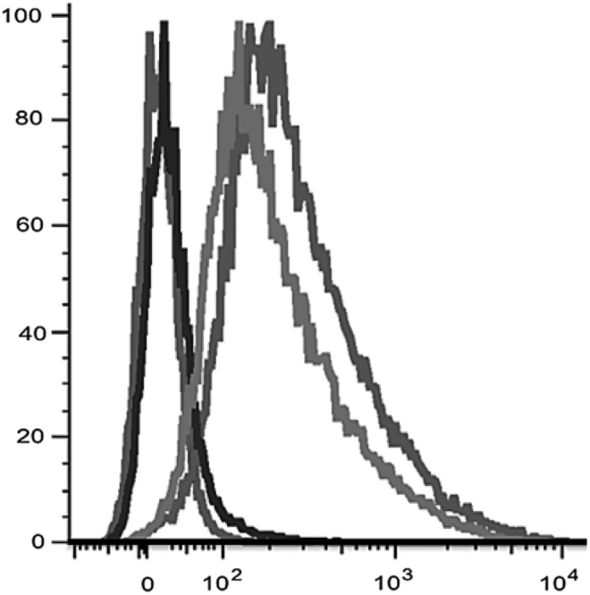
 ), round 3 (
 ), round 8 (
 ), and round 12 () of Cell-SELEX.
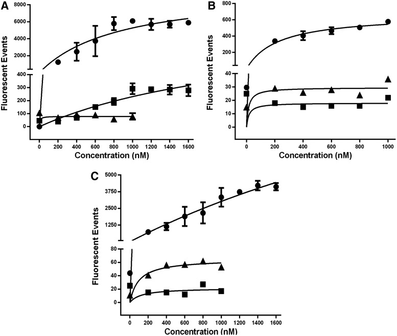

 ), 400 (
 ), and 800 nM
  ) concentrations. (B) Cy5-RLA01 incubated with indicated cell lines for 4 h. (C) Confocal imaging of indicated cell lines treated with Cy5-RLA01 imaged at 60×using a nuclear stain (DAPI as blue), a membrane stain (WGA-Alexa Fluor 488 as green), and Cy5 aptamers (Cy5 pseudocolored as yellow). (D) Binding kinetics of RLA01 when increasing molar concentrations are incubated with Caov-3 (•), SK-OV-3 (▪), and HeLa (▴). Data points represent the average fluorescent events observed (n=3, error bars±SD) at indicated nM concentrations, and individual apparent Kd values were calculated by using the equation Y=Bmax*X^h/(Kd^h+X^h).
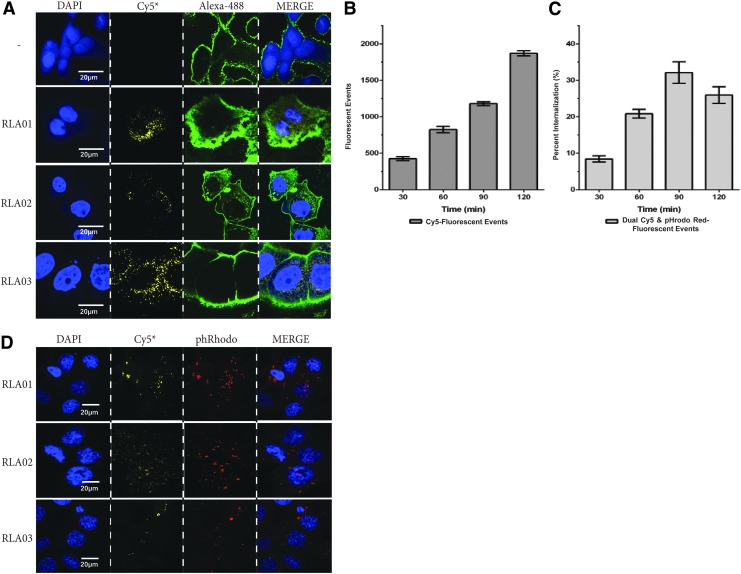
Similar articles
-
An improved SELEX technique for selection of DNA aptamers binding to M-type 11 of Streptococcus pyogenes.Methods. 2016 Mar 15;97:51-7. doi: 10.1016/j.ymeth.2015.12.005. Epub 2015 Dec 8. Methods. 2016. PMID: 26678795
-
In vitro selection of DNA aptamers recognizing drug-resistant ovarian cancer by cell-SELEX.Talanta. 2019 Mar 1;194:437-445. doi: 10.1016/j.talanta.2018.10.028. Epub 2018 Oct 12. Talanta. 2019. PMID: 30609555
-
Identification of liver cancer-specific aptamers using whole live cells.Anal Chem. 2008 Feb 1;80(3):721-8. doi: 10.1021/ac701962v. Epub 2008 Jan 5. Anal Chem. 2008. PMID: 18177018
-
Aptamers as Therapeutics.Annu Rev Pharmacol Toxicol. 2017 Jan 6;57:61-79. doi: 10.1146/annurev-pharmtox-010716-104558. Annu Rev Pharmacol Toxicol. 2017. PMID: 28061688 Free PMC article. Review.
-
Applications of aptamers in cancer cell biology.Anal Chim Acta. 2008 Jul 28;621(2):101-8. doi: 10.1016/j.aca.2008.05.031. Epub 2008 May 21. Anal Chim Acta. 2008. PMID: 18573375 Review.
Cited by
-
Aptamer Nanomaterials for Ovarian Cancer Target Theranostics.Front Bioeng Biotechnol. 2022 Mar 28;10:884405. doi: 10.3389/fbioe.2022.884405. eCollection 2022. Front Bioeng Biotechnol. 2022. PMID: 35419352 Free PMC article. Review.
-
Generating Cell Targeting Aptamers for Nanotheranostics Using Cell-SELEX.Theranostics. 2016 Jun 15;6(9):1440-52. doi: 10.7150/thno.15666. eCollection 2016. Theranostics. 2016. PMID: 27375791 Free PMC article. Review.
-
Internalized Functional DNA Aptamers as Alternative Cancer Therapies.Front Pharmacol. 2020 Jul 24;11:1115. doi: 10.3389/fphar.2020.01115. eCollection 2020. Front Pharmacol. 2020. PMID: 32848740 Free PMC article. Review.
-
Aptamers: novelty tools for cancer biology.Oncotarget. 2018 Jun 1;9(42):26934-26953. doi: 10.18632/oncotarget.25260. eCollection 2018 Jun 1. Oncotarget. 2018. PMID: 29928493 Free PMC article. Review.
-
Selection and Identification of Skeletal-Muscle-Targeted RNA Aptamers.Mol Ther Nucleic Acids. 2018 Mar 2;10:199-214. doi: 10.1016/j.omtn.2017.12.004. Epub 2017 Dec 9. Mol Ther Nucleic Acids. 2018. PMID: 29499933 Free PMC article.
References
MeSH terms
Substances
LinkOut - more resources
Full Text Sources
Other Literature Sources
Medical
