. 2015 Mar 31:10:8.
doi: 10.1186/s13064-015-0037-7.

Fgf signaling controls the telencephalic distribution of Fgf-expressing progenitors generated in the rostral patterning center

Affiliations

Fgf signaling controls the telencephalic distribution of Fgf-expressing progenitors generated in the rostral patterning center

Renée V Hoch et al. Neural Dev. .

Abstract

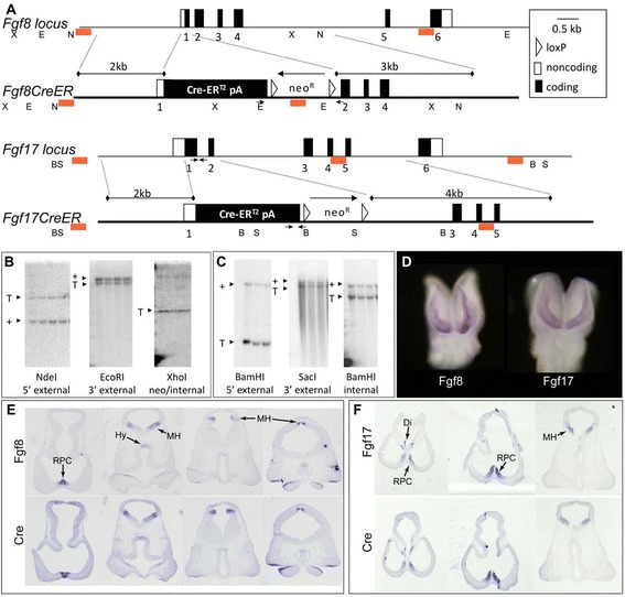
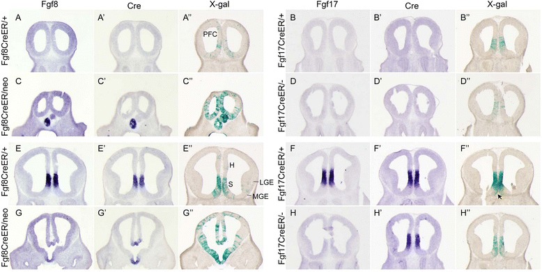
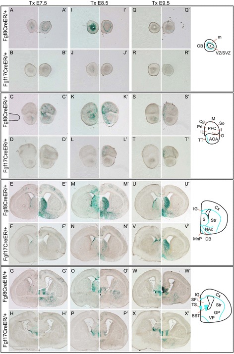
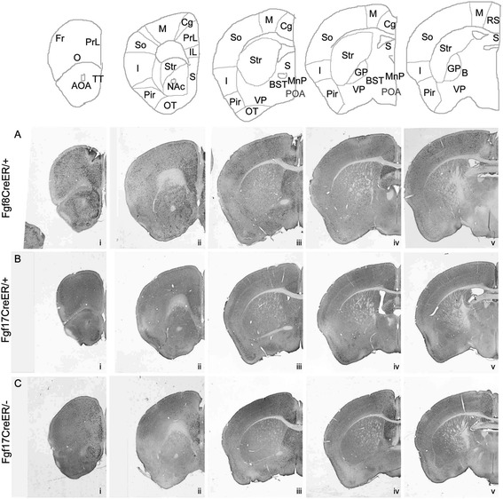
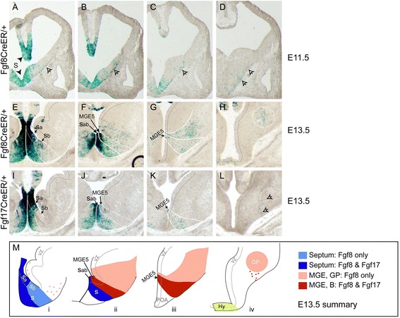
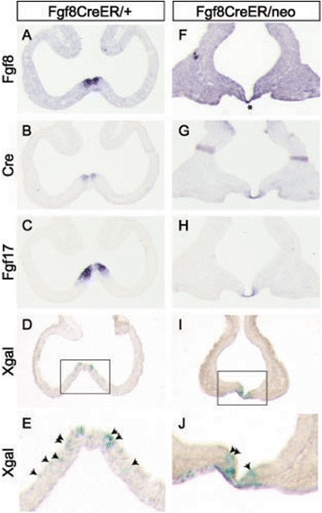
Background: The rostral patterning center (RPC) secretes multiple fibroblast growth factors (Fgfs) essential for telencephalon growth and patterning. Fgf expression patterns suggest that they mark functionally distinct RPC subdomains. We generated Fgf8(CreER) and Fgf17(CreER) mice and used them to analyze the lineages of Fgf8- versus Fgf17-expressing RPC cells.

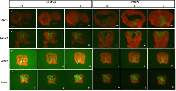
Results: Both lineages contributed to medial structures of the rostroventral telencephalon structures including the septum and medial prefrontral cortex. In addition, RPC-derived progenitors were observed in other regions of the early telencephalic neuroepithelium and generated neurons in the olfactory bulb, neocortex, and basal ganglia. Surprisingly, Fgf8(+) RPC progenitors generated the majority of basal ganglia cholinergic neurons. Compared to the Fgf8 lineage, the Fgf17 lineage was more restricted in its early dispersion and its contributions to the telencephalon. Mutant studies suggested that Fgf8 and Fgf17 restrict spread of RPC progenitor subpopulations.

Conclusions: We identified the RPC as an important source of progenitors that contribute broadly to the telencephalon and found that two molecularly distinct progenitor subtypes in the RPC make different contributions to the developing forebrain.


Figures

Figure 1
Figure 1
Generation and validation of Fgf8 CreER and Fgf17 CreER mouse lines. (A) In the Fgf8 CreER allele, the first 20 nucleotides of exon 1 coding sequence were replaced with CreER-SV40 pA-loxP-neo-loxP cassette, and the long arm of homology began 12 nucleotides upstream of the exon 1/intron 1 junction. In the Fgf17 CreER allele, all exon 1 coding sequences were replaced with CreER-SV40 pA. All Fgf17 intronic sequences were included in the targeted allele, but intron 2 was interrupted by insertion of the loxP-neo-loxP cassette and, further downstream, by a small insertion of MCS restriction enzyme sites. Symbols: small arrows, genotyping oligos; red rectangles, Southern blot probes. (B) Fgf8 CreER/+ and (C) Fgf17 CreER/+ Southern blots demonstrating correct targeting. (D) Fgf8 and Fgf17 whole mount ISHs (frontal view) showing mRNA expression in the rostral telencephalon of 12-s embryos. (E,F) E10.5 ISHs (horizontal sections) comparing Fgf8 versus Cre mRNA in an Fgf8 CreER/+ brain (E), and Fgf17 versus Cre mRNA expression in an Fgf17 CreER/+ brain (F). Sequential panels in (E) and (F) show successively more caudal planes of section. Abbreviations: B, BamHI; E, EcoRI; N, NdeI; S, SacI; X, XhoI; Di, diencephalon; MH, midbrain/hindbrain patterning center; Hy, hypothalamus. See also Additional file 1: Figure S1.
Figure 2
Figure 2
Distribution of Fgf lineage cells at E12.5. Comparison of Fgf (ISH), Cre (ISH), and βgal (Xgal stain) expression in E12.5 rostral (A-D,A′-D′,A″-D″) and caudal (E-H,E′-H′,E″-H″) coronal sections through the telencephalons of ROSA26R embryos (Tm E8.5). Note that there are Xgal+ cells rostral to the Fgf expression domains (we do not see Fgf8 or Fgf17 expression but we do see Xgal labeling in A-D″), and that in more caudal sections, Xgal+ cells are observed outside the Fgf expression domains (E-H″). Abbreviations: ob, olfactory bulb neuroepithelium; h, hippocampus; mge, medial ganglionic eminence; pfc, prefrontal cortex; s, septum. See also Additional file 2: Figure S2.
Figure 3
Figure 3
Comparison of Fgf8 and Fgf17 fate maps. Comparison of Fgf8 and Fgf17 fate maps in which progenitors were labeled at neural plate, early neurula, and late neurula stages. Xgal-stained coronal sections through E18.5 forebrains of Fgf8 CreER/+ and Fgf17 CreER/+ embryos on the (A-X) ROSA26R and (A′-X′) TauR backgrounds. Tm was administered at (A-H, A′-H′) E7.5, (I-P, I′-P′) E8.5, or (Q-X, Q′-X′) E9.5, marking cells from E8-E9.5, E9-E10.5, and E10-E11.5, respectively (see Experimental procedures). Abbreviations: m, mitral cell layer; VZ/SVZ, ventricular/subventricular zone; AOA, anterior olfactory area; TT, taenia tecta; Cx, cortex; IG, indusium griseum; S, septum; Str, striatum; NAc, nucleus accumbens; DB, diagonal band of Broca; MnP, median preoptic area; SFi, septofimbrial nucleus (septum); TS, triangular septal nucleus; BST, bed nucleus of the stria terminalis; GP, globus pallidus; VP, ventral pallidum. PFC/Cortex areas: Cg, cingulate; I, insular ; IL, infralimbic; M, motor; O, orbital; PrL, prelimbic; So, somatosensory.
Figure 4
Figure 4
Fates of Fgf8 + and Fgf17 + RPC cells in adult forebrains. Anti-βgal IHC on coronal sections through (A) Fgf8 CreER/+; TauR, (B) Fgf17 CreER/+; TauR, and (C) Fgf17 CreER/-; TauR forebrains at P40 (Tm E8.5). Abbreviations (see also Figure 2 legend): B, nucleus basalis of Meynert; POA, preoptic area; Fr, frontal cortex; Pir, piriform cortex; RS, retrosplenial cortex; OT, olfactory tubercle. Septum: Both Fgf8 + and Fgf17 + progenitors give rise to cells in the lateral septum (LS), medial septum (MS), septofimbrial and septohypothalamic nuclei, triangular septal nucleus, and indusium griseum. The MS, which receives substantial contribution from the MGE [37], contained fewer labeled cells than other septal regions. Cortex: Fgf8 + lineage cells were observed in the PFC and in most neocortical regions but were most concentrated in rostrodorsal areas (medial and dorsal frontal cortex, PrL, IL, Cg, M, RS). In contrast, Fgf17 + lineage cells only populated the medial PFC and the Cg, M, and dorsal S areas of the neocortex. Basal ganglia: The Fgf8 + lineage, but not the Fgf17 + lineage, makes prominent contributions to the striatum, ventral pallidum, accumbens, and globus pallidus. We observed left/right asymmetries in Fgf8 + and Fgf17 + fate maps that were particularly striking in the neocortex, shown in Additional file 3: Figure S3. See also Figure 3.
Figure 5
Figure 5
Origins of Fgf lineage cells in the basal ganglia. Xgal stained coronal sections through caudal telencephalons of (A-D) E11.5 Fgf8 CreER/+; ROSA26R, (E-H) E13.5 Fgf8 CreER/+; ROSA26R, and (I-L) E13.5 Fgf17 CreER/+; ROSA26R embryos (Tm E8.5). (M) Diagram illustrating similarities and differences between E13.5 fate maps. In panels (F), (J), and (Mii), Sab indicates that we are unable to distinguish Sa and Sb; the dark blue septal field extending from Sab in (Mii) likely contains cells from both progenitor domains. As shown in (H) and (L), the hypothalamus contains Fgf8 + but not Fgf17 + lineage cells at E13.5. Arrows in (A-D) indicate cells concentrated near the pial surface that appear to be migrating dorsally and caudally from the septum and vMGE. Arrowhead in (L) indicates cells from this pial migratory stream that appear to migrate radially toward the nucleus basalis of Meynert. Lateral striatum cells in the Fgf8 + lineage, indicated in pink in (Mi), are likely be derived from Sb. Abbreviations: S, septum; POA, preoptic area; Hy, hypothalamus; B, nucleus basalis of Meynert. See also Additional file 5: Figure S5.
Figure 6
Figure 6
Analysis of gene expression and fate maps in Fgf8 CreER/neo embryos. (A,F) Fgf8, (B, G) Cre, (C, H) Fgf17, and (D, E, I, J) Xgal staining in horizontal sections through E10.5 Fgf8 CreER/neo; ROSA26R and control embryos (Tm E8.5). (J,K) are high magnification images of boxed areas in (D,E); arrowheads indicate Xgal+ cells outside the Fgf8 + domain. ISH shown in (F) was left to develop for a long time in order to visualize the low level Fgf8 expression in Fgf8 CreER/neo embryos, hence the background on this section is quite high. Asterisk (F) marks the RPC, which is clearly visible in the Cre and Fgf17 ISHs. Note that Cre is expressed in the RPC but not elsewhere in the telencephalon (Horizontal lines in the caudal telencephalon in (G) are artifacts due to tissue folds).
Figure 7
Figure 7
Time lapse imaging of Fgf8 + lineage cells in brain slice culture. Fgf8 CreER/+ and Fgf8 CreER/neo mice on an mT/mG background exposed to tamoxifen at E8.5. At either E10.5 (panels in rows (A,B)) or E11.5 (panels in rows (C,D)), their brains were removed, sectioned on a vibratome and grown in slice culture [(neurobasal media (NBM)]. Slices were photographed approximately every 12 h, and representative images are shown. Cells in which the reporter had not undergone Cre-mediated recombination are red, recombined cells are green. The brains shown are from litters administered with tamoxifen and dissected in parallel. ‘Rostral’ sections, through the RPC, are in the left panels; ‘caudal’ sections, through the level of containing the MGE, are in the right panels. T0 (i, iv) = onset of culture; T1 (ii, v) = approximately 24 h in culture; T2 (iii, vi) = approximately 48 h in culture. Note that the mutant had widely distributed green cells even at T = 0, and that their distribution did not appear to change during culture.

Similar articles

Cited by

References

    1. Hoch RV, Rubenstein JL, Pleasure S. Genes and signaling events that establish regional patterning of the mammalian forebrain. Semin Cell Dev Biol. 2009;20(4):378–86. doi: 10.1016/j.semcdb.2009.02.005. - DOI - PubMed
    1. Shimamura K, Rubenstein JL. Inductive interactions direct early regionalization of the mouse forebrain. Development. 1997;124(14):2709–18. - PubMed
    1. Ye W, Shimamura K, Rubenstein JL, Hynes MA, Rosenthal A. FGF and Shh signals control dopaminergic and serotonergic cell fate in the anterior neural plate. Cell. 1998;93(5):755–66. doi: 10.1016/S0092-8674(00)81437-3. - DOI - PubMed
    1. Shanmugalingam S, Houart C, Picker A, Reifers F, Macdonald R, Barth A, et al. Ace/Fgf8 is required for forebrain commissure formation and patterning of the telencephalon. Development. 2000;127(12):2549–61. - PubMed
    1. Crossley PH, Martinez S, Ohkubo Y, Rubenstein JL. Coordinate expression of Fgf8, Otx2, Bmp4, and Shh in the rostral prosencephalon during development of the telencephalic and optic vesicles. Neuroscience. 2001;108(2):183–206. doi: 10.1016/S0306-4522(01)00411-0. - DOI - PubMed

Publication types

MeSH terms

LinkOut - more resources