Use of poly (amidoamine) dendrimer for dentinal tubule occlusion: a preliminary study
- PMID: 25885090
- PMCID: PMC4401684
- DOI: 10.1371/journal.pone.0124735
Use of poly (amidoamine) dendrimer for dentinal tubule occlusion: a preliminary study
Abstract
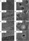
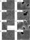
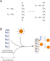
The occlusion of dentinal tubules is an effective method to alleviate the symptoms caused by dentin hypersensitivity, a significant health problem in dentistry and daily life. The in situ mineralization within dentinal tubules is a promising treatment for dentin hypersensitivity as it induces the formation of mineral on the sensitive regions and occludes the dentinal tubules. This study was carried out to evaluate the in vitro effect of a whole generation poly(amidoamine) (PAMAM) dendrimer (G3.0) on dentinal tubule occlusion by inducing mineralization within dentinal tubules. Dentin discs were treated with PAMAM dendrimers using two methods, followed by the in vitro characterization using Attenuated total reflection Fourier-transform infrared spectroscopy (ATR-FTIR), X-ray diffraction (XRD), Field emission scanning electron microscopy (FE-SEM) and Energy-Dispersive X-ray Spectroscopy (EDS). These results showed that G3.0 PAMAM dendrimers coated on dentin surface and infiltrated in dentinal tubules could induce hydroxyapatite formation and resulted in effective dentinal tubule occlusion. Moreover, crosslinked PAMAM dendrimers could induce the remineralization of demineralized dentin and thus had the potential in dentinal tubule occlusion. In this in vitro study, dentinal tubules occlusion could be achieved by using PAMAM dendrimers. This could lead to the development of a new therapeutic technique for the treatment of dentin hypersensitivity.
Conflict of interest statement
Figures


Similar articles
-
Use of phosphorylated PAMAM and carboxyled PAMAM to induce dentin biomimetic remineralization and dentinal tubule occlusion.Dent Mater J. 2021 May 29;40(3):800-807. doi: 10.4012/dmj.2020-222. Epub 2021 Mar 27. Dent Mater J. 2021. PMID: 33642446
-
Fabrication and characterization of dendrimer-functionalized nano-hydroxyapatite and its application in dentin tubule occlusion.J Biomater Sci Polym Ed. 2017 Jun;28(9):846-863. doi: 10.1080/09205063.2017.1308654. Epub 2017 Mar 31. J Biomater Sci Polym Ed. 2017. PMID: 28325103
-
8DSS peptide induced effective dentinal tubule occlusion in vitro.Dent Mater. 2018 Apr;34(4):629-640. doi: 10.1016/j.dental.2018.01.006. Epub 2018 Feb 1. Dent Mater. 2018. PMID: 29395469
-
Synthesis and Characterization of Polyamidoamine Dendrimers/Nano-hydroxyapatite and Its Role in Dentin Tubule Occlusion.Zhongguo Yi Xue Ke Xue Yuan Xue Bao. 2017 Apr 20;39(2):163-168. doi: 10.3881/j.issn.1000-503X.2017.02.001. Zhongguo Yi Xue Ke Xue Yuan Xue Bao. 2017. PMID: 28483012
-
Occlusion effects of bioactive glass and hydroxyapatite on dentinal tubules: a systematic review.Clin Oral Investig. 2022 Oct;26(10):6061-6078. doi: 10.1007/s00784-022-04639-y. Epub 2022 Jul 25. Clin Oral Investig. 2022. PMID: 35871701 Review.
Cited by
-
Research progress of biomimetic materials in oral medicine.J Biol Eng. 2023 Nov 23;17(1):72. doi: 10.1186/s13036-023-00382-4. J Biol Eng. 2023. PMID: 37996886 Free PMC article. Review.
-
Advances in biomineralization-inspired materials for hard tissue repair.Int J Oral Sci. 2021 Dec 7;13(1):42. doi: 10.1038/s41368-021-00147-z. Int J Oral Sci. 2021. PMID: 34876550 Free PMC article. Review.
-
New Advances in General Biomedical Applications of PAMAM Dendrimers.Molecules. 2018 Nov 2;23(11):2849. doi: 10.3390/molecules23112849. Molecules. 2018. PMID: 30400134 Free PMC article. Review.
-
Organic Nanomaterials and Their Applications in the Treatment of Oral Diseases.Molecules. 2016 Feb 9;21(2):207. doi: 10.3390/molecules21020207. Molecules. 2016. PMID: 26867191 Free PMC article. Review.
-
Dendritic Polymers in Tissue Engineering: Contributions of PAMAM, PPI PEG and PEI to Injury Restoration and Bioactive Scaffold Evolution.Pharmaceutics. 2023 Feb 4;15(2):524. doi: 10.3390/pharmaceutics15020524. Pharmaceutics. 2023. PMID: 36839847 Free PMC article. Review.
References
-
- Canadian Advisory Board on Dentin Hypersensitivity. Consensus-based recommendations for the diagnosis and management of dentin hypersensitivity. J Can Dent Assoc. 2002;69: 221–226. - PubMed
-
- Orchardson R, Gillam DG. Managing dentin hypersensitivity. J Am Dent Assoc. 2006;137: 990–998. - PubMed
-
- Brannstrom M, Linden LA, Johnson G. Movement of dentinal and pulpal fluid caused by clinical procedures. J Dent Res. 1968;47: 679–682. - PubMed
-
- Markowitz K, Pashley DH. Personal reflections on a sensitive subject. J Dent Res. 2007;86: 292–295. - PubMed
-
- Brannstrom M. Sensitivity of dentine. Oral Surg Oral Med Oral Pathol. 1966;21: 517–526. - PubMed
Publication types
MeSH terms
Substances
Grants and funding
LinkOut - more resources
Full Text Sources
Other Literature Sources
Miscellaneous

