. 2015 Apr 15;10(4):e0123710.
doi: 10.1371/journal.pone.0123710. eCollection 2015.

Role of the EHD2 unstructured loop in dimerization, protein binding and subcellular localization

Affiliations

Role of the EHD2 unstructured loop in dimerization, protein binding and subcellular localization

Kriti Bahl et al. PLoS One. .

Abstract

The C-terminal Eps 15 Homology Domain proteins (EHD1-4) play important roles in regulating endocytic trafficking. EHD2 is the only family member whose crystal structure has been solved, and it contains an unstructured loop consisting of two proline-phenylalanine (PF) motifs: KPFRKLNPF. In contrast, despite EHD2 having nearly 70% amino acid identity with its paralogs, EHD1, EHD3 and EHD4, the latter proteins contain a single KPF or RPF motif, but no NPF motif. In this study, we sought to define the precise role of each PF motif in EHD2's homo-dimerization, binding with the protein partners, and subcellular localization. To test the role of the NPF motif, we generated an EHD2 NPF-to-NAF mutant to mimic the homologous sequences of EHD1 and EHD3. We demonstrated that this mutant lost both its ability to dimerize and bind to Syndapin2. However, it continued to localize primarily to the cytosolic face of the plasma membrane. On the other hand, EHD2 NPF-to-APA mutants displayed normal dimerization and Syndapin2 binding, but exhibited markedly increased nuclear localization and reduced association with the plasma membrane. We then hypothesized that the single PF motif of EHD1 (that aligns with the KPF of EHD2) might be responsible for both binding and localization functions of EHD1. Indeed, the EHD1 RPF motif was required for dimerization, interaction with MICAL-L1 and Syndapin2, as well as localization to tubular recycling endosomes. Moreover, recycling assays demonstrated that EHD1 RPF-to-APA was incapable of supporting normal receptor recycling. Overall, our data suggest that the EHD2 NPF phenylalanine residue is crucial for EHD2 localization to the plasma membrane, whereas the proline residue is essential for EHD2 dimerization and binding. These studies support the recently proposed model in which the EHD2 N-terminal region may regulate the availability of the unstructured loop for interactions with neighboring EHD2 dimers, thus promoting oligomerization.


Conflict of interest statement

Competing Interests: Steve Caplan serves as an Academic Editor for PLOS ONE. This does not alter the authors' adherence to PLOS ONE Editorial policies and criteria.

Figures

Fig 1
Fig 1. EHD protein domain architecture and sequence homology.
(A) The Eps15 Homology Domain (EHD) proteins have a conserved domain architecture comprised of four domains: two helical domains, a G-domain, and a C-terminal EH domain. The G-domain of EHD2 contains an unstructured loop containing the following amino acid sequence, KPFRKLNPF, which is required for oligomerization. (B) The four EHD isoforms share 67–86% residue identity. The amino acid sequence alignment of the unstructured loop for all four EHDs (see green frame) shows that only EHD2 has two successive PF motifs: NPF and KPF, whereas the other EHD proteins have only one PF motif.
Fig 2
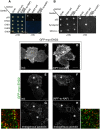
Fig 2. Modification of the EHD2 NPF motif to NAF impairs dimerization and binding with interaction partners, but does not affect EHD2 localization.
(A) S. cerevisae yeast were co-transformed with the following Gal4 binding domain (Gal4bd) fusion constructs (see Materials and Methods):-p53 (control), -Syndapin-2 and -MICAL-L1. Gal4 activating domain (Gal4ad) fusion constructs were either-SV40 (control), -EHD2 (wt) or -EHD2 (NPF-to-NAF). Co-transformants were plated on non-selective (+HIS) and selective (-HIS) agar plates. (B) As in (A), co-transformation was with Gal4bd-p53 (control), -EHD2 (wt), -EHD1 (wt), -EHD3 (wt), and -EHD4 (wt) and with Gal4ad-SV40 (control), -EHD2 (wt) and -EHD2 (NPF-to-NAF). (C–H) HeLa cells were grown on coverslips, transfected with GFP-myc-EHD2 (wt) (C,E,G) or with GFP-myc-EHD2 (NAF-to-NPF) (D,F,H) and fixed. (C–D) are transfected cells imaged by confocal microscopy, whereas (E–H) are micrographs obtained by Structured Illumination Microscopy (SIM). Asterisks indicate transfected cells. Bar; 10 μm.
Fig 3
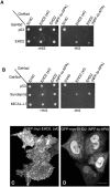
Fig 3. Modification of the EHD2 NPF motif to APA induces loss of plasma membrane localization, but does not affect interactions with binding partners.
(A) S. cerevisae yeast were co-transformed with the following Gal4bd fusion constructs: Gal4bd-p53 (control), -Syndapin-2 and -MICAL-L1 along with Gal4ad fusion constructs Gal4ad-SV40 (control), -EHD2 (wt.) and -EHD2 (NPF-to-APA). (B) As in (A) co-transformation was with Gal4bd fusion constructs: Gal4bd-p53 (control), -EHD2 (wt) along with Gal4ad fusion constructs: Gal4ad-SV40 (control), -EHD2 (wt) and -EHD2 (NPF-to-APA). Co-transformants from A-B were plated on non-selective (+HIS) and selective (-HIS) agar plates. (C–D) HeLa cells were grown on coverslips, transfected with GFP-myc-EHD2 (C) or GFP-myc-EHD2 NPF-to-APA (D). Bar; 10 μm.
Fig 4
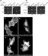
Fig 4. The NPF phenylalanine residue is responsible for the plasma membrane localization of EHD2.
(A) S. cerevisae yeast were co-transformed with the following Gal4bd fusion constructs: Gal4bd-p53 (control), -Syndapin-2 and -MICAL-L1 along with Gal4ad fusion constructs: Gal4ad-SV40 (control), -EHD2 (wt) and -EHD2 (NPF-to-NPA). (B) As in (A), yeast were co-transformed with the Gal4bd fusion constructs: Gal4bd-p53 (control), -EHD2 (wt) along with Gal4ad fusion constructs: Gal4ad-SV40 (control), -EHD2 (wt.) and -EHD2 (NPF-to-NPA). Co-transformants were plated on non-selective (+HIS) and selective (-HIS) agar plates. (C–I) HeLa cells were grown on coverslips and transfected with: (C) GFP-myc-EHD2 (wt), (D) GFP-myc-EHD2 (NPF-to-NPA), (E) GFP-myc-EHD2 (NPF-to-NPY), (F) GFP-myc-EHD2 (NPF-to-NPA), (G) GFP-myc-EHD2 (NPF-to-NFP). Bar; 10 μm.
Fig 5
Fig 5. Disruption of the EHD2 KPF motif induces relocalization of EHD2 to the nucleus, but does not alter its binding ability.
(A–D) S. cerevisae yeast were co-transformed with the following Gal4bd fusion constructs: Gal4bd-p53 (control), -Syndapin-2 and -MICAL-L1 along with Gal4ad fusion constructs: Gal4ad-SV40 (control), -EHD2 (wt), -EHD2 (KPF-to-APA) and—EHD2 (KPF-to-KAF). Co-transformants were plated on non-selective (+HIS) and selective (-HIS) media. (E–H) HeLa cells were grown on coverslips, transfected with GFP-myc-EHD2 (wt) (E), GFP-myc-EHD2 (KPF-to-APA) (F), or GFP-myc-EHD2 (KPF-to-KAF) (G) and analyzed by confocal microscopy. Bar; 10 μm.
Fig 6
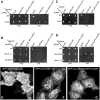
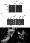
Fig 6. A single EHD1 PF motif (RPF) controls its homo- and hetero-dimerization, binding to interaction partners, and localization to Tubular Recycling Endosomes (TRE).
(A) S. cerevisae yeast were co-transformed with Gal4bd fusion constructs: Gal4bd-p53 (control), -Syndapin-2 and -MICAL-L1 along with Gal4ad-SV40 (control), -EHD1 (wt.) and -EHD1 (RPF-to-APA). (B) As in (A), yeast were co-transformed with Gal4bd-p53 (control), -EHD1 (wt), -EHD3 (wt), -EHD4 (wt), along with Gal4ad-SV40 (control), -EHD1 (wt) and EHD1 (RPF-to-APA). Co-transformants from A-B were plated on non-selective (+HIS) and selective (-HIS) agar plates. (C–D) HeLa cells were grown on coverslips, transfected with GFP-myc-EHD1 (wt) (C), GFP-myc-EHD1 (RPF-to-APA) (D, arrows point to TRE). Bar; 10 μm.
Fig 7
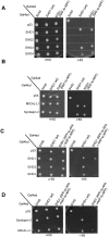
Fig 7. The NAF motif of EHD1 is dispensable for homo- or hetero-oligomerization, and for its association with binding partners.
(A) S. cerevisae yeast were co-transformed with the following Gal4bd fusion constructs: Gal4bd-p53 (control), -MICAL-L1, and -Syndapin-2 along with Gal4ad-SV40 (control), -EHD1 (wt.) and -EHD1 (NAF to NPF). (B) As in (A), yeast were co-transformed with Gal4bd-p53 (control), -EHD1 (wt), -EHD2 (wt), -EHD3 (wt) and -EHD4 (wt), along with Gal4ad-SV40 (control), -EHD1 (wt) and -EHD1 (NAF-to-NPF). (C) As in (A), yeast were co-transformed with Gal4bd-p53 (control), -EHD1 (wt), -EHD3 (wt), and -EHD4 (wt), along with Gal4ad-SV40 (control), -EHD1 (wt) and -EHD1 (NAF-to-NPF). (D) Yeast were co-transformed with Gal4bd-p53 (control), -Syndapin-2, and -MICAL-L1, along with Gal4ad-SV40 (control), EHD1 (wt) and EHD1 (NAF-to-NPF). Co-transformants in A-D were plated on non-selective (+HIS) and selective (-HIS) agar plates.
Fig 8
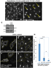
Fig 8. The EHD1 RPF motif is essential for receptor recycling.
(A–B) HeLa cells grown on coverslips were mock-treated (A) or treated with EHD1-siRNA (B–F). For A–B, After a 30 min. serum-starvation, cells were allowed to uptake Tf-568 for 15 min., and then chased in complete media for 15 min. to permit Tf-568 recycling to the PM. (C) Immunoblot analysis of Mock- or EHD1-siRNA-treated cells depicting levels of actin (control) or EHD1. (D–G) During the EHD1-siRNA treatment, cells were transfected with the siRNA-resistant EHD1 constructs: siRNA-res-GFP-myc-EHD1 (wt) in (D), and siRNA-res-GFP-myc-EHD1 (RPF-to-APA) in (F). Following serum-starvation, Tf-568 was internalized for 15 min., and chased in complete media for 15 min. Note the typical concentration of Tf in the ERC in untransfected cells treated with EHD1-siRNA. (H) Quantitative analysis of 100 cells from 3 independent experiments as in (E and G). Cells that recycled Tf-568 (“empty of Tf”) were scored and calculated as % of total cell number, and portrayed with standard error bars. Stars represent significance of p < 0.01 (one star) or p < 0.05 (three stars) for one-way ANOVA tests. Bar; 10 μm.
Fig 9
Fig 9. Model for the role of the EHD2 unstructured KPFRKLNPF motif in subcellular localization.
(A) In the absence of PIP2-containing membrane, the N-termini interact with regions flanking the unstructured loops (U.L.) of EHD2 dimer pairs. (B) PIP2-binding by the N-terminus facilitates a-8 helical binding to the membrane and frees the U.L. (C) the U.L. NPF motif is now capable of interacting with an EH domain of a neighboring EHD2 dimer pair, inducing oligomerization. (D) For EHD2 NPF-to-NPA mutants, we hypothesize that the N-terminus may not insert into membranes, thus preventing the U.L. from oligomerizing and maintaining the lysine residues on α-8 helix free to serve as a nuclear localization sequence.

Similar articles

Cited by

References

    1. Naslavsky N, Caplan S (2011) EHD proteins: key conductors of endocytic transport. Trends Cell Biol 21: 122–131. 10.1016/j.tcb.2010.10.003 - DOI - PMC - PubMed
    1. Kieken F, Jovic M, Naslavsky N, Caplan S, Sorgen PL (2007) EH domain of EHD1. J Biomol NMR 39: 323–329. - PubMed
    1. Kieken F, Jovic M, Tonelli M, Naslavsky N, Caplan S, et al. (2009) Structural insight into the interaction of proteins containing NPF, DPF, and GPF motifs with the C-terminal EH-domain of EHD1. Protein Sci 18: 2471–2479. 10.1002/pro.258 - DOI - PMC - PubMed
    1. Kieken F, Sharma M, Jovic M, Giridharan SS, Naslavsky N, Caplan S, et al. (2010) Mechanism for the selective interaction of C-terminal Eps15 homology domain proteins with specific Asn-Pro-Phe-containing partners. The Journal of biological chemistry 285: 8687–8694. 10.1074/jbc.M109.045666 - DOI - PMC - PubMed
    1. Henry GD, Corrigan DJ, Dineen JV, Baleja JD (2010) Charge effects in the selection of NPF motifs by the EH domain of EHD1. Biochemistry 49: 3381–3392. 10.1021/bi100065r - DOI - PMC - PubMed

Publication types

MeSH terms

LinkOut - more resources