Genetic Evidence Supports a Major Role for Akt1 in VSMCs During Atherogenesis
- PMID: 25868464
- PMCID: PMC4561531
- DOI: 10.1161/CIRCRESAHA.116.305895
Genetic Evidence Supports a Major Role for Akt1 in VSMCs During Atherogenesis
Abstract
Rationale: Coronary artery disease, the direct result of atherosclerosis, is the most common cause of death in Western societies. Vascular smooth muscle cell (VSMC) apoptosis occurs during the progression of atherosclerosis and in advanced lesions and promotes plaque necrosis, a common feature of high-risk/vulnerable atherosclerotic plaques. Akt1, a serine/threonine protein kinase, regulates several key endothelial cell and VSMC functions including cell growth, migration, survival, and vascular tone. Although global deficiency of Akt1 results in impaired angiogenesis and massive atherosclerosis, the specific contribution of VSMC Akt1 remains poorly characterized.
Objective: To investigate the contribution of VSMC Akt1 during atherogenesis and in established atherosclerotic plaques.
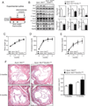
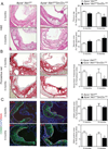
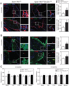
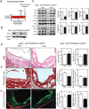
Methods and results: We generated 2 mouse models in which Akt1 expression can be suppressed specifically in VSCMs before (Apoe(-/-)Akt1(fl/fl)Sm22α(CRE)) and after (Apoe(-/-)Akt1(fl/fl)SM-MHC-CreER(T2E)) the formation of atherosclerotic plaques. This approach allows us to interrogate the role of Akt1 during the initial and late steps of atherogenesis. The absence of Akt1 in VSMCs during the progression of atherosclerosis results in larger atherosclerotic plaques characterized by bigger necrotic core areas, enhanced VSMC apoptosis, and reduced fibrous cap and collagen content. In contrast, VSMC Akt1 inhibition in established atherosclerotic plaques does not influence lesion size but markedly reduces the relative fibrous cap area in plaques and increases VSMC apoptosis.
Conclusions: Akt1 expression in VSMCs influences early and late stages of atherosclerosis. The absence of Akt1 in VSMCs induces features of plaque vulnerability including fibrous cap thinning and extensive necrotic core areas. These observations suggest that interventions enhancing Akt1 expression specifically in VSMCs may lessen plaque progression.
Keywords: atherogenesis; atherosclerosis; proto-oncogene proteins c-akt.
© 2015 American Heart Association, Inc.
Figures


Similar articles
-
Absence of Akt1 reduces vascular smooth muscle cell migration and survival and induces features of plaque vulnerability and cardiac dysfunction during atherosclerosis.Arterioscler Thromb Vasc Biol. 2009 Dec;29(12):2033-40. doi: 10.1161/ATVBAHA.109.196394. Epub 2009 Sep 17. Arterioscler Thromb Vasc Biol. 2009. PMID: 19762778 Free PMC article.
-
C/EBP-Homologous Protein (CHOP) in Vascular Smooth Muscle Cells Regulates Their Proliferation in Aortic Explants and Atherosclerotic Lesions.Circ Res. 2015 May 22;116(11):1736-43. doi: 10.1161/CIRCRESAHA.116.305602. Epub 2015 Apr 14. Circ Res. 2015. PMID: 25872946 Free PMC article.
-
Effects of DNA damage in smooth muscle cells in atherosclerosis.Circ Res. 2015 Feb 27;116(5):816-26. doi: 10.1161/CIRCRESAHA.116.304921. Epub 2014 Dec 18. Circ Res. 2015. PMID: 25524056
-
Vascular smooth muscle cell death, autophagy and senescence in atherosclerosis.Cardiovasc Res. 2018 Mar 15;114(4):622-634. doi: 10.1093/cvr/cvy007. Cardiovasc Res. 2018. PMID: 29360955 Review.
-
Vascular Smooth Muscle Cells in Atherosclerosis.Circ Res. 2016 Feb 19;118(4):692-702. doi: 10.1161/CIRCRESAHA.115.306361. Circ Res. 2016. PMID: 26892967 Free PMC article. Review.
Cited by
-
Role of PCK2 in the proliferation of vascular smooth muscle cells in neointimal hyperplasia.Int J Biol Sci. 2022 Aug 8;18(13):5154-5167. doi: 10.7150/ijbs.75577. eCollection 2022. Int J Biol Sci. 2022. PMID: 35982907 Free PMC article.
-
Shifting the Focus of Preclinical, Murine Atherosclerosis Studies From Prevention to Late-Stage Intervention.Circ Res. 2017 Mar 3;120(5):775-777. doi: 10.1161/CIRCRESAHA.116.310101. Circ Res. 2017. PMID: 28254801 Free PMC article.
-
Long Non-Coding RNA PCAT19 Suppresses Cell Proliferation and Angiogenesis in Coronary Artery Disease through Interaction with GCNT2.Cell Biochem Biophys. 2024 Sep;82(3):2237-2248. doi: 10.1007/s12013-024-01335-4. Epub 2024 Jun 7. Cell Biochem Biophys. 2024. PMID: 38849695
-
PI3Kγ (Phosphoinositide 3-Kinase γ) Regulates Vascular Smooth Muscle Cell Phenotypic Modulation and Neointimal Formation Through CREB (Cyclic AMP-Response Element Binding Protein)/YAP (Yes-Associated Protein) Signaling.Arterioscler Thromb Vasc Biol. 2019 Mar;39(3):e91-e105. doi: 10.1161/ATVBAHA.118.312212. Arterioscler Thromb Vasc Biol. 2019. PMID: 30651001 Free PMC article.
-
Endothelial Cell Autonomous Role of Akt1: Regulation of Vascular Tone and Ischemia-Induced Arteriogenesis.Arterioscler Thromb Vasc Biol. 2018 Apr;38(4):870-879. doi: 10.1161/ATVBAHA.118.310748. Epub 2018 Feb 15. Arterioscler Thromb Vasc Biol. 2018. PMID: 29449333 Free PMC article.
References
-
- Glass CK, Witztum JL. Atherosclerosis. the road ahead. Cell. 2001;104:503–516. - PubMed
-
- Libby P. Inflammation in atherosclerosis. Nature. 2002;420:868–874. - PubMed
-
- Clarke MC, Figg N, Maguire JJ, Davenport AP, Goddard M, Littlewood TD, Bennett MR. Apoptosis of vascular smooth muscle cells induces features of plaque vulnerability in atherosclerosis. Nature medicine. 2006;12:1075–1080. - PubMed
-
- Allard D, Figg N, Bennett MR, Littlewood TD. Akt regulates the survival of vascular smooth muscle cells via inhibition of FoxO3a and GSK3. J Biol Chem. 2008;283:19739–19747. - PubMed
Publication types
MeSH terms
Substances
Grants and funding
LinkOut - more resources
Full Text Sources
Medical
Molecular Biology Databases
Research Materials
Miscellaneous

