. 2015 Apr 8;35(14):5680-92.
doi: 10.1523/JNEUROSCI.4953-14.2015.

Muscarinic receptors modulate dendrodendritic inhibitory synapses to sculpt glomerular output

Affiliations

Muscarinic receptors modulate dendrodendritic inhibitory synapses to sculpt glomerular output

Shaolin Liu et al. J Neurosci. .

Abstract

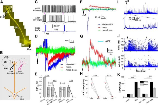
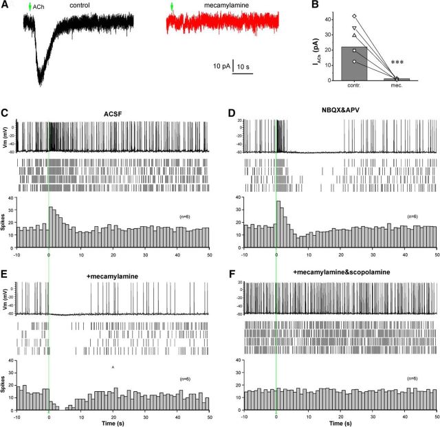
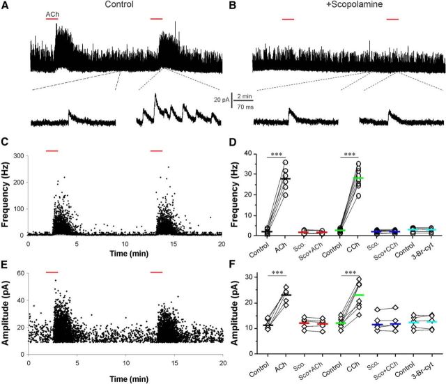
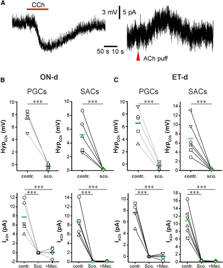
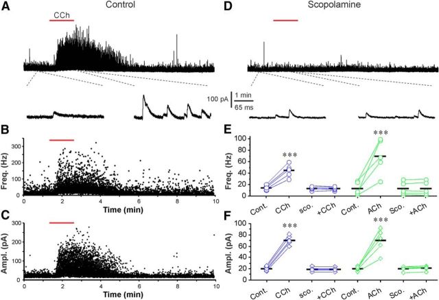
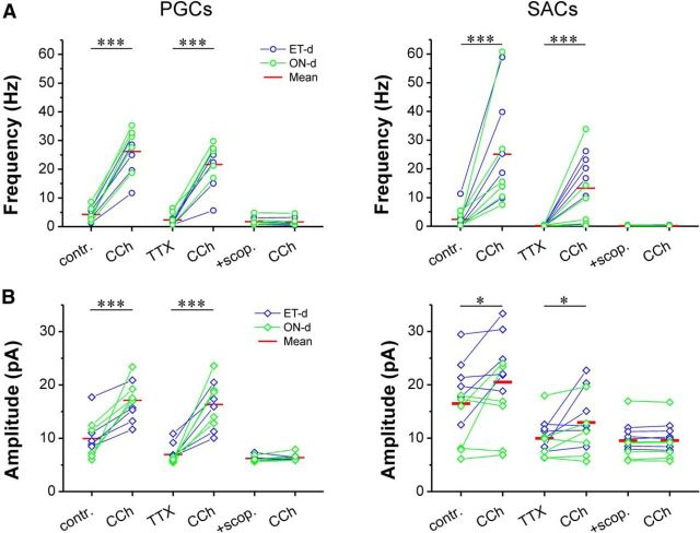
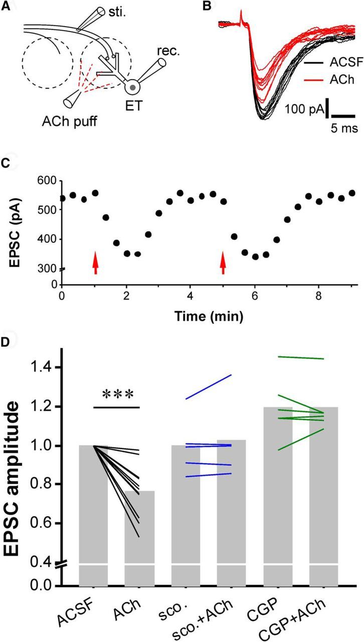
Cholinergic [acetylcholine (ACh)] axons from the basal forebrain innervate olfactory bulb glomeruli, the initial site of synaptic integration in the olfactory system. Both nicotinic acetylcholine receptors (nAChRs) and muscarinic acetylcholine receptors (mAChRs) are expressed in glomeruli. The activation of nAChRs directly excites both mitral/tufted cells (MTCs) and external tufted cells (ETCs), the two major excitatory neurons that transmit glomerular output. The functional roles of mAChRs in glomerular circuits are unknown. We show that the restricted glomerular application of ACh causes rapid, brief nAChR-mediated excitation of both MTCs and ETCs in the mouse olfactory bulb. This excitation is followed by mAChR-mediated inhibition, which is blocked by GABAA receptor antagonists, indicating the engagement of periglomerular cells (PGCs) and/or short axon cells (SACs), the two major glomerular inhibitory neurons. Indeed, selective activation of glomerular mAChRs, with ionotropic GluRs and nAChRs blocked, increased IPSCs in MTCs and ETCs, indicating that mAChRs recruit glomerular inhibitory circuits. Selective activation of glomerular mAChRs in the presence of tetrodotoxin increased IPSCs in all glomerular neurons, indicating action potential-independent enhancement of GABA release from PGC and/or SAC dendrodendritic synapses. mAChR-mediated enhancement of GABA release also presynaptically suppressed the first synapse of the olfactory system via GABAB receptors on sensory terminals. Together, these results indicate that cholinergic modulation of glomerular circuits is biphasic, involving an initial excitation of MTC/ETCs mediated by nAChRs followed by inhibition mediated directly by mAChRs on PGCs/SACs. This may phasically enhance the sensitivity of glomerular outputs to odorants, an action that is consistent with recent in vivo findings.

Keywords: cholinergic modulation; dendrodendritic synapses; glomerular circuits; glomerular output; inhibitory interneurons; olfactory bulb.


Figures

Figure 1.
Figure 1.
Cholinergic modulation of the glomerular circuit shapes OB output. A, Epifluorescent image showing a Lucifer yellow labeled MC. GL, Glomerular layer; MC, mitral cell; puff, puffing pipette; rec., recording electrode. B, Schematic of experimental setup showing the pipette for puffing ACh to the off-target (magenta) or on-target (black) glomerulus receiving the apical dendrite of the recorded MTC. ONL, Olfactory nerve layer. C, Current-clamp recording showing MTC responses to (arrows, 1 mm, 1 s) the “off-target” (top trace) or “on-target” (middle and bottom traces) micropuffing ACh, as illustrated in B before ACSF, and after NBQX (10 μm) and APV (50 μm). Note: off-target puffing of ACh showed no effect, while on-target puffing of ACh elicited a brief enhancement of firing followed by hyperpolarization and firing termination; and this effect was not affected by NBQX and APV. D, Typical current-clamp recording traces from one MTC showing that the nAChR blocker mecamylamine (mec.; 10 μm, red) abolished the ACh-evoked firing and reduced the following hyperpolarization in the presence of NBQX and APV (blue); the residual hyperpolarization is eliminated by the addition of scopolamine (sco.; 10 μm, green). E, Quantification data from five MTCs showing the following: (1) that the ACh-induced hyperpolarization in the presence of NBQX and APV (Control) is partially reduced by scopolamine, GBZ (10 μm), or mecamylamine, and was completely eliminated by scopolamine plus mecamylamine; and (2) that the residual hyperpolarization in the presence of scopolamine or GBZ is not affected by the combination of scopolamine and GBZ. F, Voltage-clamp recording traces from the same MTC showing that glomerular micropuffing of ACh to the target glomeruli produces an inward current in the MTC held at −60 mV in the presence of NBQX and APV (blue). The addition of mecamylamine (10 μm) in the bath blocks this inward current but reveals an outward current (red). This outward current is abolished by the further addition of scopolamine (10 μm). G, Voltage-clamp recording traces showing that glomerular micropuffed ACh-induced outward current in the presence of NBQX, APV, and mecamylamine (red) is blocked by the further addition of either scopolamine (green) or GBZ (cyan). H, Quantification data from five MTCs showing that the ACh-induced outward current shown in E is abolished by either scopolamine or GBZ. I, Typical voltage-clamp recordings showing that glomerular micropuffing of ACh (arrows) reproducibly enhances spontaneous IPSCs in MTCs (top trace). The bottom trace is a blow-up from the top trace showing the initial changes in sIPSCs in response to ACh application. J, Scatter plots showing that both the frequency (top graph) and amplitude (bottom graph) of sIPSCs in MTCs are increased by glomerular micropuffed ACh. K, Quantification data from five MTCs showing that the enhancement of both frequency (top graph) and amplitude (bottom graph) of sIPSCs by glomerular-applied ACh is eliminated by scopolamine (10 μm), but not by mecamylamine (10 μm). One-way ANOVA was performed in E, between control and sco. Ampl., Amplitude; Freq., frequency. Groups in K; repeated-measures ANOVA was performed in comparison among sco., sco. + GBZ, and sco. + GBZ + mec. groups in E. Paired t tests were performed among GBZ and GBZ + sco., mec., and mec. + sco. groups in E, as well as in H. ***p < 0.001.
Figure 2.
Figure 2.
Glomerular ACh produces a direct and nAChR-mediated excitation followed by an mAChR-mediated inhibition in ETCs. A, Voltage-clamp traces showing that micropuffing of ACh (arrows, 1 mm, 1 s) to the glomeruli receiving apical dendrites of the recorded ETC held at −60 mV produces an inward current (left, black) in the presence of NBQX (10 μm), APV (50 μm), and GBZ (10 μm). This ACh-induced inward current is abolished by bath-applied mecamylamine (10 μm, right, red). B, Quantification data from five ETCs showing that the glomerular applied ACh-induced inward current is abolished by mecamylamine. C, Top, Current-clamp trace showing that glomerular micropuffing of ACh (1 mm, 1 s, red line) induces a brief firing enhancement in an ETC from the OB slice perfused with ACSF. Middle, Raster graph showing the ACh effect on ETC firing in four repeated traces from the same cell. Bottom, Averaged histogram from six ETCs showing the ACh-induced enhancement of ETC firing (bin size, 1 s). D, Data collected from the same cell shown in C showing that bath-applied NBQX (10 μm) and APV (50 μm) do not alter the ACh-induced brief enhancement of ETC firing but reveal a following spiking termination/inhibition. E, Data collected from the same cell shown in C and D showing that the addition of mecamylamine (10 μm) abolishes the ACh-induced firing enhancement, but leaves the inhibition intact. F, Data collected from the same cell shown in C, D, and E showing that the ACh-elicited inhibition in the presence of NBQX, APV, and mecamylamine is eliminated by scopolamine (10 μm). Paired t tests were performed in B. **p < 0.01. Vm, Membrane voltage.
Figure 3.
Figure 3.
ACh via mAChRs enhances sIPSCs in ETCs. A, Top, Voltage-clamp trace showing that bath-applied ACh (200 μm, 2 min, red bar) enhances sIPSCs in ETCs. Bottom, Blow-up from A showing sIPSCs before and after ACh. B, Voltage-clamp trace from the same cell showing the ACh-induced sIPSC enhancement is eliminated by bath-applied scopolamine (10 μm). C, E, Scatter plots from the top trace in A showing that both the frequency (C) and amplitude (E) of sIPSCs are reproducibly enhanced by ACh. D, F, Quantification data showing that the ACh-induced or 20 μm CCh-induced enhancement of both frequency (D) and amplitude (F) of sIPSCs is eliminated by scopolamine (Sco., 10 μm), and that the nAChR activator 3-Br-cytisine (5 μm) does not alter sIPSCs in ETCs. Paired t tests were performed in D and F. ***p < 0.001.
Figure 4.
Figure 4.
Activation of mAChRs inhibits PGCs and SACs. A, Somatic recordings in current clamp (left) or voltage clamp (right) from a TH-GFP-labeled SAC showing that bath-applied CCh (20 μm) produces hyperpolarization and glomerular micropuffing of ACh (red arrowhead, 1 mm, 1 s), produced an outward current when the cell was held at −60 mV in the presence NBQX (10 μm), APV (50 μm), and GBZ (10 μm) to eliminate the circuit effect. B, C, Quantification data showing that CCh-induced hyperpolarization or ACh-induced outward current was observed in all tested ONd (B) and ETd (C) PGCs and SACs, and these effects were completely eliminated by scopolamine (10 μm). Paired t test and repeated-measures ANOVA were performed for current-clamp results (top graphs in B) and voltage-clamp results (bottom graphs in B), respectively. ***p < 0.001. contr., Control; sco., scopolamine; mec., mecamylamine.
Figure 5.
Figure 5.
Activation of mAChRs enhances mIPSCs in ETCs. A, Top, Voltage-clamp trace showing that bath-applied CCh (20 μm, red bar) enhances mIPSCs when the ETC was held at 0 mV in the presence of NBQX (10 μm), APV (50 μm), and TTX (1 μm). Bottom, Blow up from the top trace showing changes in mIPSCs before and after CCh administration. B, C, Scatter plots from the top trace in A showing that both frequency (Freq.; B) and amplitude (Ampl.; C) of mIPSCs were enhanced by CCh. D, Voltage-clamp traces from the same cell in A showing that the CCh-induced enhancement of mIPSCs was completely blocked by bath-applied scopolamine (scop.; 10 μm). E, F, Quantification data from five cells showing that either CCh or ACh (200 μm) enhances both the frequency (E) and amplitude (F) of mIPSCs in all tested ETCs, and that these effects were completely eliminated by scopolamine. Repeated-measures ANOVA was performed in E. ***p < 0.001. Cont., Control.
Figure 6.
Figure 6.
Activation of mAChRs enhances sIPSCs and mIPSCs in both PGCs and SACs. A, Bath-applied CCh enhances the frequency (top graphs) of sIPSCs (contr.) and mIPSCs (TTX) in both PGCs (left graphs) and SACs (right graphs), and these effects were completely blocked by bath-applied scopolamine (scop.; 10 μm). B, Bath-applied CCh enhances amplitude (bottom graphs) of sIPSCs (contr.) and mIPSCs (TTX) in both PGCs (left graphs) and SACs (right graphs), and these effects were completely blocked by bath-applied scopolamine (10 μm). Paired t test was applied. ***p < 0.001, *p < 0.05.
Figure 7.
Figure 7.
Glomerular ACh enhances the presynaptic inhibition of ON-ETC transmission. A, Simplified sketch showing experimental design. B, ON-evoked EPSCs in ETCs before (black) and after (red) micropuffing of ACh (1 mm, 1 s). C, Scatter plot showing that ACh reversibly and reproducibly reduces the amplitude of ON-ETC EPSCs. D, Quantification data showing the ACh-induced reduction of ON-ETC EPSC amplitude was completely blocked by bath application of either the mAChR antagonist scopolamine (scop.; 10 μm) or GABAB receptor antagonist CGP55845 (10 μm). Paired t test was applied. sti., Stimulus; rec. receptor. ***p < 0.001.

Similar articles

Cited by

References

    1. Antal M, Eyre M, Finklea B, Nusser Z. External tufted cells in the main olfactory bulb form two distinct subpopulations. Eur J Neurosci. 2006;24:1124–1136. doi: 10.1111/j.1460-9568.2006.04988.x. - DOI - PMC - PubMed
    1. Arrigoni E, Saper CB. What optogenetic stimulation is telling us (and failing to tell us) about fast neurotransmitters and neuromodulators in brain circuits for wake-sleep regulation. Curr Opin Neurobiol. 2014;29:165–171. doi: 10.1016/j.conb.2014.07.016. - DOI - PMC - PubMed
    1. Aungst JL, Heyward PM, Puche AC, Karnup SV, Hayar A, Szabo G, Shipley MT. Centre-surround inhibition among olfactory bulb glomeruli. Nature. 2003;426:623–629. doi: 10.1038/nature02185. - DOI - PubMed
    1. Boyd AM, Sturgill JF, Poo C, Isaacson JS. Cortical feedback control of olfactory bulb circuits. Neuron. 2012;76:1161–1174. doi: 10.1016/j.neuron.2012.10.020. - DOI - PMC - PubMed
    1. Castillo PE, Carleton A, Vincent JD, Lledo PM. Multiple and opposing roles of cholinergic transmission in the main olfactory bulb. J Neurosci. 1999;19:9180–9191. - PMC - PubMed

Publication types

MeSH terms