

Hepatic serum amyloid A1 aggravates T cell-mediated hepatitis by inducing chemokines via Toll-like receptor 2 in mice
- PMID: 25847238
- PMCID: PMC4432296
- DOI: 10.1074/jbc.M114.635763
Hepatic serum amyloid A1 aggravates T cell-mediated hepatitis by inducing chemokines via Toll-like receptor 2 in mice
Abstract
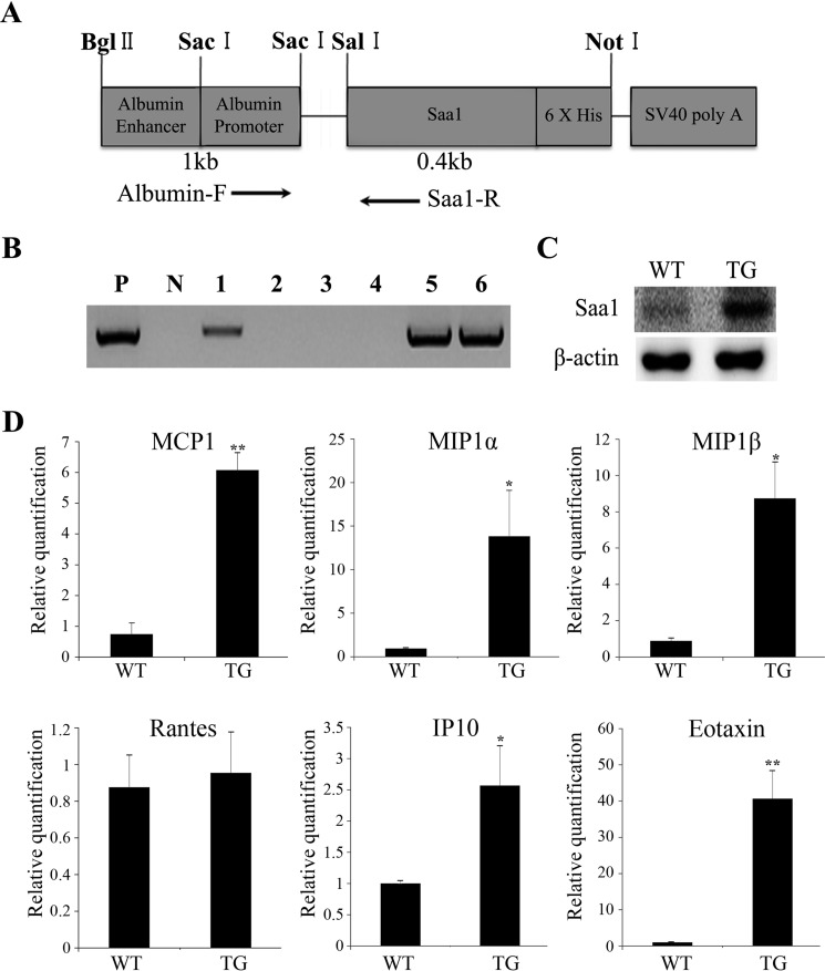
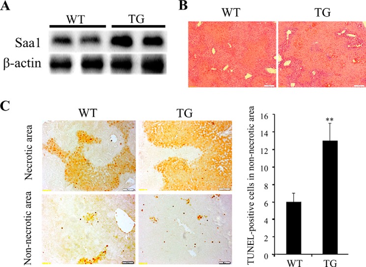
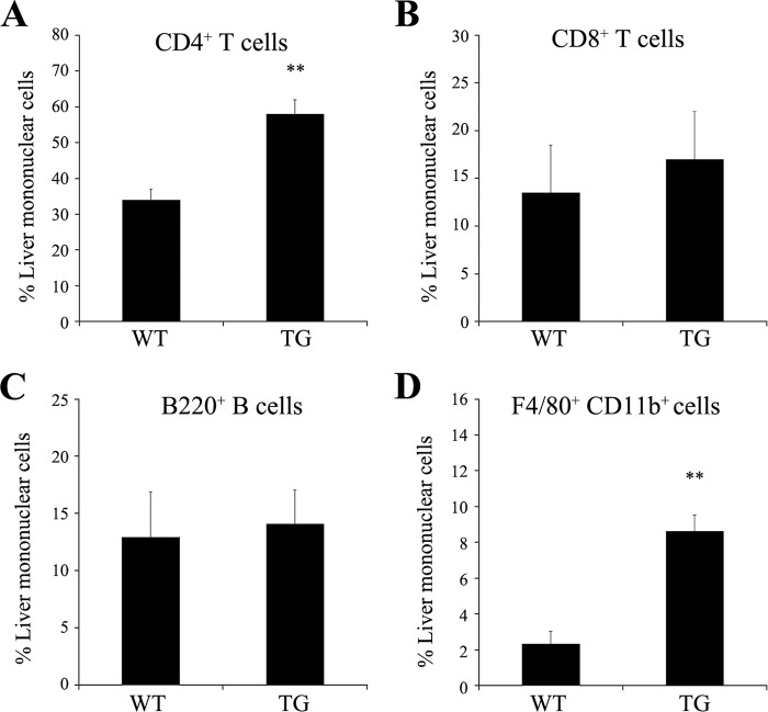
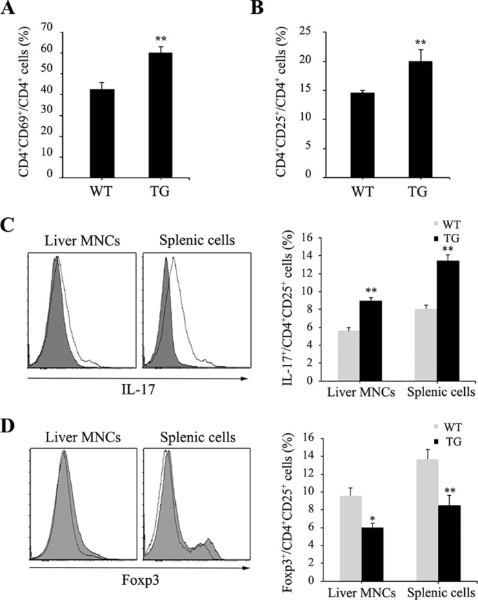
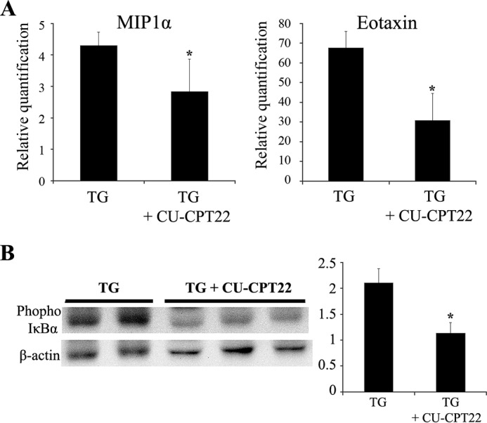
Serum amyloid A is a proinflammatory molecule that induces leukocyte infiltration and promotes neutrophil adhesion to endothelial cells under inflammatory conditions. The aim of this study was to examine whether Saa1 aggravates T cell-mediated hepatitis by inducing chemokines in a liver-specific, Saa1-overexpressing, transgenic (TG) mouse model. We generated TG mice in which Saa1 was overexpressed specifically in liver tissue. The chemokines monocyte chemotactic protein 1 (MCP1), MIP1α, MIP1β, interferon γ-induced protein 10 (IP-10), and eotaxin were induced in Saa1 TG mice. After concanavalin A treatment, Saa1 expression was higher in Saa1 TG mice than in WT mice. More severe liver injury, increased hepatocyte apoptosis, and higher levels of hepatic enzymes were observed in Saa1 TG mice than in WT mice. Liver infiltration of CD4(+) T cells and macrophages increased after inducing hepatitis. Activation of T cells was higher in Saa1 TG mice than in WT mice, and the populations of Th17 cells and regulatory T cells were altered by overexpressing Saa1 in TG mice. Secretion of various cytokines, such as interferon γ, tumor necrosis factor α, and interleukin 6, increased in Saa1 TG mice. Injecting a Toll-like receptor 2 (TLR2) antagonist in vivo inhibited chemokine expression and IκBα phosphorylation and showed that the induction of chemokines by Saa1 was dependent on TLR2. Hepatic Saa1 accelerated T cell-mediated hepatitis by inducing chemokine production and activating T cells by TLR2. Therefore, Saa1 might be a novel inflammatory factor that acts as a chemokine modulator in hepatitis.
Keywords: Saa1; T cell activation; Toll like receptor; chemokine; concanavalin A; cytokine; inflammation; liver injury; transgenic mice.
© 2015 by The American Society for Biochemistry and Molecular Biology, Inc.
Figures

Similar articles
-
Over-expression of Roquin aggravates T cell mediated hepatitis in transgenic mice using T cell specific promoter.Biochem Biophys Res Commun. 2014 Sep 26;452(3):822-7. doi: 10.1016/j.bbrc.2014.09.001. Epub 2014 Sep 6. Biochem Biophys Res Commun. 2014. PMID: 25201726
-
Hepatic serum amyloid A1 upregulates interleukin-17 (IL-17) in γδ T cells through Toll-like receptor 2 and is associated with psoriatic symptoms in transgenic mice.Scand J Immunol. 2019 Jun;89(6):e12764. doi: 10.1111/sji.12764. Epub 2019 Apr 4. Scand J Immunol. 2019. PMID: 30892738
-
Ablation of interaction between IL-33 and ST2+ regulatory T cells increases immune cell-mediated hepatitis and activated NK cell liver infiltration.Am J Physiol Gastrointest Liver Physiol. 2016 Aug 1;311(2):G313-23. doi: 10.1152/ajpgi.00097.2016. Epub 2016 Jun 23. Am J Physiol Gastrointest Liver Physiol. 2016. PMID: 27340126
-
Role of IL-17 and Th17 cells in liver diseases.Clin Dev Immunol. 2011;2011:345803. doi: 10.1155/2011/345803. Epub 2010 Dec 15. Clin Dev Immunol. 2011. PMID: 21197451 Free PMC article. Review.
-
Functional role of chemokines in liver disease models.Nat Rev Gastroenterol Hepatol. 2010 Dec;7(12):682-90. doi: 10.1038/nrgastro.2010.168. Epub 2010 Oct 26. Nat Rev Gastroenterol Hepatol. 2010. PMID: 20975742 Review.
Cited by
-
Toll-like receptor 2 activation and up-regulation by high mobility group box-1 contribute to post-operative neuroinflammation and cognitive dysfunction in mice.J Neurochem. 2021 Jul;158(2):328-341. doi: 10.1111/jnc.15368. Epub 2021 May 5. J Neurochem. 2021. PMID: 33871050 Free PMC article.
-
Serum amyloid A1 is involved in amyloid plaque aggregation and memory decline in amyloid beta abundant condition.Transgenic Res. 2019 Dec;28(5-6):499-508. doi: 10.1007/s11248-019-00166-x. Epub 2019 Aug 12. Transgenic Res. 2019. PMID: 31407125
-
Serum SAA1 and APOE are novel indicators for human cytomegalovirus infection.Sci Rep. 2017 Oct 17;7(1):13407. doi: 10.1038/s41598-017-13591-x. Sci Rep. 2017. PMID: 29042594 Free PMC article.
-
The JAK inhibitor tofacitinib ameliorates immune‑mediated liver injury in mice.Mol Med Rep. 2019 Dec;20(6):4883-4892. doi: 10.3892/mmr.2019.10750. Epub 2019 Oct 16. Mol Med Rep. 2019. PMID: 31638166 Free PMC article.
-
Characterization of Hepatic Dysfunction in Subjects Diagnosed With Chronic GVHD by NIH Consensus Criteria.Transplant Cell Ther. 2022 Nov;28(11):747.e1-747.e10. doi: 10.1016/j.jtct.2022.07.017. Epub 2022 Jul 22. Transplant Cell Ther. 2022. PMID: 35878742 Free PMC article.
References
-
- Eggink H. F., Houthoff H. J., Huitema S., Gips C. H., Poppema S. (1982) Cellular and humoral immune reactions in chronic active liver disease: I: lymphocyte subsets in liver biopsies of patients with untreated idiopathic autoimmune hepatitis, chronic active hepatitis B and primary biliary cirrhosis. Clin. Exp. Immunol. 50, 17–24 - PMC - PubMed
-
- Kanellopoulos P. N., Pavlou K., Perrakis A., Agianian B., Vorgias C. E., Mavrommatis C., Soufi M., Tucker P. A., Hamodrakas S. J. (1996) The crystal structure of the complexes of concanavalin A with 4′-nitrophenyl-α-d-mannopyranoside and 4′-nitrophenyl-α-d-glucopyranoside. J. Struct. Biol. 116, 345–355 - PubMed
-
- Knolle P. A., Gerken G., Loser E., Dienes H. P., Gantner F., Tiegs G., Meyer zum Buschenfelde K. H., Lohse A. W. (1996) Role of sinusoidal endothelial cells of the liver in concanavalin A-induced hepatic injury in mice. Hepatology 24, 824–829 - PubMed
-
- Tsui T. Y., Obed A., Siu Y. T., Yet S. F., Prantl L., Schlitt H. J., Fan S. T. (2007) Carbon monoxide inhalation rescues mice from fulminant hepatitis through improving hepatic energy metabolism. Shock 27, 165–171 - PubMed
-
- Ray A., Schatten H., Ray B. K. (1999) Activation of Sp1 and its functional co-operation with serum amyloid A-activating sequence binding factor in synoviocyte cells trigger synergistic action of interleukin-1 and interleukin-6 in serum amyloid A gene expression. J. Biol. Chem. 274, 4300–4308 - PubMed
Publication types
MeSH terms
Substances
LinkOut - more resources
Full Text Sources
Medical
Molecular Biology Databases
Research Materials
Miscellaneous
