. 2015 Apr 2;5(6):1129-43.
doi: 10.1534/g3.115.017707.

The Drosophila melanogaster Mutants apblot and apXasta Affect an Essential apterous Wing Enhancer

Affiliations

The Drosophila melanogaster Mutants apblot and apXasta Affect an Essential apterous Wing Enhancer

Dimitri Bieli et al. G3 (Bethesda). .

Abstract

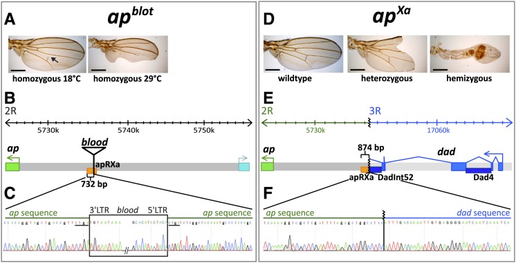
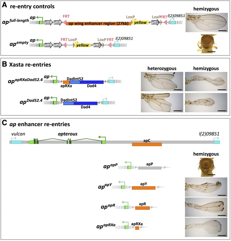
The selector gene apterous (ap) plays a key role during the development of the Drosophila melanogaster wing because it governs the establishment of the dorsal-ventral (D-V) compartment boundary. The D-V compartment boundary is known to serve as an important signaling center that is essential for the growth of the wing. The role of Ap and its downstream effectors have been studied extensively. However, very little is known about the transcriptional regulation of ap during wing disc development. In this study, we present a first characterization of an essential wing-specific ap enhancer. First, we defined an 874-bp fragment about 10 kb upstream of the ap transcription start that faithfully recapitulates the expression pattern of ap in the wing imaginal disc. Analysis of deletions in the ap locus covering this element demonstrated that it is essential for proper regulation of ap and formation of the wing. Moreover, we showed that the mutations ap(blot) and ap(Xasta) directly affect the integrity of this enhancer, leading to characteristic wing phenotypes. Furthermore, we engineered an in situ rescue system at the endogenous ap gene locus, allowing us to investigate the role of enhancer fragments in their native environment. Using this system, we were able to demonstrate that the essential wing enhancer alone is not sufficient for normal wing development. The in situ rescue system will allow us to characterize the ap regulatory sequences in great detail at the endogenous locus.

Keywords: Drosophila; apterous; boundary; compartment.


Figures

Figure 1
Figure 1
LacZ reporter assay and deletion analysis at the apterous locus. (A) Diagrammatic representation of the ap locus. As drawn at the top of the panel, it extends over roughly 50 kb. Its transcribed part is shown in green. ap is flanked by two genes indicated in blue: vulcan on the proximal and l(2)09851 on its distal side. Arrows above the genomic interval specify the direction of transcription of the three genes. Fragment apC, indicated in orange, has been reported to drive reporter expression in the dorsal compartment of the pouch, the hinge and the notum of the wing imaginal disc, where ap is normally expressed. Below, the relative positions and dimensions of nine fragments tested with our LacZ reporter assay are depicted. Fragments colored in orange (apO, apR, apOR, apOR3, and apRXa) elicit the same expression pattern as apC. Fragments depicted in gray (apP, apQ, apS, apOR2) do not drive reporter gene expression in the wing disc. (B) X-Gal staining in the wing disc of an apC-LacZ transgenic fly. Scale bar: 100 µm. (C) Deletions generated at the endogenous ap locus with FRT-containing inserts. At the top of the panel, triangles along the ap locus indicate the position of six different inserts. Pink arrowheads within them mark the orientation of the FRT sites according to the definition of Thibault et al. (2004). The location of the apRXa fragment is shown in orange. apDG3 deletes approximately 44 kb between inserts apf08090 to ape01573, thereby removing most of ap ORF and upstream sequences. apDG8 is a 20-kb deficiency that deletes the complete ap ORF from apf00878 to apD5f.1. apDG1 removes the complete intergenic spacer between apMM to ape01573. apDG11 deletes an 11-kb fragment from apMM to apDD35.34. Note that apD5f.1 and apMM have exactly the same insertion site. (D) Notum pictures of a wild-type fly and trans-heterozygous ap mutants. In the wild type, the wing and the haltere (arrowhead) are well formed and clearly visible. Df(2R)BSC696 is a large deletion at the base of 2R, deleting approximately 360 kb, including the whole ap locus. When Df(2R)BSC696 is crossed to apDG3 all wing and haltere structures are lost. Only small stumps of amorphic tissue remain at the actual attachment site of the wing (see arrow).Very similar phenotypes are observed in apDG8/apDG3, apDG1/apDG3 and apDG11/apDG3 flies. Scale bar: 25 µm.
Figure 2
Figure 2
The mutations apblot and apXa affect the ap wing enhancer region. (A) Temperature-sensitive wing phenotypes obtained for the homozygous apblot allele. At 18°, less than 30% of the wings are affected and most of them only show a disruption of the posterior crossvein (arrow). At 29°, ∼70% of the wings have a phenotype. In many of them, the posterior compartment is severely affected. (B) At the top of the panel, the coordinates of the apterous locus are indicated. The insertion site of blood, a retrotransposable element, within the apRXa wing enhancer is depicted. (C) Sequence data close to the insertion site of the blood element in apblot. The insertion causes a four bp duplication (CTGA, underlined). Exact coordinates of the 4 bp duplication: 2R:5735176.0.5735179 (Flybase Release FB2014_06). (D) Preparations of wild type and apXa mutant wings. All apXa/+ flies show a dominant phenotype: the distal part of the wing blade is lost and the characteristic mitten phenotype is formed. In hemizygous condition, the wing tissue of apXa/apDG3 flies forms a short tube-like structure. Margin bristles are absent except for sometimes a few at the tip. All scale bars are 50 µm. (E) Molecular characteristics of the apXa mutation. A reciprocal translocation involving the right arms of the second and third chromosome causes a breakpoint just upstream of the apRXa wing enhancer (indicated in orange) and juxtaposes the daughters against dpp (dad) locus (indicated in blue) next to the ap gene. The dark blue rectangles represent the well-studied cis-regulatory elements Dadint52 and Dad4 which are active in the wing disc (Weiss et al. 2010). (F) Chromatograph of the apXa sequence across the rearrangement break point. The coordinates of the breakpoints are: 2R:5375319 and 3R:17065902 (Flybase Release FB2014_06).
Figure 3
Figure 3
Wing disc phenotypes in apblot and apXa. All discs are shown anterior to the left and dorsal side up. (A) in situ hybridization against ap mRNA in late 3rd instar larval wing discs. In wild type, the dorsal compartment of the wing pouch is filled and outlined by the ap transcript. apblot discs show reduced ap mRNA levels. At 18°, the ap expression pattern remains very similar as that in wild type. At 29°, expression of ap in the posterior compartment is disturbed and the tissue is deformed (see arrow). In heterozygous apXa discs, ectopic ap expression is seen in the ventral part of the wing disc, with the strongest signal in median regions. The black arrows point to the edges of the disc where ap transcript is absent. In hemizygous apXa/apDG3 larvae, a similar pattern is observed. Note the change in shape of the wing disc. (B) α-Wg antibody staining of 3rd instar wing discs. In wild type, a characteristic thin stripe of Wg traverses the wing pouch along the D-V compartment boundary. In apblot, Wg expression is normal at 18°C. At 29°C, the Wg stripe is much weaker and less well defined in posterior regions of the wing pouch. In apXa/+ discs, the Wg stripe is interrupted in the median pouch region. In hemizygous apXa discs, the Wg stripe is lost and only a dot of Wg expression in the middle of the pouch is visible. In addition, the size of the pouch is reduced. (C) GFP expression driven by the Dad4 enhancer is detected in the central part of an apXa/+ wing disc. Note that absence of Wg stripe correlates well with higher GFP levels. Therefore, stripe formation is more affected in the anterior than in the posterior compartment. (D) α-GFP and α-Wg antibody staining of an ap::GFP/apXa wing disc. GFP expression is restricted to the dorsal compartment of the wing pouch. In particular, Ap-GFP fusion protein does not spread ventrally where the Wg stripe is interrupted. This indicates that dad enhancers on the apXasta chromosome are unable to activate ap::GFP located on the homologous chromosome. (E) Expression of Beadex- and fringe-Gal4 enhancer trap lines in wild type and apXa/+ discs. Note that ectopic expression (white arrows) of these two validated Ap targets in the ventral compartment is only detected where the Wg stripe is interrupted. All scale bars are 100 µm.
Figure 4
Figure 4
Margin formation in adult wings depends on well-defined On-Off Apterous expression levels during larval development. All discs are shown anterior to the left and dorsal side up. (A−E) 3rd instar imaginal wing discs showing UAS-GFP patterns (in green) elicited by the five Gal4 drivers indicated at the top of the panel. α-Wg antibody staining (in red) outlines the pouch and the position of the D−V compartment boundary. (A´−E´) α-Wg antibody staining. The effect of ectopic Ap production as a consequence of Gal4 > EY03046 on D-V boundary formation is shown. (A´´−E´´) Adult wings as obtained after ectopic Ap expression in (A´´) actin > EY03046, (B´´) Dad4 > EY03046, (C´´) salE > EY03046, (D´´) dpp > EY03046, and (E´´) ptc > EY03046 animals. In (D´´), the arrowhead points to a small lesion near the tip of the wing. Scale bars in (A−F) and (A´−F´) are 100 µm. Scale bars in (A´´−F´´) are 50 µm. (F) Insertion site of P{EPgy2}EY03046 relative to the ap TSS is shown. The triangle depicts the structure of the transgene. The red box corresponds to the mini-white marker, the yellow box to the yellow marker and the blue oval to an array of UAS sites. Arrows specify the transcriptional direction of mini-white, yellow, and the UAS-driven promoter. P{EPgy2} transgenes are intended for regulated expression of genes proximate to the site of the insertion: genes in direct orientation with respect to the UAS-controlled promoter can be conditionally expressed via transgene-derived Gal4 activity (Bellen et al. 2004). Note that at apterous, the UAS-driven promoter is at a considerable distance from and in opposite orientation to the apterous promoter (shown in green). We propose that in Gal4 > EY03046 flies, Gal4 activates ap transcription in much the same way as the eye-specific GMR-Gal4{w-} driver boosts mini white expression in GMR-Gal4{w-}/EY03046 flies. These have red eyes while the eye pigmentation in EY03046/+ flies is faint yellow (M. Müller, unpublished data). Drawing not to scale.
Figure 5
Figure 5
Generation of the in situ rescue system at the endogenous ap locus. (A−B) Direct gene conversion at apterous. P-element insertion apMM located ∼400 bp upstream of the ap TSS was previously isolated. By mobilization of apMM and concomitant injection of plasmid pLAPGPRA, fly line apc1.4b could be isolated. It contains two inverted attP sites flanking a GFP reporter. (B−C) ΦC31-integrase mediated site-specific recombination. By injection of plasmid pBSattBattPLoxFRTy, new attP, LoxP, and FRT sites were introduced into the ap locus. Note that pBSattBattPLoxFRTy can insert in two different attP sites leading to oppositely oriented insertions. apattBPFRTy2 is the appropriate one for our purpose. (C−D) Flipase-mediated deletion. Trans-heterozygous apattBPFRTy2/ape01573 animals were repeatedly treated with Flipase during larval stages. Among the progeny of these flies, apattPΔEnh could be isolated. It lacks the 27kb intergenic spacer but retains a strategically positioned attP site. (D−E) apattPΔEnh serves as a platform to reinsert enhancer fragments. These are cloned into pEnh-Reentry. This plasmid is injected into young embryos and integrates into the ap locus by ΦC31-integrase mediated recombination. Transgenics of the type apEnh-Reentry can be isolated thanks to the yellow marker. If desired, yellow can be removed by Cre-treatment. In addition, the complete insert can be excised by Flipase treatment.
Figure 6
Figure 6
Testing dad and ap enhancers in the endogenous apterous locus. (A) Positive control: the whole 27-kb ap wing enhancer region was re-inserted and apfull-length flies obtained. In apfull-length/apDG3 animals, perfectly wild-type wings are formed. Negative control: the empty pEnh-Reentry plasmid gave rise to apempty flies. No wing tissue is formed in apempty/apDG3 adults. (B) Xasta phenocopies are obtained with apapRXaDad52.4 and apDad52.4 alleles. apapRXaDad52.4 contains three enhancer elements: apRXa, DadInt52, and Dad4. Heterozygous flies only produce rather weak phenocopies. The junction between wing vein L2 and the margin (see arrow head) is present in almost 100% of the wings. apDad52.4 contains only the 2 dad enhancers. Faithful phenocopies of apXa/+ wings where the junction between vein L2 and the margin is missing are often observed. The wing phenotypes of hemizygous apapRXaDad52.4, apDad52.4 and apXa are similar: tube-like wing stumps of variable length are formed. Margin bristles are absent except for sometimes a few at the tip of the wing. (C) Testing the wing enhancer activity of four apC derivatives. At the top of the panel, the ap locus is depicted. Below, the positions of fragments apP, apY, apR and apRXa are shown relative to apC. The respective wing phenotypes in hemizygous condition are shown to the right of the corresponding fragments. Flies transgenic for the gray apP fragment behave like a true null allele: no wings are formed. Fragments drawn in orange have partial rescue activity: inflated wings are formed, where most of the margin and the alula are missing. The hinge is poorly formed. Note that in B and C, for space reasons, parts of the reentry plasmid have been omitted. All scale bars are 50 µm.

Similar articles

Cited by

References

    1. Affolter M., Basler K., 2007. The decapentaplegic morphogen gradient: from pattern formation to growth regulation. Nat. Rev. Genet. 8: 663–674. - PubMed
    1. Ahmad K., Henikoff S., 2001. Modulation of a transcription factor counteracts heterochromatic gene silencing in Drosophila. Cell 104: 839–847. - PubMed
    1. Alexandre C., Jacinto A., Ingham P. W., 1996. Transcriptional activation of hedgehog target genes in Drosophila is mediated directly by the cubitus interruptus protein, a member of the GLI family of zinc finger DNA-binding proteins. Genes Dev. 10: 2003–2013. - PubMed
    1. Alexandre C., Baena-Lopez A., Vincent J.-P., 2014. Patterning and growth control by membrane-tethered Wingless. Nature 505: 180–185. - PMC - PubMed
    1. Ashburner M., 1989. Drosophila: A Laboratory Handbook and Manual (two volumes) Cold Spring Harbor Laboratory Press, Cold Spring Harbor, New York.

Publication types

LinkOut - more resources