Review
. 2015 Jun;79(2):193-224.
doi: 10.1128/MMBR.00052-14.

Regulation of bacterial virulence by Csr (Rsm) systems

Affiliations
Review

Regulation of bacterial virulence by Csr (Rsm) systems

Christopher A Vakulskas et al. Microbiol Mol Biol Rev. 2015 Jun.

Abstract

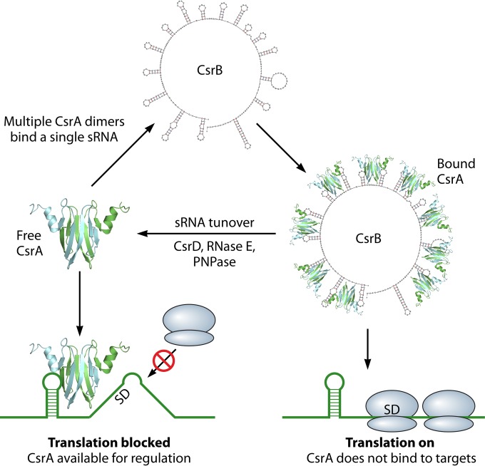
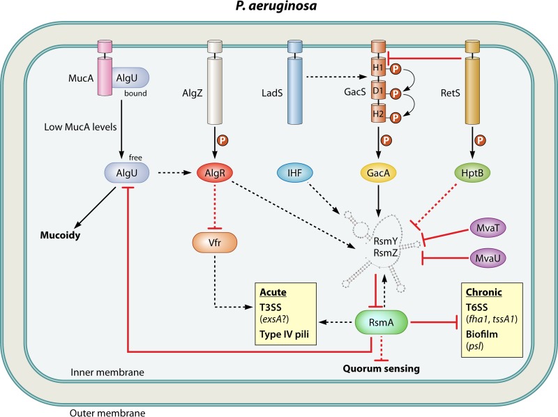
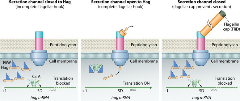
Most bacterial pathogens have the remarkable ability to flourish in the external environment and in specialized host niches. This ability requires their metabolism, physiology, and virulence factors to be responsive to changes in their surroundings. It is no surprise that the underlying genetic circuitry that supports this adaptability is multilayered and exceedingly complex. Studies over the past 2 decades have established that the CsrA/RsmA proteins, global regulators of posttranscriptional gene expression, play important roles in the expression of virulence factors of numerous proteobacterial pathogens. To accomplish these tasks, CsrA binds to the 5' untranslated and/or early coding regions of mRNAs and alters translation, mRNA turnover, and/or transcript elongation. CsrA activity is regulated by noncoding small RNAs (sRNAs) that contain multiple CsrA binding sites, which permit them to sequester multiple CsrA homodimers away from mRNA targets. Environmental cues sensed by two-component signal transduction systems and other regulatory factors govern the expression of the CsrA-binding sRNAs and, ultimately, the effects of CsrA on secretion systems, surface molecules and biofilm formation, quorum sensing, motility, pigmentation, siderophore production, and phagocytic avoidance. This review presents the workings of the Csr system, the paradigm shift that it generated for understanding posttranscriptional regulation, and its roles in virulence networks of animal and plant pathogens.


Figures

FIG 1
FIG 1
Structure of CsrA/RsmA/RsmE orthologs and the RsmE-hcnA RNA complex. (A) Protein secondary structure is shown at the top, with β-strands and α-helices depicted as arrows and cylinders, respectively, and amino acid sequence comparisons are shown immediately below. Sequence alignments from the Gammaproteobacteria (top) and from species containing the csrA gene in proximity to fliW (bottom) are depicted. Red boxes indicate the locations of conserved residues that are important for RNA binding and in vivo regulation in E. coli (24). (B) Ribbon diagram of the RsmE dimer (P. fluorescens) in a 1:2 complex with a 20-nucleotide segment of hcnA RNA. Individual RsmE polypeptides are colored with green or cyan, and the hcnA ribose-phosphate backbone is shown in orange. The critical R44 and L4 (red boxes) residues of RsmE and the GGA (blue boxes) recognition motif present on each hcnA molecule are indicated. The PDB structure file for RsmE-hcnA (2JPP) was downloaded from the RCSB Protein Data Bank (http://www.rcsb.org/), and the protein structure was rendered using PyMOL. (Adapted from reference by permission from Macmillan Publishers Ltd., copyright 2007.) (C) Two-dimensional interpretation of the interaction of RsmE with hcnA RNA. Amino acid residues contributed by an individual RsmE polypeptide are shown in green or cyan. Hydrogen bonds and hydrophobic interactions are indicated with dashed blue lines and orange highlights, respectively. The effects of alanine substitution on in vivo regulatory activities (% of wild type [WT], top) and in vitro binding affinities (Kd, bottom) for the E. coli CsrA protein were determined by Mercante et al. (24). Asterisks indicate amino acid positions that differ between E. coli CsrA and P. fluorescens RsmE. (Adapted from reference by permission from Macmillan Publishers Ltd., copyright 2007.)
FIG 2
FIG 2
Models for repression and activation by CsrA/RsmA. (A) E. coli CsrA represses glgC translation by competing with ribosome (30S) binding. (Top) CsrA homodimer first binds to a high-affinity site present in the single-stranded region of an RNA hairpin, located in the 5′ untranslated leader of the glgC transcript (20, 21, 38). The tethered CsrA homodimer can then bind via its available RNA binding surface to a low-affinity site that overlaps the SD sequence, thus blocking ribosome binding. (Bottom) In the absence of free CsrA, the ribosome can bind to the SD sequence, and translation can proceed. (B) P. aeruginosa RsmA represses translation of psl by stabilizing a stem-loop structure that sequesters the RBS. (Top) RsmA can bind to a single site (GGA) present in the 5′ untranslated leader of the psl transcript (40). RsmA binding stabilizes an RNA hairpin formed between the SD and anti-SD sequences, thus blocking ribosome access. (Bottom) In the absence of RsmA, the predicted hairpin structure is unstable, and ribosome binding and translation can proceed. (C) E. coli CsrA binding promotes Rho-dependent transcription termination of pgaA. (Top) CsrA binds to six sites in the pgaA mRNA (29), two of which are located in a segment that forms a hairpin in the absence of CsrA (53). CsrA binding prevents hairpin formation and exposes rut sites for entry of Rho transcription termination protein. Rho binding leads to premature termination (dashed line) of pgaA transcription. (Bottom) In the absence of CsrA, rut sites are shielded by RNA base pairing, Rho is unable to bind pgaA mRNA, and transcription can proceed. (D) P. aeruginosa RsmA binding to the phz2 untranslated leader prevents the formation of secondary structure that blocks translation (45). (E) E. coli activates translation of moaA by altering RNA structure. (Top) CsrA binds to two sites (GGA) present within the predicted moaA riboswitch aptamer that overlaps the SD (35). CsrA binding alters the aptamer structure, which reveals the SD for ribosome binding. (Bottom) In the absence of CsrA, the riboswitch aptamer sequesters the SD, thus blocking ribosome access. (F) E. coli CsrA stabilizes flhDC by preventing endonuclease cleavage by RNase E. (Top) CsrA binds to sites (GGA) present in the single-stranded region of two RNA hairpins located at the 5′ end of flhDC (47). CsrA binding prevents 5′ end-dependent cleavage by RNase E. (Bottom) In the absence of CsrA, RNase E binds to the 5′ monophosphorylated end of flhDC and performs several cleavages that initiate turnover of the transcript (47).
FIG 3
FIG 3
Outline of the Csr system in E. coli. CsrA binding to target mRNAs can have several regulatory outcomes: blocking translation initiation (as shown), stabilizing or destabilizing mRNA, or resulting in premature transcriptional termination. The concentration of free CsrA and therefore its regulatory activity depends on the levels of inhibitory small RNAs (CsrB is shown here). These sRNAs can bind to multiple CsrA dimers with high affinity and prevent them from binding target mRNAs. sRNA levels are regulated at the level of transcription (not shown) and turnover. Ribosomes are depicted in blue.
FIG 4
FIG 4
Csr regulatory circuitry in E. coli. The inner and outer membranes are indicated. Solid lines indicate regulatory connections with molecular mechanisms supported by experimental evidence, whereas dashed lines show regulatory effects for which a mechanism is lacking. Activation or repression is indicated by a black arrowhead or red T-bar, respectively. A phosphoryl group is indicated by “P.” See the accompanying text for details.
FIG 5
FIG 5
Rsm regulatory circuitry in P. aeruginosa, depicted as in Fig. 4.
FIG 6
FIG 6
Structural comparison between RsmE of P. fluorescens and RsmF (RsmN) of P. aeruginosa. (A and C) Cartoons depicting the NMR solution structure of RsmE and crystal structure of RsmF (157, 175). Individual polypeptides of the RsmE homodimer are shown in green or cyan, RsmF polypeptides are shown in blue or maroon, and secondary structural elements are labeled with subscript “A” or “B,” as shown. (B and D) Structures of RsmE (top) and RsmF (RsmN) (bottom) rotated 90°. The β-strands that form the RNA interaction surfaces are highlighted using the coloring scheme from panels A and C. Protein structures were rendered using PyMOL. The PDB structure files for RsmE-hcnA (2JPP) and RsmF (4K59) were downloaded from the RCSB Protein Data Bank (http://www.rcsb.org/).
FIG 7
FIG 7
Csr regulatory circuitry in in L. pneumophila, depicted as in Fig. 4. Pentagons depict the autoinducer LAI-1.
FIG 8
FIG 8
Csr regulatory circuitry in V. cholerae, depicted as in Fig. 4. Autoinducers CAI-1 and AI-2 are shown as pentagons and triangles, respectively. (Adapted from reference with permission of the publisher.)
FIG 9
FIG 9
Model for CsrA-FliW-Hag control of flagellin homeostasis in B. subtilis. Flagellin homeostasis is controlled by a partner-switching mechanism involving FliW (dark blue triangles), CsrA, and Hag (orange barbells). The basal body (blue and pink), flagellar hook (cyan), filament (orange), cytoplasmic membrane, and peptidoglycan layer are indicated. (Adapted from reference with permission of the publisher.)
FIG 10
FIG 10
The csrA and fliW genes tend to cluster in the genomes of many taxonomically diverse species, with the notable exception of the Gammaproteobacteria, which lack FliW. Gene neighborhoods around csrA (blue) and/or fliW (dark green) in Clostridium botulinum A strain ATCC 19397, Clostridium difficile 630, Borrelia burgdorferi B31, Treponema pallidum subsp. pallidum strain Nichols, Petrotoga mobilis SJ95, Thermotoga maritima MSB8, Geobacter sulfurreducens PCA, Campylobacter jejuni NCTC 11168, Helicobacter pylori J99, Escherichia coli MG1655, Pseudomonas aeruginosa PAO1 and Vibrio cholerae MZO-2 are shown. The associated phylum and class taxonomic information is shown to the left of and immediately beneath the species names. Protein-coding genes are represented as arrows, and tRNA genes are represented with gray bars. Flagellin genes are colored orange, and other motility-associated genes are colored pale green. Slashes indicate that csrA and fliW are not within the same genomic neighborhood of a species. Annotation and gene position information was determined using MicrobesOnline (http://www.microbesonline.org/) and reference .
None
None
None
None
None

Similar articles

Cited by

References

    1. Carmichael GG, Weber K, Niveleau A, Wahba AJ. 1975. The host factor required for RNA phage Qβ RNA replication in vitro. Intracellular location, quantitation, and purification by polyadenylate-cellulose chromatography. J Biol Chem 250:3607–3612. - PubMed
    1. Babitzke P, Baker CS, Romeo T. 2009. Regulation of translation initiation by RNA binding proteins. Annu Rev Microbiol 63:27–44. doi:10.1146/annurev.micro.091208.073514. - DOI - PMC - PubMed
    1. Romeo T, Vakulskas CA, Babitzke P. 2013. Post-transcriptional regulation on a global scale: form and function of Csr/Rsm systems. Environ Microbiol 15:313–324. doi:10.1111/j.1462-2920.2012.02794.x. - DOI - PMC - PubMed
    1. Heroven AK, Bohme K, Dersch P. 2012. The Csr/Rsm system of Yersinia and related pathogens: a post-transcriptional strategy for managing virulence. RNA Biol 9:379–391. doi:10.4161/rna.19333. - DOI - PubMed
    1. Lucchetti-Miganeh C, Burrowes E, Baysse C, Ermel G. 2008. The post-transcriptional regulator CsrA plays a central role in the adaptation of bacterial pathogens to different stages of infection in animal hosts. Microbiology 154:16–29. doi:10.1099/mic.0.2007/012286-0. - DOI - PubMed

Publication types

MeSH terms

LinkOut - more resources