. 2015 Sep;72(17):3355-73.
doi: 10.1007/s00018-015-1891-5. Epub 2015 Apr 1.

Intracellular EP2 prostanoid receptor promotes cancer-related phenotypes in PC3 cells

Affiliations

Intracellular EP2 prostanoid receptor promotes cancer-related phenotypes in PC3 cells

Ana Belén Fernández-Martínez et al. Cell Mol Life Sci. 2015 Sep.

Abstract

Prostaglandin E2 (PGE2) and hypoxia-inducible factor-1α (HIF-1α) affect many mechanisms that have been involved in the pathogenesis of prostate cancer (PC). HIF-1α, which is up-regulated by PGE2 in LNCaP cells and PC3 cells, has been shown to contribute to metastasis and chemo-resistance of castrate-resistant PC (a lethal form of PC) and to promote in PC cells migration, invasion, angiogenesis and chemoresistance. The selective blockade of PGE2-EP2 signaling pathway in PC3 cells results in inhibition of cancer cell proliferation and invasion. PGE2 affects many mechanisms that have been shown to play a role in carcinogenesis such as proliferation, apoptosis, migration, invasion and angiogenesis. Recently, we have found in PC3 cells that most of these PGE2-induced cancer-related features are due to intracellular PGE2 (iPGE2). Here, we aimed to study in PC3 cells the role of iPGE2-intracellular EP2 (iEP2)-HIF-1α signaling in several events linked to PC progression using an experimental approach involving pharmacological inhibition of the prostaglandin uptake transporter and EGFR and pharmacological and genetic modulation of EP2 receptor and HIF-1α. We found that iPGE2 increases HIF-1α expression through iEP2-dependent EGFR transactivation and that inhibition of any of the axis iEP2-EGFR-HIF-1α in cells treated with PGE2 or EP2 agonist results in prevention of the increase in PC3 cell proliferation, adhesion, migration, invasion and angiogenesis in vitro. Of note, PGE2 induced EP2 antagonist-sensitive DNA synthesis in nuclei isolated from PC3 cells, which indicates that they have functional EP2 receptors. These results suggest that PGE2-EP2 dependent intracrine mechanisms involving EGFR and HIF-1α play a role in PC.


Figures

Fig. 1
Fig. 1
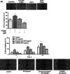
Role of EP2 receptor in PGE2-induced HIF-1α up-regulation. a Left treatment with PGE2 results in increased HIF-1α expression and its prevention by 1.9 μM EP2 antagonist PF04419948. PC3 cells were pre-incubated for 1 h with 1 μM PF04419948 and then, treated for 8 h with 1 µM PGE2. Afterwards, HIF-1α expression was assessed by Western blot. Right expression of HIF-1α expression increases upon treatment with EP2 receptor selective agonist ONO-AE1-259-01. Preventive effect of AH6809, a selective antagonist of EP2 receptors, and bromocresol green, an inhibitor of the PGT transporter. PC3 cells were pre-incubated for 1 h with 1 μM EP2 antagonist AH6809 or with 50 µM PGT inhibitor bromocresol green and then, incubated for 8 h with 0.5 µM EP2 agonist ONO-AE1-259-01. Afterwards, HIF-1α expression was assessed by Western blot. All experiments were repeated three times. b Left panel treatment with EP2 receptor agonist results in increased EGFR tyrosine phosphorylation. PC3 cells were incubated for up to 30 min with a selective agonist of EP2 receptor, 0.5 μM ONO-AE1-259-01 and tyrosine phosphorylation of EGFR was assessed by Western blot analysis. Right panel the inhibitor of the EGFR phosphorylation AG1478 prevents EP2 agonist-induced increase in EGFR tyrosine phosphorylation. PC3 cells were pre-treated with 1 µM AG1478 for 1 h and incubated with 0.5 μM EP2 agonist for 8 h and phosphorylation of EGFR was assessed by Western blot analysis. All experiments were repeated three times. c EP2 agonist determined a BG-sensitive increase in EP2 receptor expression. PC3 cells were pretreated with 50 μM bromocresol green for 1 h and the treated with 0.5 μM EP2 agonist for 8 h and the expression and location of EP2 receptor was assayed for immunocytochemistry
Fig. 2
Fig. 2
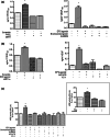
Intracellular EP2 receptors, in an AG1478-, YC-1-sensitive manner, mediate the increase in the proliferation of PC3 cells upon treatment with PGE2. a Intracellular EP2 receptors mediate in PGE2-induced proliferation of PC3 cells. PC3 cells were transfected with EP2 siRNA or control siRNA (scramble) as indicated in section “Materials and methods” and were treated with 1 µM PGE2 agonist for 24 h (upper panel left). Alternatively, PC3 cells were treated with 0.5 μM EP2 agonist ONO-AE1-259-01 or 1 μM alternative EP2 agonist butaprost for 24 h (upper panel right), and then cell proliferation was assessed by MTT assay as described in “Materials and methods”. Other set of cells were pre-treated with 50 µM PGT inhibitor bromocresol green or 1 µM EP2 antagonist AH6809 and then cell proliferation was assessed by MTT assay (middle panel) or BrdU incorporation assay (lower panel). b Nuclear EP receptors participate in the synthesis of DNA required for PGE2-induced cell proliferation. Upper panel nuclei of PC3 cells were isolated as indicated in “Materials and methods” section (the inset shows the purity of the nuclear fraction as assessed by immunoblotting with annexin II, hsp60 or nucleoporin p62; which are, respectively plasma membrane-, cytosol- or nucleus-specific proteins) and the expression of EP receptors was assayed by immunofluorescence assay. Lower panel PGE2 increased the DNA synthesis in isolated nuclei of PC3 cells. PC3 nuclei were isolated and pre-incubated with 1 µM EP2 antagonist AH6809 for 1 h and then, incubated with 1 µM PGE2 for 3 h. The incorporation of BrdU was analysed by immunofluorescence assay as indicated in section “Materials and methods”. c EGFR transactivation and HIF-1α participated in mediating the effect of intracellular PGE2 on PC3 cell proliferation. Upper panel cells were pretreated with 1 µM EGFR inhibitor AG1478 or 10 μM HIF-1α inhibitor YC-1 for 1 h and treated with 0.5 μM EP2 agonist ONO-AE1-259-01 for 24 h. Cell proliferation was assayed by MTT assay (left panel) or incorporation of BrdU (right panel). Lower panel PC3 cells were transfected with HIF-1α siRNA or control siRNA (scramble) as indicated in section “Materials and methods” and then, they were treated with 0.5 µM EP2 agonist ONO-AE1-259-01 for 24 h. Cell proliferation was assessed by MTT assay (left panel) or incorporation of BrdU (right panel). All experiments were repeated three times. Statistical analysis: *P < 0.01 vs all groups
Fig. 2
Fig. 2
Intracellular EP2 receptors, in an AG1478-, YC-1-sensitive manner, mediate the increase in the proliferation of PC3 cells upon treatment with PGE2. a Intracellular EP2 receptors mediate in PGE2-induced proliferation of PC3 cells. PC3 cells were transfected with EP2 siRNA or control siRNA (scramble) as indicated in section “Materials and methods” and were treated with 1 µM PGE2 agonist for 24 h (upper panel left). Alternatively, PC3 cells were treated with 0.5 μM EP2 agonist ONO-AE1-259-01 or 1 μM alternative EP2 agonist butaprost for 24 h (upper panel right), and then cell proliferation was assessed by MTT assay as described in “Materials and methods”. Other set of cells were pre-treated with 50 µM PGT inhibitor bromocresol green or 1 µM EP2 antagonist AH6809 and then cell proliferation was assessed by MTT assay (middle panel) or BrdU incorporation assay (lower panel). b Nuclear EP receptors participate in the synthesis of DNA required for PGE2-induced cell proliferation. Upper panel nuclei of PC3 cells were isolated as indicated in “Materials and methods” section (the inset shows the purity of the nuclear fraction as assessed by immunoblotting with annexin II, hsp60 or nucleoporin p62; which are, respectively plasma membrane-, cytosol- or nucleus-specific proteins) and the expression of EP receptors was assayed by immunofluorescence assay. Lower panel PGE2 increased the DNA synthesis in isolated nuclei of PC3 cells. PC3 nuclei were isolated and pre-incubated with 1 µM EP2 antagonist AH6809 for 1 h and then, incubated with 1 µM PGE2 for 3 h. The incorporation of BrdU was analysed by immunofluorescence assay as indicated in section “Materials and methods”. c EGFR transactivation and HIF-1α participated in mediating the effect of intracellular PGE2 on PC3 cell proliferation. Upper panel cells were pretreated with 1 µM EGFR inhibitor AG1478 or 10 μM HIF-1α inhibitor YC-1 for 1 h and treated with 0.5 μM EP2 agonist ONO-AE1-259-01 for 24 h. Cell proliferation was assayed by MTT assay (left panel) or incorporation of BrdU (right panel). Lower panel PC3 cells were transfected with HIF-1α siRNA or control siRNA (scramble) as indicated in section “Materials and methods” and then, they were treated with 0.5 µM EP2 agonist ONO-AE1-259-01 for 24 h. Cell proliferation was assessed by MTT assay (left panel) or incorporation of BrdU (right panel). All experiments were repeated three times. Statistical analysis: *P < 0.01 vs all groups
Fig. 3
Fig. 3
Intracellular EP2 receptors, in an 1 μM EGFR inhibitor AG1478-, 10 μM HIF-1μ inhibitor YC-1-sensitive manner, promote PC3 cell adhesion. PC3 cells in suspension were pre-incubated for 1 h with 50 µM PGT inhibitor bromocresol green, 1 µM EGFR inhibitor AG1478, 1 µM EP2 antagonist AH6809 or 10 µM HIF-1α inhibitor YC-1 and treated with 0.5 µM EP2 agonist ONO-AE1-259-01. Then cells were plated in collagen coated 96-well plates (25 × 103 cells per well) and incubated for up to 120 min. The number of adherent cells was assessed by the MTT assay. All experiments were repeated three times. Statistical analysis: *P < 0.01 vs all groups
Fig. 4
Fig. 4
Intracellular EP2 receptors, in an EGFR inhibitor AG1478-sensitive manner, promote HIF-1α-dependent PC3 cell motility. a PGE2-induced tumour cell motility is dependent on intracellular EP2 receptors. PC3 cells were transfected with EP2 siRNA or control siRNA (scramble) as indicated in section “Materials and methods” and were seeded in 24-well plates until confluent (upper panel) and treated with or without 1 μM PGE2 (upper panel). Other set of cells was cultured in 24-well plates until confluent and then were pre-treated with 50 µM PGT inhibitor bromocresol green or 1 µM EP2 antagonist AH6809. Afterwards, PC3 cells were incubated with or without a selective 0.5 μM EP2 agonist ONO-AE1-259-01 (lower panel). Confluent cells were carefully wounded using sterile pipette tips. Cell migration or wound repair was photographed (0, 4, 8 and 20 h) and measured vs time 0 h. All experiments were repeated three times and a representative photograph is shown. Statistical analysis: *P < 0.01 vs control. b EGFR and HIF-1α mediate EP2 agonist-induced increase PC3 cell motility. Cells were pretreated with 1 µM EGFR inhibitor AG1478 or 10 μM HIF-1α YC-1 for 1 h and treated with 0.5 μM EP2 ONO-AE1-259-01 agonist and cell motility was measured as a. Inset PC3 cells were transfected with HIF-1α siRNA or control siRNA (scramble), were seeded in 24-well plates until confluent and treated with or without 1 μM PGE2. Cell motility was analysed as a
Fig. 4
Fig. 4
Intracellular EP2 receptors, in an EGFR inhibitor AG1478-sensitive manner, promote HIF-1α-dependent PC3 cell motility. a PGE2-induced tumour cell motility is dependent on intracellular EP2 receptors. PC3 cells were transfected with EP2 siRNA or control siRNA (scramble) as indicated in section “Materials and methods” and were seeded in 24-well plates until confluent (upper panel) and treated with or without 1 μM PGE2 (upper panel). Other set of cells was cultured in 24-well plates until confluent and then were pre-treated with 50 µM PGT inhibitor bromocresol green or 1 µM EP2 antagonist AH6809. Afterwards, PC3 cells were incubated with or without a selective 0.5 μM EP2 agonist ONO-AE1-259-01 (lower panel). Confluent cells were carefully wounded using sterile pipette tips. Cell migration or wound repair was photographed (0, 4, 8 and 20 h) and measured vs time 0 h. All experiments were repeated three times and a representative photograph is shown. Statistical analysis: *P < 0.01 vs control. b EGFR and HIF-1α mediate EP2 agonist-induced increase PC3 cell motility. Cells were pretreated with 1 µM EGFR inhibitor AG1478 or 10 μM HIF-1α YC-1 for 1 h and treated with 0.5 μM EP2 ONO-AE1-259-01 agonist and cell motility was measured as a. Inset PC3 cells were transfected with HIF-1α siRNA or control siRNA (scramble), were seeded in 24-well plates until confluent and treated with or without 1 μM PGE2. Cell motility was analysed as a
Fig. 5
Fig. 5
Intracellular EP2 receptors, in an EGFR inhibitor AG1478-, HIF-1α inhibitor YC-1-sensitive manner, mediate the increase of PC3 cells invasion upon treatment with PGE2. a Intracellular EP2 receptors mediate PGE2-induced PC3 cell invasion. Cells in suspension (5 × 105 cells/ml) were transfected with EP2 siRNA or scramble and treated with 1 μM PGE2 (upper panel). Other set of cells was treated for 1 h with 50 µM PGT inhibitor bromocresol green or 1 µM PGE2 antagonist AH6809 and incubated with 0.5 μM EP2 agonist ONO-AE1-259-01 (lower panel). Then cells were seeded into transwell inserts which had been previously coated with Matrigel basement membrane matrix and invasive activity was assessed as indicated in “Materials and methods”. All experiments were repeated three times and a representative photograph (×10 magnification) is shown. Statistical analysis: *P < 0.01 vs scramble/control. b HIF-1α and EGFR play a relevant role in both basal and EP2 agonist-induced increase PC3 cell invasion. Cells were pretreated with 1 µM EP2 antagonist AG1478 or 10 μM HIF-1α inhibitor YC-1 for 1 h and treated with 0.5 μM EP2 agonist ONO-AE1-259-01 and invasive activity was measured as a. Inset PC3 cells were transfected with HIF-1α siRNA or control siRNA (scramble) and treated with or without 1 μM PGE2. Invasive activity was analysed as a
Fig. 5
Fig. 5
Intracellular EP2 receptors, in an EGFR inhibitor AG1478-, HIF-1α inhibitor YC-1-sensitive manner, mediate the increase of PC3 cells invasion upon treatment with PGE2. a Intracellular EP2 receptors mediate PGE2-induced PC3 cell invasion. Cells in suspension (5 × 105 cells/ml) were transfected with EP2 siRNA or scramble and treated with 1 μM PGE2 (upper panel). Other set of cells was treated for 1 h with 50 µM PGT inhibitor bromocresol green or 1 µM PGE2 antagonist AH6809 and incubated with 0.5 μM EP2 agonist ONO-AE1-259-01 (lower panel). Then cells were seeded into transwell inserts which had been previously coated with Matrigel basement membrane matrix and invasive activity was assessed as indicated in “Materials and methods”. All experiments were repeated three times and a representative photograph (×10 magnification) is shown. Statistical analysis: *P < 0.01 vs scramble/control. b HIF-1α and EGFR play a relevant role in both basal and EP2 agonist-induced increase PC3 cell invasion. Cells were pretreated with 1 µM EP2 antagonist AG1478 or 10 μM HIF-1α inhibitor YC-1 for 1 h and treated with 0.5 μM EP2 agonist ONO-AE1-259-01 and invasive activity was measured as a. Inset PC3 cells were transfected with HIF-1α siRNA or control siRNA (scramble) and treated with or without 1 μM PGE2. Invasive activity was analysed as a
Fig. 6
Fig. 6
Intracellular EP2 receptors mediate the increase in VEGF-A production induced by PGE2 in PC3 cells through HIF-1α-dependent activation of the HRE in the promoter of the VEGF-A gene. a iEP2 receptors mediate the increase in the production of VEGF-A induced by PGE2 in PC3 cells. Cells were transfected with EP2 siRNA or scramble and treated with 1 µM PGE2 for 24 h (left panel). Other set of cells were treated for 1 h with 50 µM bromocresol green or 1 µM AH6809 and incubated with 0.5 mM EP2 agonist ONO-AE1-259-01 for 24 h (right panel). The extracellular medium was collected to analysed the VEGF-A levels by ELISA assay.*P < 0.01 vs all groups. b HIF-1α, in an EGFR inhibitor AG1478-sensitive manner, mediates the iEP2-dependent increase in the production of VEGF-A upon treatment of PC3 cells with PGE2. Cells were pretreated with 1 µM EGFR inhibitor AG1478 or 10 µM HIF-1α inhibitor YC-1 for 1 h and treated with 0.5 µM EP2 agonist ONO-AE1-259-01 and VEGF production was measured as a. Inset PC3 cells were transfected with HIF-1α siRNA or control siRNA (scramble) and treated with or without 1 µM PGE2. VEGF levels were analysed as a, c EP2 agonist-induced increased activity of a hypoxia-responsive element (HRE)-driven reporter construct is abolished by an inhibitor of the PGT transporter, an inhibitor of the EGFR phosphorylation and a selective antagonist of EP2 receptor. PC3 cells were transiently transfected with the plasmid as described in section “Materials and methods”. Then, cells were pre-incubated with 50 µM PGT inhibitor bromocresol green, 1 µM EGFR inhibitor AG1478 or 1 µM EP2 antagonist AH6809 for 1 h and incubated with 1 µM PGE2 (left panel) or 0.5 µM EP2 agonist ONO-AE1-259-01 (center and right panel) for 4 h and luciferase activity in cell lysates was measured and expressed as Relative Luminescence Units (RLU/s). All experiments were repeated three times. Statistical analysis: *P < 0.01 vs all groups
Fig. 7
Fig. 7
Conditioned media collected from EP2 agonist treated PC3 cells increases tube formation by HUVECs in a bromocresol green-, AG1478-, AH6809- and YC-1-dependent manner. HUVECs cells were seeded on Matrigel-coated wells and cultured in conditioned media collected from PC3 cells after pre-treatment with 50 µM PGT inhibitor bromocresol green, 1 µM EGFR inhibitor AG1478, 1 µM EP2 antagonist AH6809 or 10 µM HIF-1α inhibitor YC-1, and incubated with 0.5 μM EP2 agonist ONO-AE1-259-01 for 24 h. Representative photographs of tube-like structures were showed

Similar articles

Cited by

References

    1. Badawi AF. The role of prostaglandin synthesis in prostate cancer. BJU Int. 2000;85:451–462. doi: 10.1046/j.1464-410x.2000.00507.x. - DOI - PubMed
    1. Wang D, Dubois RN. Eicosanoids and cancer. Nat Rev Cancer. 2010;10:181–193. doi: 10.1038/nrc2809. - DOI - PMC - PubMed
    1. Greenhough A, Smartt HJ, Moore AE, Roberts HR, Williams AC, Paraskeva C, Kaidi A. The COX-2/PGE2 pathway: key roles in the hallmarks of cancer and adaptation to the tumour microenvironment. Carcinogenesis. 2009;30:377–386. doi: 10.1093/carcin/bgp014. - DOI - PubMed
    1. Jiang J, Dingledine R. Role of prostaglandin receptor EP2 in the regulations of cancer cell proliferation, invasion, and inflammation. J Pharmacol Exp Ther. 2013;344:360–367. doi: 10.1124/jpet.112.200444. - DOI - PMC - PubMed
    1. Jain S, Chakraborty G, Raja R, Kale S, Kundu GC. Prostaglandin E2 regulates tumor angiogenesis in prostate cancer. Cancer Res. 2008;68:7750–7759. doi: 10.1158/0008-5472.CAN-07-6689. - DOI - PubMed

Publication types

MeSH terms