P2Y12 expression and function in alternatively activated human microglia
- PMID: 25821842
- PMCID: PMC4370387
- DOI: 10.1212/NXI.0000000000000080
P2Y12 expression and function in alternatively activated human microglia
Erratum in
-
Erratum: P2Y12 expression and function in alternatively activated human microglia.Neurol Neuroimmunol Neuroinflamm. 2015 May 7;2(3):e106. doi: 10.1212/NXI.0000000000000106. eCollection 2015 Jun. Neurol Neuroimmunol Neuroinflamm. 2015. PMID: 25977933 Free PMC article.
Abstract
Objective: To investigate and measure the functional significance of altered P2Y12 expression in the context of human microglia activation.
Methods: We performed in vitro and in situ experiments to measure how P2Y12 expression can influence disease-relevant functional properties of classically activated (M1) and alternatively activated (M2) human microglia in the inflamed brain.
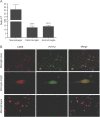
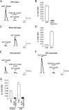
Results: We demonstrated that compared to resting and classically activated (M1) human microglia, P2Y12 expression is increased under alternatively activated (M2) conditions. In response to ADP, the endogenous ligand of P2Y12, M2 microglia have increased ligand-mediated calcium responses, which are blocked by selective P2Y12 antagonism. P2Y12 antagonism was also shown to decrease migratory and inflammatory responses in human microglia upon exposure to nucleotides that are released during CNS injury; no effects were observed in human monocytes or macrophages. In situ experiments confirm that P2Y12 is selectively expressed on human microglia and elevated under neuropathologic conditions that promote Th2 responses, such as parasitic CNS infection.
Conclusion: These findings provide insight into the roles of M2 microglia in the context of neuroinflammation and suggest a mechanism to selectively target a functionally unique population of myeloid cells in the CNS.
Figures


Similar articles
-
Cyclic AMP is a key regulator of M1 to M2a phenotypic conversion of microglia in the presence of Th2 cytokines.J Neuroinflammation. 2016 Jan 13;13:9. doi: 10.1186/s12974-015-0463-9. J Neuroinflammation. 2016. PMID: 26757726 Free PMC article.
-
Comparison of polarization properties of human adult microglia and blood-derived macrophages.Glia. 2012 May;60(5):717-27. doi: 10.1002/glia.22298. Epub 2012 Jan 30. Glia. 2012. PMID: 22290798
-
Alternatively activated microglia and macrophages in the central nervous system.Prog Neurobiol. 2015 Aug;131:65-86. doi: 10.1016/j.pneurobio.2015.05.003. Epub 2015 Jun 8. Prog Neurobiol. 2015. PMID: 26067058 Review.
-
Microglia control the spread of neurotropic virus infection via P2Y12 signalling and recruit monocytes through P2Y12-independent mechanisms.Acta Neuropathol. 2018 Sep;136(3):461-482. doi: 10.1007/s00401-018-1885-0. Epub 2018 Jul 19. Acta Neuropathol. 2018. PMID: 30027450 Free PMC article.
-
Diversity and plasticity of microglial cells in psychiatric and neurological disorders.Pharmacol Ther. 2015 Oct;154:21-35. doi: 10.1016/j.pharmthera.2015.06.010. Epub 2015 Jun 27. Pharmacol Ther. 2015. PMID: 26129625 Review.
Cited by
-
Inhibition of REV-ERBs stimulates microglial amyloid-beta clearance and reduces amyloid plaque deposition in the 5XFAD mouse model of Alzheimer's disease.Aging Cell. 2020 Feb;19(2):e13078. doi: 10.1111/acel.13078. Epub 2019 Dec 4. Aging Cell. 2020. PMID: 31800167 Free PMC article.
-
Advances in Visualizing Microglial Cells in Human Central Nervous System Tissue.Biomolecules. 2022 Apr 19;12(5):603. doi: 10.3390/biom12050603. Biomolecules. 2022. PMID: 35625531 Free PMC article. Review.
-
An overview on microglial origin, distribution, and phenotype in Alzheimer's disease.J Cell Physiol. 2024 Jun;239(6):e30829. doi: 10.1002/jcp.30829. Epub 2022 Jul 13. J Cell Physiol. 2024. PMID: 35822939 Review.
-
Microglia in Alzheimer's Disease: Exploring How Genetics and Phenotype Influence Risk.J Mol Biol. 2019 Apr 19;431(9):1805-1817. doi: 10.1016/j.jmb.2019.01.045. Epub 2019 Feb 7. J Mol Biol. 2019. PMID: 30738892 Free PMC article. Review.
-
LILRB2-mediated TREM2 signaling inhibition suppresses microglia functions.Mol Neurodegener. 2022 Jun 18;17(1):44. doi: 10.1186/s13024-022-00550-y. Mol Neurodegener. 2022. PMID: 35717259 Free PMC article.
References
-
- Moore CS, Rao VT, Durafourt BA, et al. miR-155 as a multiple sclerosis-relevant regulator of myeloid cell polarization. Ann Neurol 2013;74:709–720. - PubMed
-
- Durafourt BA, Moore CS, Zammit DA, et al. Comparison of polarization properties of human adult microglia and blood-derived macrophages. Glia 2012;60:717–727. - PubMed
-
- Lambert C, Ase AR, Seguela P, Antel JP. Distinct migratory and cytokine responses of human microglia and macrophages to ATP. Brain Behav Immun 2010;24:1241–1248. - PubMed
-
- Williams K, Ulvestad E, Antel JP. B7/BB-1 antigen expression on adult human microglia studied in vitro and in situ. Eur J Immunol 1994;24:3031–3037. - PubMed
-
- Williams K, Bar-Or A, Ulvestad E, Olivier A, Antel JP, Yong VW. Biology of adult human microglia in culture: comparisons with peripheral blood monocytes and astrocytes. J Neuropathol Exp Neurol 1992;51:538–549. - PubMed
Grants and funding
LinkOut - more resources
Full Text Sources
Other Literature Sources
