Review
. 2015 May 5;34(9):1164-79.
doi: 10.15252/embj.201490386. Epub 2015 Mar 26.

Switching roles: the functional plasticity of adult tissue stem cells

Affiliations
Review

Switching roles: the functional plasticity of adult tissue stem cells

Agnieszka Wabik et al. EMBO J. .

Abstract

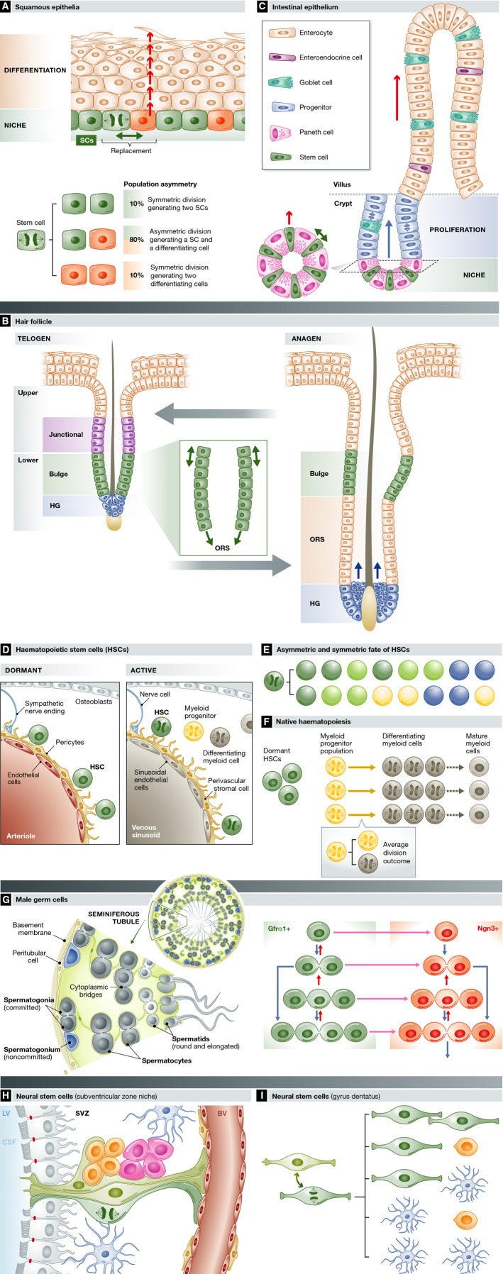
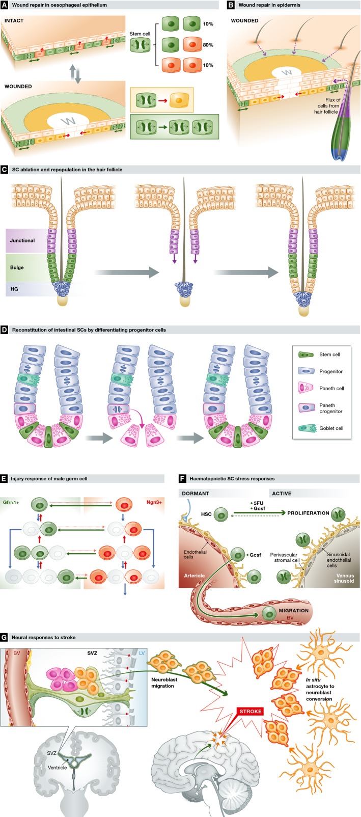
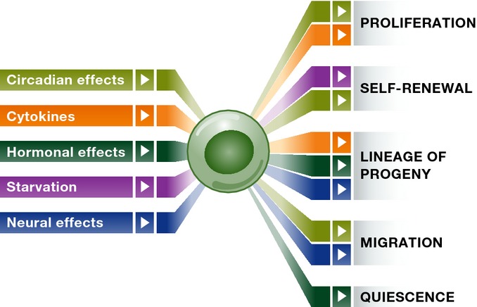
Adult organisms have to adapt to survive, and the same is true for their tissues. Rates and types of cell production must be rapidly and reversibly adjusted to meet tissue demands in response to both local and systemic challenges. Recent work reveals how stem cell (SC) populations meet these requirements by switching between functional states tuned to homoeostasis or regeneration. This plasticity extends to differentiating cells, which are capable of reverting to SCs after injury. The concept of the niche, the micro-environment that sustains and regulates stem cells, is broadening, with a new appreciation of the role of physical factors and hormonal signals. Here, we review different functions of SCs, the cellular mechanisms that underlie them and the signals that bias the fate of SCs as they switch between roles.

Keywords: differentiation; niche; regeneration; signal transduction; stem cells.


Figures

Figure 1
Figure 1. SC turnover across mouse tissues
Average cycle times for SCs in the lineages indicated.
Figure 2
Figure 2. SC dynamics across lineages
(A) Squamous epithelia consist of layers of keratinocytes, SCs (green) reside in the basal cell layer, along with post-mitotic cells waiting to stratify (red). When a differentiating cell leaves the niche (arrow), a nearby SC divides with one of the three division outcomes shown to maintain constant cell density in the niche. The probabilities of each outcome (expressed as per cent, oesophageal epithelium shown) are balanced, so equal numbers of SCs and differentiated cells are produced across the population. (B) The hair follicle cycles between a resting stage (telogen) and expansion of the lower follicle (anagen). Multiple SCs have been identified, in the junctional zone (purple), the bulge (green) and the hair germ (blue), which lie in contact with the mesenchymal cells of the dermal papilla (blue). The hair shaft (black) is surrounded by concentric layers of inner root sheath cells that have been omitted for clarity. In the transition into anagen, hair germ and upper bulge cells self-duplicate. In the bulge, divisions are aligned parallel with the axis of the hair shaft (inset). Lower bulge cells contribute differentiating progeny to greatly expand the root sheath. Later in anagen, hair germ cells assemble around the dermal papilla and then generate inner root sheath cells (arrows). Self-renewal and differentiation are balanced, so the numbers of SCs in each compartment is maintained at a constant level across multiple hair cycles. (C) Intestinal epithelium contains four lineages sustained by SCs (green) that lie between Paneth cells in the crypt base. Differentiating cells migrate through a progenitor compartment in the upper crypt from which post-mitotic cells populate the villus, from which they are shed. Inset shows a simplified top-down view of the niche. As a differentiating SC exits the niche, it is replaced by the self-duplicating division of an immediately adjacent SC. (D) Heamatopoietic SCs reside close to blood vessels in the bone marrow. Dormant SCs (green) lie close to arterioles, receiving paracrine signals from endothelial cells (red), perivascular cells expressing Ng2 and sympathetic nerve endings (blue). On activation, SCs migrate to be close to venous sinusoids that support Lepr-expressing perivascular cells. (E) Asymmetric fate and symmetric fate of haematopoietic SCs. Transplantation of daughter cells of a single SC reveals self-duplicating and asymmetric divisions: dark green: long-term-reconstituting SCs; light green: short- or intermediate-term-reconstituting SCs; yellow: megakaryocyte progenitor; and blue: common myeloid progenitor. (F) Native haematopoiesis. Lineage tracing in homoeostasis suggests that myeloid lineages may be maintained by self-sustaining progenitor cells (yellow) exhibiting ‘population asymmetry’ by generating equal proportions of progenitor and differentiating cells (grey) (see inset). Haematopoietic SCs (green) make negligible contribution to myelopoiesis in homoeostasis, but function as ‘reserve’ cells. (G) Male germ cell SCs are diverse in appearance but functionally equivalent. Male germ cells expressing GFRα1 reside in the outermost layer of the seminiferous tubule. SCs (green) exist as singles or 2–4 cell syncytia connected by cytoplasmic bridges, which may self-duplicate (blue arrows) to generate two SCs or undergo fragmentation (short red arrows). Upon differentiation (pink arrow) into Ngn3-positive cells (red), the same behaviour continues, but Ngn3+ cells are unlikely to revert to GFRα1+ SCs in homoeostasis. Once in the Ngn3 compartment, syncytia larger than 4 cells form which may undergo further differentiation into cKit-expressing cells (not shown), which are even less likely to revert to GFRα1-positive cells. (H) Neural SCs exhibit symmetric and asymmetric cell divisions. Quiescent neural SCs (light green) reside in the subventricular zone niche, extending processes into cerebrospinal fluid (blue) that fills the lateral ventricles (LV) and underlying endothelial cells (red) lining blood vessels (BV). Endothelial cells that line the LV are shown in grey. Differentiating transit amplifying cells (pink) and neural precursor cells (orange) lie adjacent to the SCs. Figure after Silva-Vargas et al (2013). (I) Division outcomes of neural SCs in the dentate gyrus inferred from lineage tracing. Neural SCs interconvert between quiescent (light green) and proliferating (dark green) states. Division of SCs has a range of symmetric and asymmetric outcomes as shown, generating neural SCs, neuroblasts (orange) or differentiated astrocytes (blue).
Figure 3
Figure 3. Stem cell responses to stress
(A) Wound repair in oesophageal epithelium. Following wounding, progenitor cells next to the wound (w) exit the cell cycle and migrate towards the defect (yellow area and inset). Behind this ‘migrating front’, cycling progenitors undergo divisions heavily biased to self-duplication (green area and inset), expanding the progenitor compartment to generate the excess cells required to repair the epithelium. Once the wound has closed, progenitor behaviour switches back to homoeostasis. (B) Wound repair in epidermis. In the epidermis, progenitor cells change their behaviour as in the oesophagus, but wound repair is supported by a flux of cells into the epidermis from hair follicles (purple arrows) and, in tail skin, mobilisation of quiescent reserve cells in the epidermis. Migration explains the appearance of radial clones around a healed wound seen in lineage-tracing experiments. (C) HF ablation and repopulation. Following ablation of SCs in the bulge (green) of telogen HFs, cells in the upper follicle (purple) migrate and proliferate (purple arrows) to reconstitute the bulge SC population, enabling the hair cycle to proceed normally. (D) Reconstitution of intestinal SCs by differentiating progenitor cells. Following ablation of Lgr5-expressing SCs in the crypt base (green), Paneth cell precursors (blue with pink border) dedifferentiate and colonise the crypt base niche with SCs functionally equivalent to the original population. (E) Injury response of male germ SCs. Following treatment with the cytotoxic agent busulphan, the GFRα1+ SC population is depleted (grey outlines) but then reconstituted by surviving Ngn3+ cells reverting into GFRα1+ cells (green arrows) and a decrease in the probability of GFRα1+ cells transferring to the Ngn3+ compartment (pink dotted arrows). (F) Haematopoietic SC stress responses. Dormant SCs are recruited into cycle by treatment after damage to the haematopoietic system from a cytotoxic drug (5FU) or cytokines, such as Gcsf, released after infection. Gcsf also triggers dormant SCs to migrate into the circulation. (G) Neural SC responses to stroke. Following a CNS stroke, which results in cell death due to ischaemia, clusters of neuroblasts (orange) are seen in the injured region. Lineage tracing argues these derive both from mobilisation of neural SCs in the SVC followed by migration to the site of injury (orange with green border) and from differentiated astrocytes within the area of the stroke entering a neurogenic programme (orange with red borders).
Figure 4
Figure 4. SC regulation from outside the niche
Summary of systemic influences on SC fate. Fate outcome is mapped by colour to distinct systemic factors. See the text for more detail.

Similar articles

Cited by

References

    1. Aimone JB, Li Y, Lee SW, Clemenson GD, Deng W, Gage FH. Regulation and function of adult neurogenesis: from genes to cognition. Physiol Rev. 2014;94:991–1026. - PMC - PubMed
    1. Alcolea MP, Greulich P, Wabik A, Frede J, Simons BD, Jones PH. Differentiation imbalance in single oesophageal progenitor cells causes clonal immortalization and field change. Nat Cell Biol. 2014;16:615–622. - PMC - PubMed
    1. Alcolea MP, Jones PH. Tracking cells in their native habitat: lineage tracing in epithelial neoplasia. Nat Rev Cancer. 2013;13:161–171. - PubMed
    1. Alcolea MP, Jones PH. Lineage analysis of epidermal stem cells. Cold Spring Harb Perspect Med. 2014;4:a015206. - PMC - PubMed
    1. Amoyel M, Simons BD, Bach EA. Neutral competition of stem cells is skewed by proliferative changes downstream of Hh and Hpo. EMBO J. 2014;33:2295–2313. - PMC - PubMed

Publication types