. 2015 Feb 27:12:23.
doi: 10.1186/s12977-015-0140-1.

Myocyte enhancer factor (MEF)-2 plays essential roles in T-cell transformation associated with HTLV-1 infection by stabilizing complex between Tax and CREB

Myocyte enhancer factor (MEF)-2 plays essential roles in T-cell transformation associated with HTLV-1 infection by stabilizing complex between Tax and CREB

Pooja Jain et al. Retrovirology. .

Abstract

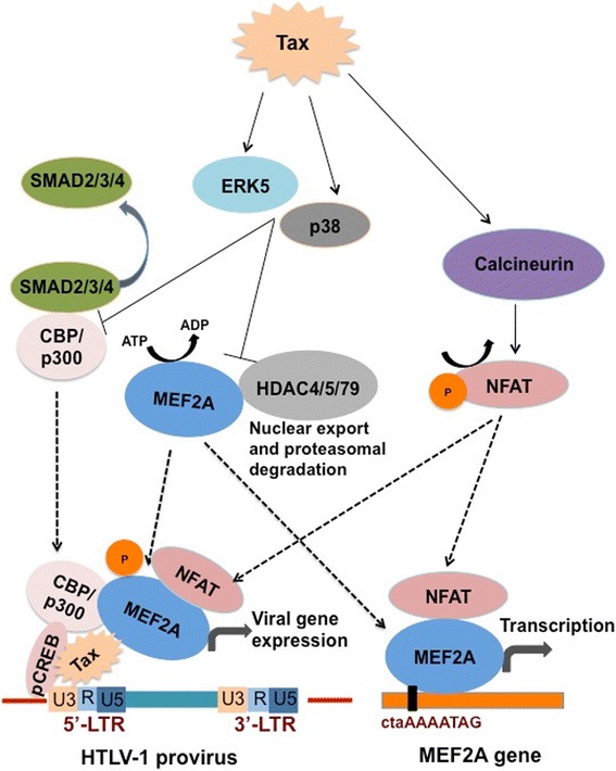
Background: The exact molecular mechanisms regarding HTLV-1 Tax-mediated viral gene expression and CD4 T-cell transformation have yet to be fully delineated. Herein, utilizing virus-infected primary CD4+ T cells and the virus-producing cell line, MT-2, we describe the involvement and regulation of Myocyte enhancer factor-2 (specifically MEF-2A) during the course of HTLV-1 infection and associated disease syndrome.

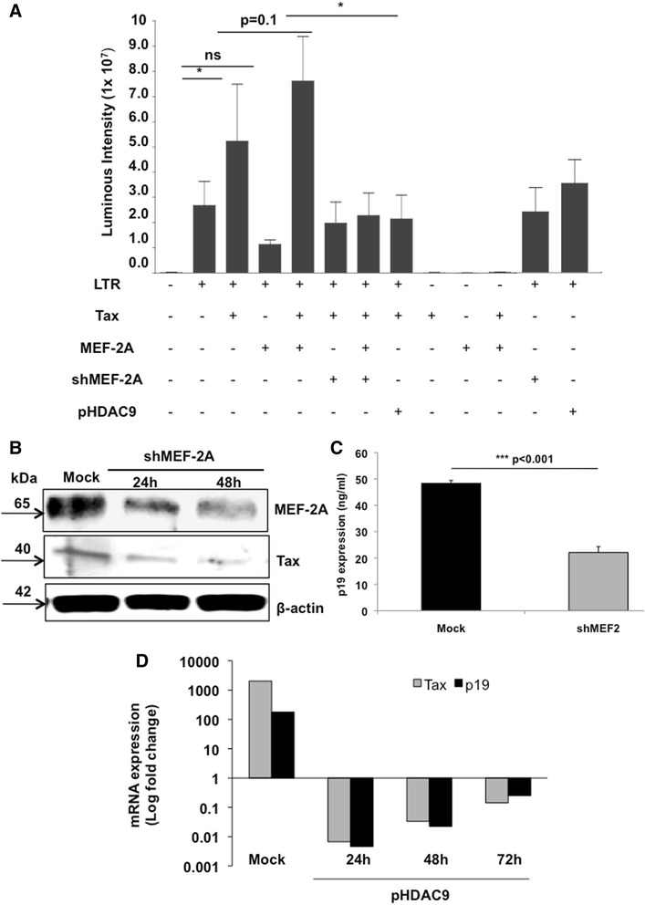
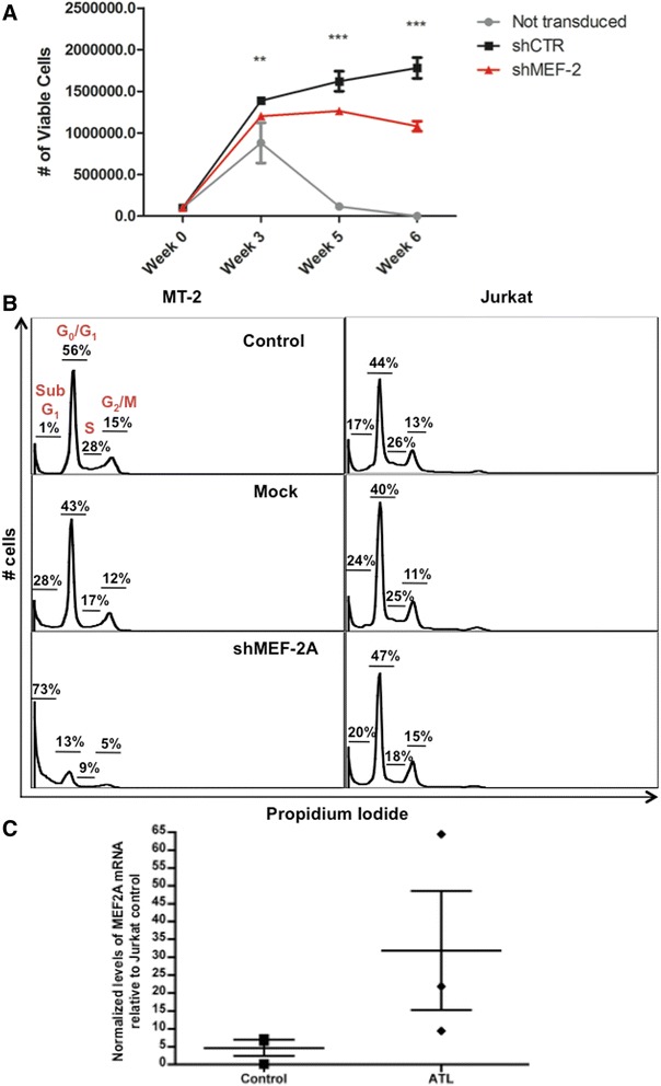
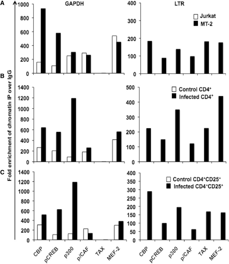
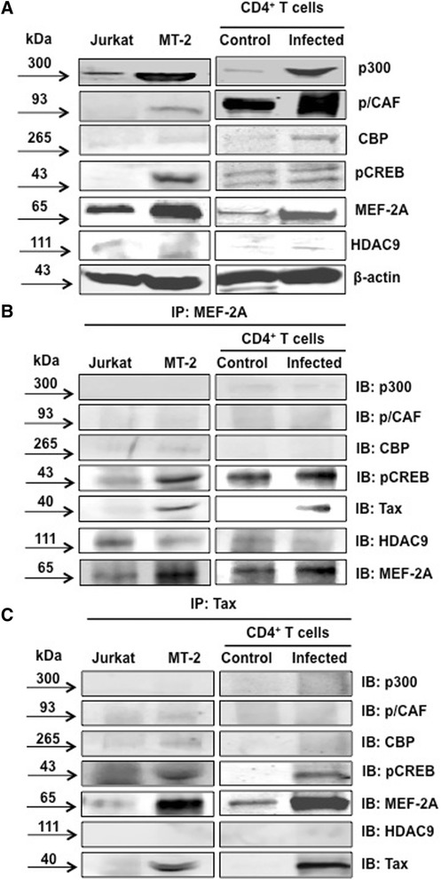
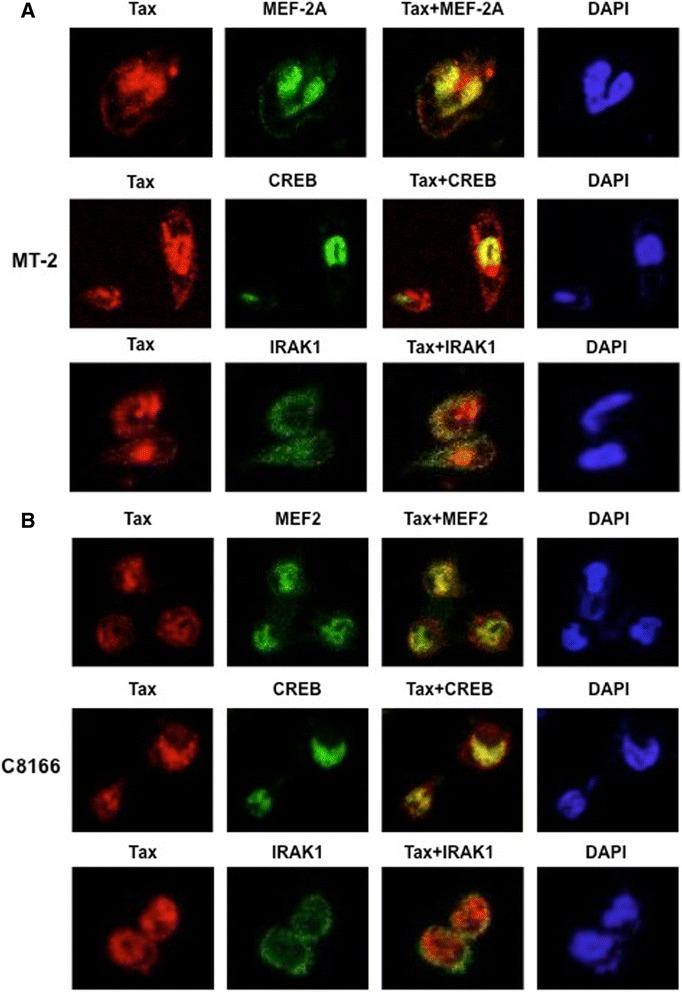
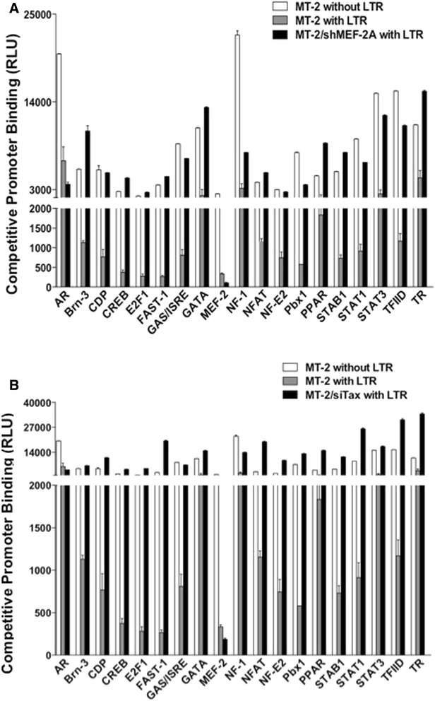
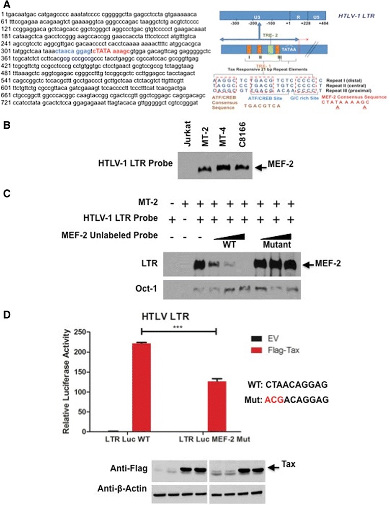
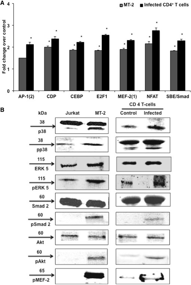
Results: Inhibition of MEF-2 expression by shRNA and its activity by HDAC9 led to reduced viral replication and T-cell transformation in correlation with a heightened expression of MEF-2 in ATL patients. Mechanistically, MEF-2 was recruited to the viral promoter (LTR, long terminal repeat) in the context of chromatin, and constituted Tax/CREB transcriptional complex via direct binding to the HTLV-1 LTR. Furthermore, an increase in MEF-2 expression was observed upon infection in an extent similar to CREB (known Tax-interacting transcription factor), and HATs (p300, CBP, and p/CAF). Confocal imaging confirmed MEF-2 co-localization with Tax and these proteins were also shown to interact by co-immunoprecipitation. MEF-2 stabilization of Tax/CREB complex was confirmed by a novel promoter-binding assay that highlighted the involvement of NFAT (nuclear factor of activated T cells) in this process via Tax-mediated activation of calcineurin (a calcium-dependent serine-threonine phosphatase). MEF-2-integrated signaling pathways (PI3K/Akt, NF-κB, MAPK, JAK/STAT, and TGF-β) were also activated during HTLV-1 infection of primary CD4+ T cells, possibly regulating MEF-2 activity.

Conclusions: We demonstrate the involvement of MEF-2 in Tax-mediated LTR activation, viral replication, and T-cell transformation in correlation with its heightened expression in ATL patients through direct binding to DNA within the HTLV-1 LTR.


Figures

Figure 1
Figure 1
MEF-2 inhibition reduces HTLV-1 LTR transactivation, Tax expression, and viral replication. (A) Transient transfection of Jurkat cells with pU3R-luc (HTLV-1 LTR luciferase reporter construct) as well as plasmids that express Tax, MEF-2A, HDAC9 and MEF-2A shRNA, was done as described in Methods. Before co-transfecting two or more plasmids, each of these plasmids was transfected alone to establish the background levels of luciferase activity. Cells were collected 24 hr post-transfection, lysed and assayed using the dual luciferase assay system. Firefly luciferase activity was normalized with that of Renilla luciferase expressed from phRL/CMV. Each bar represents the average of triplicate samples. Significance among groups was derived by student’s t-test to determine the p-value. (*p < 0.05). (B) MT-2 cells were transfected with either scrambled or shMEF-2 plasmid. Western blot analysis was performed at 24 hr and 48 hr to determine protein levels of MEF-2, Tax, and beta-actin. Data represent one of two separate experiments. (C) To analyze effects of shMEF-2A on virus production, transfected MT-2 cells were washed at 48 hr and incubated in fresh medium for another 24–36 hr. Thereafter, supernatants were assessed for HTLV-1 core protein levels (pg/ml) by the p19-specific ELISA (ZeptoMetrix, Buffalo, NY). (D) MT-2 cells were transfected either with a mock plasmid or MITR/HDAC9 plasmids followed by cell collection at every 24 hr over a 72 hr period. Real-time PCR analyses were performed to determine relative mRNA levels of Tax and p19. Data is representative of at least three independent experiments.
Figure 2
Figure 2
MEF-2 inhibition perturbs HTLV-1-mediated T-cell transformation in correlation with a heightened MEF-2 expression in ATL patients. (A) PBMCs were transduced with scrambled or shMEF-2 expressing lentivirus by spinoculation. Viable cell proliferation of PBMCs was determined by trypan blue exclusion assay, after co-culture with lethally irradiated MT-2 cells for the indicated times. Error bars represent standard deviation of triplicate samples with high significance (***p < 0.001) between shMEF-2 versus shControl samples. (B) MT-2 and Jurkat cells were collected at 24 hr post a 3-day transfection followed by staining with propidium iodide (PI-25 μg/ml, RNAase- 40 μg/ml, sodium citrate-0.1% and Triton-100 × −0.03%). Cell cycle progression of MT-2 and Jurkat cells were observed via flow cytometry with no transfection (upper panel), mock transfected (middle panel) and shMEF2 plasmid (2.5 μg/1×106 cells) transfection (lower panel). The percentage of sub-G1 cells, G0/G1, S and G2-M cells was analyzed using the FLowjo software. (C) MEF-2A mRNA levels was determined by the quantitative real-time PCR as described in Methods. At least two replicates per donor were processed and MEF-2 levels were compared between seronegative controls and ATL patients (n = 3, each). Each point represents average mRNA expression in individual donors. Bars represent mean with Standard Error (SEM) derived by a two-tailed, unpaired nonparametric t-test (Mann–Whitney).
Figure 3
Figure 3
Tax and MEF-2 are recruited to the HTLV-1 LTR. Chromatin immunoprecipitation of Tax protein and transcription factors bound to cellular and viral promoters during HTLV-1 infection in (A) cell lines, (B) primary CD4+ T cells, and (C) primary CD4+CD25+ T cells was performed using the ChIP-IT Express kit. Cells were lysed in a dounce homogenizer to obtain sheared chromatin following formaldehyde fixation. The sheared chromatin was immunoprecipitated at 4°C overnight using 2 μg of antibodies against the Tax protein, indicated cellular factors and controls. The immunoprecipitated chromatin was then subjected to PCR using primers for HTLV-1 LTR and human GAPDH. Data is presented as average fold change over control IgG immunoprecipitation, and is representative of three independent experiments.
Figure 4
Figure 4
MEF-2 physically interacts with Tax. (A) Control (Jurkat), infected (MT-2) cell lines, control primary CD4+ T cells and HTLV-infected primary CD4+ T cells were lysed, sonicated and protein concentration was determined by Bradford assay. Equal protein quantities were then resolved by SDS-PAGE and transferred to PVDF membrane. Following a 1 hr block membranes were incubated with antibodies against the transcription factors. Western blot shows the expression of transcription factors in control and infected cell lines and primary cells. (B) MEF-2 complex formation with Tax and transcription factors was analyzed using an immunoprecipitation assay. Cells were lysed using an immunoprecipitation lysis buffer and then incubated with MEF-2 antibody overnight at 4°C as described in Methods. Western immunoblot analysis was performed to confirm immunoprecipitation. (C) Control and infected cell lines and primary cells were enriched for Tax and immunoblotted to determine complex formation with MEF-2 and transcription factors. Data is representative of multiple individual experiments.
Figure 5
Figure 5
MEF-2 co-localizes with Tax in the nucleus of HTLV-1 transformed cell lines. MT-2 cells (A) and C8166 cells (B) were cultured overnight on poly-L-lysine coated glass coverslips. The cells were then fixed, permeabilized and then stained with anti-Tax, anti-MEF-2, anti-CREB or anti-IRAK1 as indicated. Nuclei were stained with DAPI and cells were subjected to confocal microscopy.
Figure 6
Figure 6
HTLV-1 LTR transcriptional activation complexes contain MEF-2. Binding of various transcription factors present in the nuclear extract isolated from MT-2 cells to the HTLV-1 LTR was assessed using the Promoter-Binding Transcription Factor Profiling Array I upon knocking down MEF-2A (A) and Tax (B) as described in Methods. The nuclear extract was incubated with oligonucleotide probe mix with the HTLV-1 LTR or a control DNA. The binding of each transcription factor to LTR was indicated by average reduction in chemiluminescence of transcription factor-specific oligonucleotide probe specific to each factor from triplicate samples.
Figure 7
Figure 7
Assessment of MEF-2 binding site(s) within the HTLV-1 LTR. (A) HTLV-1 LTR nucleotide sequence with two putative MEF-2 binding sites shown in blue and red. The HTLV-1 LTR comprises U3 (unique 3′), R (Repeated), and U5 (unique 5′) regions. The U3 region regulates viral gene expression via three 21 bp repeats known as Tax-responsive element - 1 (TRE - 1), which confers Tax-based trans-activation. These repeats contain 3 conserved domains labeled A, B and C. The location for ATF/CREB binding and putative MEF-2 binding are illustrated. (B) EMSA was performed with a probe corresponding to the MEF-2 site in the HTLV-1 LTR using nuclear extracts from Jurkat, MT-2, MT-4 and C8166 cells. (C) EMSA competition assay was performed using nuclear extracts from MT-2 cells, with increasing amounts of unlabeled consensus MEF-2 specific probe (200, 300 and 400 fold molar excess respectively) or a mutated MEF-2 specific probe. Oct-1 was used as a loading control. (D) HTLV-1 LTR luciferase assays in 293 T cells transfected with empty vector (EV) or Flag-Tax using LTR luciferase WT plasmid (LTR Luc WT) or a MEF-2-specific binding mutant (LTR Luc MEF-2 Mut). Luciferase values are presented as “fold induction” relative to the control (EV). Two-tailed unpaired t-test was performed with Prism software. Error bars represent the standard deviation of triplicate samples. The level of significance was defined as: ***p < 0.001. Western blots were performed with anti-Flag and anti-β-actin using whole-cell lysates.
Figure 8
Figure 8
MEF-2-associated signaling pathways are triggered during HTLV-1 infection. (A) A protein-DNA array was used to determine the activation of the various eukaryotic transcription factors. Nuclear extracts from control and HTLV-infected primary cells were mixed with biotinylated DNA binding oligonucleotides for the formation of protein-DNA complexes. These probes were then hybridized to pretreated array membranes and the bound probe was detected as described in Methods. Densitometric analysis was used to quantify the spots and data was normalized to their respective controls after background subtraction. Fold change in expression of selected transcription factors from the array data compared to relevant uninfected cells. Significance was determined using the Student’s t-test (*P ≤ 0.05). (B) Cells were lysed and Western blotting was performed to confirm the array data.
Figure 9
Figure 9
Model explaining MEF-2 activity on Tax-mediated transactivation of HTLV-1 LTR. Type II HDACs (HDAC4/5/7/9) bind to MEF-2A and repress its transcriptional activity. Upon HTLV-1 infection, Tax activates p38 and ERK5, which phosphorylate MEF-2 leading to its dissociation from the MEF-2A: HDAC repressive complex. On the other hand, Tax also binds to Smad2/3/4 to prevent their constitutive binding to transcription co-activators CBP/p300. This leads to increased availability of CBP/p300 to bind Tax/pCREB complex bound to the 5′ LTR region of the provirus. Along with Tax/pCREB/CBP/p300 complex, recruitment of MEF-2A to the 5′ LTR promotes viral gene expression. Tax also activates Calcineurin (a calcium-dependent serine-threonine phosphatase), which dephosphorylates NFAT. Upon dephosphorylation, NFAT translocates to nucleus and is recruited to the HTLV-1 5′ LTR along with the Tax/pCREB/CBP/p300 complex. NFAT is also recruited to the MEF-2A gene promoter where it binds to MEF-2A and turns on transcription resulting in upregulation of MEF-2A expression. HDAC, Histone deacetylase; MEF-2A, Myocyte-specific enhancer factor 2A; ERK5, Extracellular-signal-regulated kinase 5; Smad, Sma- and Mad-Related Protein; CREB, cAMP response element-binding protein; CBP, CREB-binding protein; NFAT, Nuclear factor of activated T cells.

Similar articles

Cited by

References

    1. Gallo RC. The discovery of the first human retrovirus: HTLV-1 and HTLV-2. Retrovirology. 2005;2:17. doi: 10.1186/1742-4690-2-17. - DOI - PMC - PubMed
    1. Gessain A, Barin F, Vernant JC, Gout O, Maurs L, Calender A, et al. Antibodies to human T-lymphotropic virus type-I in patients with tropical spastic paraparesis. Lancet. 1985;2:407–10. doi: 10.1016/S0140-6736(85)92734-5. - DOI - PubMed
    1. Osame M, Usuku K, Izumo S, Ijichi N, Amitani H, Igata A, et al. HTLV-I associated myelopathy, a new clinical entity. Lancet. 1986;1:1031–2. doi: 10.1016/S0140-6736(86)91298-5. - DOI - PubMed
    1. Palutke M, Patt DJ, Weise R, Varadachari C, Wylin RF, Bishop CR, et al. T cell leukemia–lymphoma in young adults. Am J Clin Pathol. 1977;68:429–39. - PubMed
    1. Poiesz BJ, Ruscetti FW, Gazdar AF, Bunn PA, Minna JD, Gallo RC. Detection and isolation of type C retrovirus particles from fresh and cultured lymphocytes of a patient with cutaneous T-cell lymphoma. Proc Natl Acad Sci U S A. 1980;77:7415–9. doi: 10.1073/pnas.77.12.7415. - DOI - PMC - PubMed

Publication types

MeSH terms

Substances