. 2015 May;22(5):578-85.
doi: 10.1128/CVI.00080-15. Epub 2015 Mar 25.

Induction of potent adaptive immunity by the novel polyion complex nanoparticles

Affiliations

Induction of potent adaptive immunity by the novel polyion complex nanoparticles

Tomofumi Uto et al. Clin Vaccine Immunol. 2015 May.

Abstract

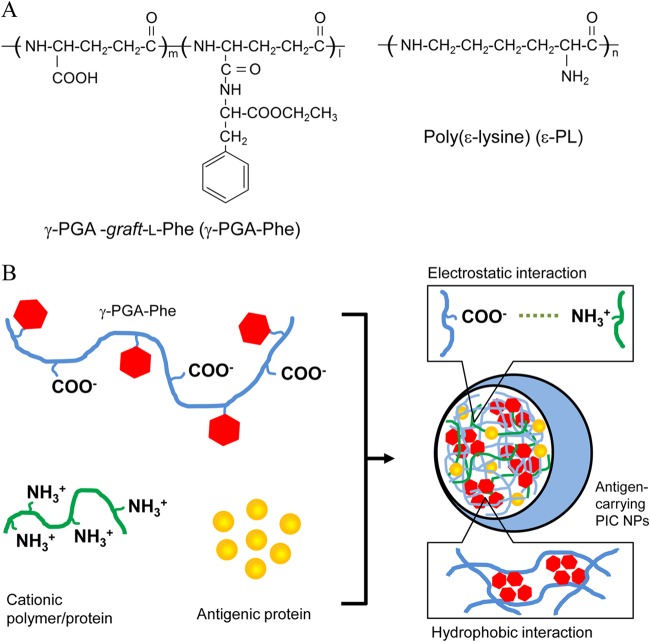
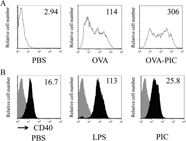
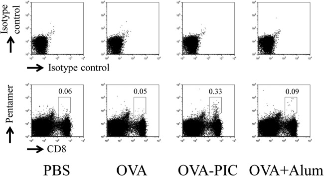
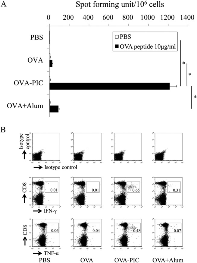
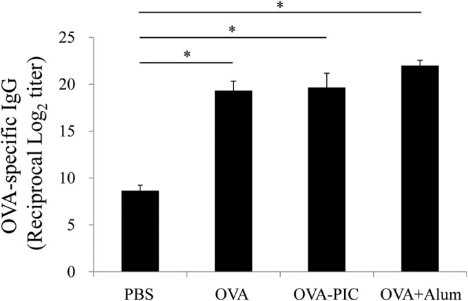
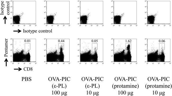
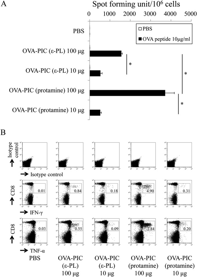
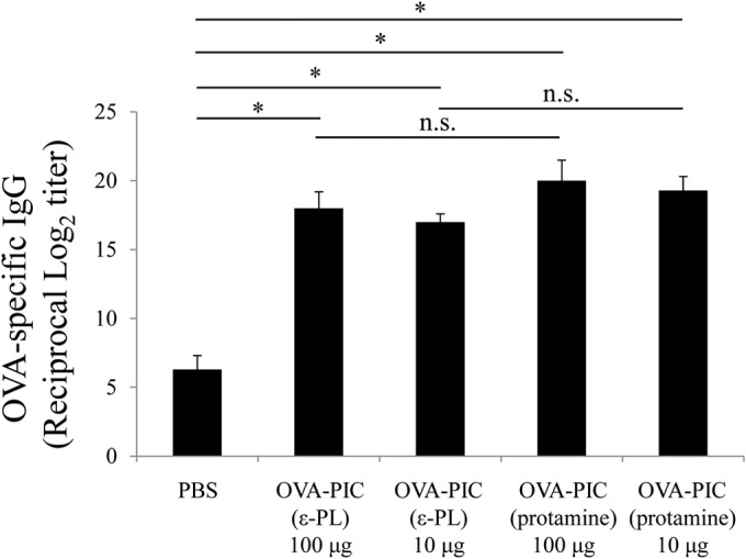
The development of effective and simple methods of vaccine preparation is desired for the prophylaxis and treatment of a variety of infectious diseases and cancers. We have created novel polyion complex (PIC) nanoparticles (NPs) composed of amphiphilic anionic biodegradable poly(γ-glutamic acid) (γ-PGA) and cationic polymers as a vaccine adjuvant. PIC NPs can be prepared by mixing γ-PGA-graft-l-phenylalanine ethylester (γ-PGA-Phe) polymer with cationic polymer in phosphate-buffered saline. We examined the efficacy of PIC NPs for antigen delivery and immunostimulatory activity in vitro and in vivo. PIC NPs enhanced the uptake of ovalbumin (OVA) by dendritic cells (DCs) and subsequently induced DC maturation. The immunization of mice with OVA-carrying PIC NPs induced potent and antigen-specific cellular and humoral immunity. Since PIC NPs can be created with water-soluble anionic γ-PGA-Phe and a cationic polymer by simple mixing in the absence of any organic solvents, PIC NPs may have potential as a novel candidate for an effective antigen carrier and vaccine adjuvant.


Figures

FIG 1
FIG 1
(A) Chemical structures of Phe-grafted γ-PGA (γ-PGA-Phe) and ε-PL. (B) Schematic representation of antigen-carrying PIC NP formation. PIC NPs were formed and stabilized by hydrophobic interaction of γ-PGA-Phe and electrostatic interaction between γ-PGA and cationic polymers.
FIG 2
FIG 2
Uptake of PIC (ε-PL) NPs by DCs and their maturation. (A) iDCs were incubated in the absence (PBS) or presence of Alexa Fluor 488-labeled OVA (10 μg/ml) or Alexa Fluor 488-labeled OVA (10 μg/ml) in PIC NPs (OVA-PIC) for 1 h at 37°C. After incubation, cell-associated fluorescence was assessed by flow cytometry. (B) iDCs were incubated in the absence (PBS) or presence of LPS (1 μg/ml) or PIC NPs (100 μg/ml γ-PGA-Phe and 20 μg/ml ε-PL). After incubation for 24 h, the cells were collected, stained with an anti-CD40–FITC MAb, and analyzed by flow cytometry. Representative results of three separate experiments are shown.
FIG 3
FIG 3
Antigen-specific CD8+ T cells induced by OVA-PIC (ε-PL) NPs. Mice (n = 3) were subcutaneously immunized twice with PBS, OVA (100 μg), OVA-PIC NPs (OVA-PIC) (1 mg of γ-PGA-Phe, 200 μg ε-PL, and 100 μg of OVA), or OVA plus alum (100 μg of OVA and 1 mg of alum). Spleen cells were isolated on day 14 after the final immunization. The cells were stained with an anti-CD8 MAb and the H-2Kb/OVA257–264 pentamer. The number in each panel indicates the percentage of CD8+ pentamer+ T cells. A representative result for one mouse in each group is shown.
FIG 4
FIG 4
Functional analysis of antigen-specific cytokine-producing T cells induced by OVA-PIC (ε-PL) NPs. Mice (n = 3) were subcutaneously immunized twice with PBS, OVA, OVA-PIC NPs (OVA-PIC), or OVA plus alum. Spleen cells were isolated on day 14 after the final immunization. (A) Spleen cells were stimulated with no peptide (PBS) or the OVA257–264 CTL epitope peptide (10 μg/ml) and evaluated for their IFN-γ production by ELISPOT. Data are expressed as the number of IFN-γ-positive spots per million cells and represent the mean result ± standard deviation (SD) for each group. Statistical analysis was carried out among the 4 groups stimulated with the OVA257–264 epitope peptide (10 μg/ml) (*, P < 0.05). (B) Spleen cells were stimulated with the OVA257–264 peptide and examined for their production of IFN-γ and TNF-α by intracellular cytokine staining. The number in each panel indicates the percentage of cytokine-positive CD8+ T cells.
FIG 5
FIG 5
Induction of antigen-specific IgG antibody responses in mice immunized with OVA-PIC (ε-PL) NPs. Mice (n = 3) were subcutaneously immunized twice with PBS, OVA, OVA-PIC NPs (OVA-PIC), or OVA plus alum. Sera were collected on day 14 after the final immunization. The levels of OVA-specific IgG antibody were measured by ELISA. Data represent the means ± SD of the endpoint titers for groups of three mice. Statistical analysis was carried out among the results from 4 groups (*, P < 0.05).
FIG 6
FIG 6
Effects of different cationic proteins on PIC NPs for induction of antigen-specific CD8+ T cells. Mice (n = 3) were subcutaneously immunized twice with ε-PL-based OVA-PIC NPs [OVA-PIC (ε-PL) 100 μg (1 mg of γ-PGA-Phe, 200 μg of ε-PL, and 100 μg of OVA) or OVA-PIC (ε-PL) 10 μg (100 μg of γ-PGA-Phe, 20 μg of ε-PL, and 10 μg of OVA)] or protamine-based OVA-PIC NPs [OVA-PIC (protamine) 100 μg (1 mg of γ-PGA-Phe, 200 μg of protamine, and 100 μg of OVA) or OVA-PIC (protamine) 10 μg (100 μg of γ-PGA-Phe, 20 μg of protamine, and 10 μg of OVA)]. Spleen cells were isolated on day 14 after the final immunization. The cells were stained with an anti-CD8 MAb and the H-2Kb/OVA257–264 pentamer. The number in each panel indicates the percentage of CD8+ pentamer+ T cells. A representative result for one mouse in each group is shown.
FIG 7
FIG 7
Functional analysis of antigen-specific cytokine-producing T cells induced by OVA-PIC NPs containing different cationic proteins. Mice (n = 3) were subcutaneously immunized twice with PBS, OVA-PIC (ε-PL) 100 μg (1 mg of γ-PGA-Phe, 200 μg of ε-PL, and 100 μg of OVA), OVA-PIC (ε-PL) 10 μg (100 μg of γ-PGA-Phe, 20 μg of ε-PL, and 10 μg of OVA), OVA-PIC (protamine) 100 μg (1 mg of γ-PGA-Phe, 200 μg of protamine, and 100 μg of OVA), or OVA-PIC (protamine) 10 μg (100 μg of γ-PGA-Phe, 20 μg of protamine, and 10 μg of OVA). Spleen cells were isolated on day 14 after the final immunization. (A) Spleen lymphocytes were stimulated with no peptide (PBS) or the OVA257–264 CTL epitope peptide (10 μg/ml) and evaluated for their IFN-γ production by ELISPOT. Data are expressed as the numbers of IFN-γ-positive spots per million cells. Data represent the mean result ± SD for each group. Statistical analysis was carried out among the 4 groups stimulated with the OVA257–264 epitope peptide (10 μg/ml) (*, P < 0.05). (B) Spleen lymphocytes were stimulated with the OVA257–264 peptide and examined for their production of IFN-γ and TNF-α by intracellular cytokine staining. The number in each panel indicates the percentage of cytokine-positive CD8+ T cells.
FIG 8
FIG 8
Induction of antigen-specific IgG antibody in mice immunized with OVA-PIC NPs containing different cationic proteins. Mice (n = 3) were subcutaneously immunized twice with PBS, OVA-PIC (ε-PL) 100 μg (1 mg of γ-PGA-Phe, 200 μg of ε-PL, and 100 μg of OVA), OVA-PIC (ε-PL) 10 μg (100 μg of γ-PGA-Phe, 20 μg of ε-PL, and 10 μg of OVA), OVA-PIC (protamine) 100 μg (1 mg of γ-PGA-Phe, 200 μg of protamine, and 100 μg of OVA), or OVA-PIC (protamine) 10 μg (100 μg of γ-PGA-Phe, 20 μg of protamine, and 10 μg of OVA). Sera were collected on day 14 after the final immunization. The levels of OVA-specific IgG antibody were measured by ELISA. Data represent the means ± SD of the endpoint titers for groups of three mice. Statistical analysis was carried out among the results for the 5 groups (*, P < 0.05). n.s., not statistically significant.

Similar articles

Cited by

References

    1. Pulendran B, Ahmed R. 2011. Immunological mechanisms of vaccination. Nat Immunol 12:509–517. doi:10.1038/ni.2039. - DOI - PMC - PubMed
    1. Reed SG, Orr MT, Fox CB. 2013. Key roles of adjuvants in modern vaccines. Nat Med 19:1597–1608. doi:10.1038/nm.3409. - DOI - PubMed
    1. Reed SG, Bertholet S, Coler RN, Friede M. 2009. New horizons in adjuvants for vaccine development. Trends Immunol 30:23–32. doi:10.1016/j.it.2008.09.006. - DOI - PubMed
    1. Sahdev P, Ochyl LJ, Moon JJ. 2014. Biomaterials for nanoparticle vaccine delivery systems. Pharm Res 31:2563–2582. doi:10.1007/s11095-014-1419-y. - DOI - PMC - PubMed
    1. De Gregorio E, Caproni E, Ulmer JB. 2013. Vaccine adjuvants: mode of action. Front Immunol 31:214. doi:10.3389/fimmu.2013.00214. - DOI - PMC - PubMed

Publication types

LinkOut - more resources