

Renal endothelial injury and microvascular dysfunction in acute kidney injury
- PMID: 25795503
- PMCID: PMC4476528
- DOI: 10.1016/j.semnephrol.2015.01.010
Renal endothelial injury and microvascular dysfunction in acute kidney injury
Abstract
The kidney is comprised of heterogeneous cell populations that function together to perform a number of tightly controlled, complex and interdependent processes. Renal endothelial cells contribute to vascular tone, regulation of blood flow to local tissue beds, modulation of coagulation and inflammation, and vascular permeability. Both ischemia and sepsis have profound effects on the renal endothelium, resulting in microvascular dysregulation resulting in continued ischemia and further injury. In recent years, the concept of the vascular endothelium as an organ that is both the source of and target for inflammatory injury has become widely appreciated. Here we revisit the renal endothelium in the light of ever evolving molecular advances.
Keywords: AKI; Kidney; endothelium.
Copyright © 2015 Elsevier Inc. All rights reserved.
Figures
Similar articles
-
The microcirculation of the septic kidney.Semin Nephrol. 2015 Jan;35(1):75-84. doi: 10.1016/j.semnephrol.2015.01.008. Semin Nephrol. 2015. PMID: 25795501 Review.
-
Alteration of microvascular permeability in acute kidney injury.Microvasc Res. 2009 Jan;77(1):4-7. doi: 10.1016/j.mvr.2008.09.004. Epub 2008 Sep 25. Microvasc Res. 2009. PMID: 18938184 Free PMC article. Review.
-
Endothelial injury and dysfunction: role in the extension phase of acute renal failure.Kidney Int. 2004 Aug;66(2):496-9. doi: 10.1111/j.1523-1755.2004.761_5.x. Kidney Int. 2004. PMID: 15253696 Review.
-
Microvasculopathy in ischemic acute kidney injury: consequences and therapeutic implications.Panminerva Med. 2012 Mar;54(1):45-52. Panminerva Med. 2012. PMID: 22278116 Review.
-
Sepsis-associated acute kidney injury: macrohemodynamic and microhemodynamic alterations in the renal circulation.Semin Nephrol. 2015 Jan;35(1):64-74. doi: 10.1016/j.semnephrol.2015.01.007. Semin Nephrol. 2015. PMID: 25795500 Review.
Cited by
-
Delayed Mitogen-Activated Protein Kinase/Extracellular Signal-Regulated Kinase Inhibition by Trametinib Attenuates Systemic Inflammatory Responses and Multiple Organ Injury in Murine Sepsis.Crit Care Med. 2016 Aug;44(8):e711-20. doi: 10.1097/CCM.0000000000001672. Crit Care Med. 2016. PMID: 27031380 Free PMC article.
-
The roles of hydrogen sulfide in renal physiology and disease states.Ren Fail. 2022 Dec;44(1):1289-1308. doi: 10.1080/0886022X.2022.2107936. Ren Fail. 2022. PMID: 35930288 Free PMC article. Review.
-
High Preoperative Serum Syndecan-1, a Marker of Endothelial Glycocalyx Degradation, and Severe Acute Kidney Injury after Valvular Heart Surgery.J Clin Med. 2020 Jun 10;9(6):1803. doi: 10.3390/jcm9061803. J Clin Med. 2020. PMID: 32531891 Free PMC article.
-
Malaria-Associated Acute Kidney Injury in African Children: Prevalence, Pathophysiology, Impact, and Management Challenges.Int J Nephrol Renovasc Dis. 2021 Jul 8;14:235-253. doi: 10.2147/IJNRD.S239157. eCollection 2021. Int J Nephrol Renovasc Dis. 2021. PMID: 34267538 Free PMC article. Review.
-
Prevention and treatment of sepsis-induced acute kidney injury: an update.Ann Intensive Care. 2015 Dec;5(1):51. doi: 10.1186/s13613-015-0095-3. Epub 2015 Dec 21. Ann Intensive Care. 2015. PMID: 26690796 Free PMC article.
References
-
- Casellas D, Mimran A. Shunting in renal microvasculature of the rat: a scanning electron microscopic study of corrosion casts. Anat Rec. 1981;201(2):237–48. - PubMed
-
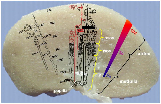
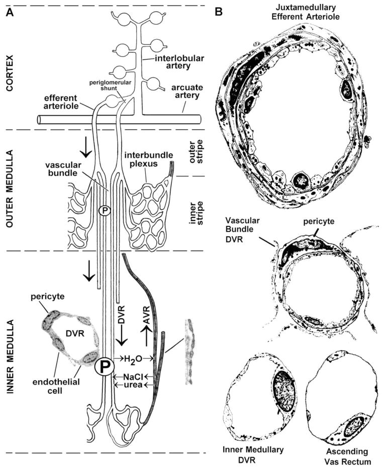
- Pallone TL, et al. Countercurrent exchange in the renal medulla. Am J Physiol Regul Integr Comp Physiol. 2003;284(5):R1153–75. - PubMed
-
- Rops AL, et al. Isolation and characterization of conditionally immortalized mouse glomerular endothelial cell lines. Kidney Int. 2004;66(6):2193–201. - PubMed
-
- Roberts WG, Palade GE. Increased microvascular permeability and endothelial fenestration induced by vascular endothelial growth factor. J Cell Sci. 1995;108( Pt 6):2369–79. - PubMed
Publication types
MeSH terms
Grants and funding
LinkOut - more resources
Full Text Sources
Other Literature Sources
Medical
