. 2015 Mar 12:9:1555-84.
doi: 10.2147/DDDT.S74197. eCollection 2015.

Pro-apoptotic and pro-autophagic effects of the Aurora kinase A inhibitor alisertib (MLN8237) on human osteosarcoma U-2 OS and MG-63 cells through the activation of mitochondria-mediated pathway and inhibition of p38 MAPK/PI3K/Akt/mTOR signaling pathway

Affiliations

Pro-apoptotic and pro-autophagic effects of the Aurora kinase A inhibitor alisertib (MLN8237) on human osteosarcoma U-2 OS and MG-63 cells through the activation of mitochondria-mediated pathway and inhibition of p38 MAPK/PI3K/Akt/mTOR signaling pathway

Ning-Kui Niu et al. Drug Des Devel Ther. .

Abstract

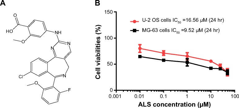
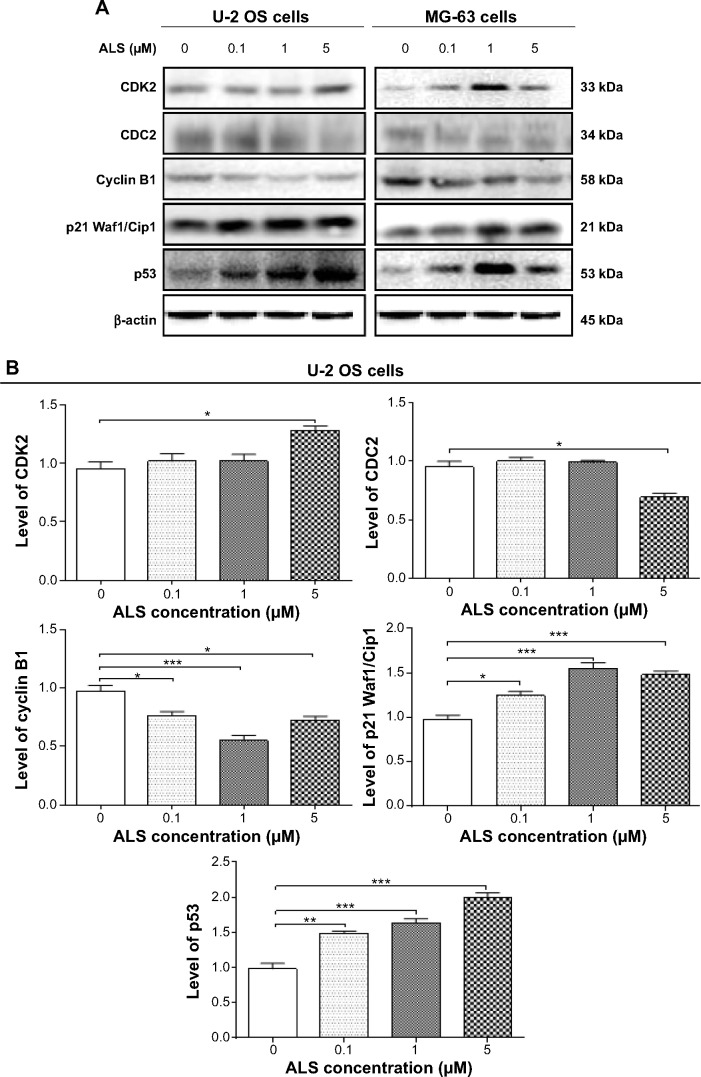
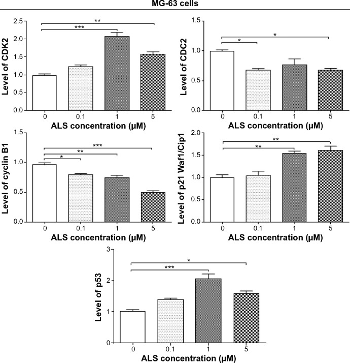
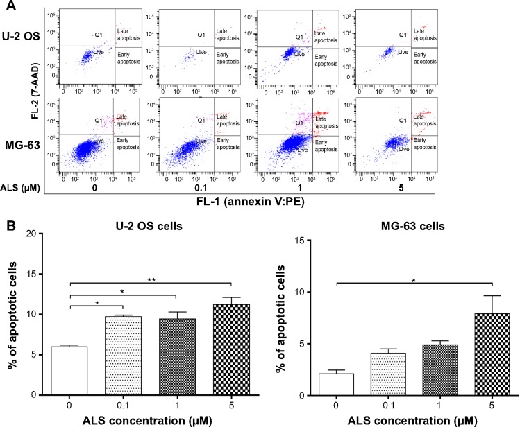
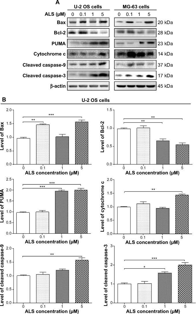
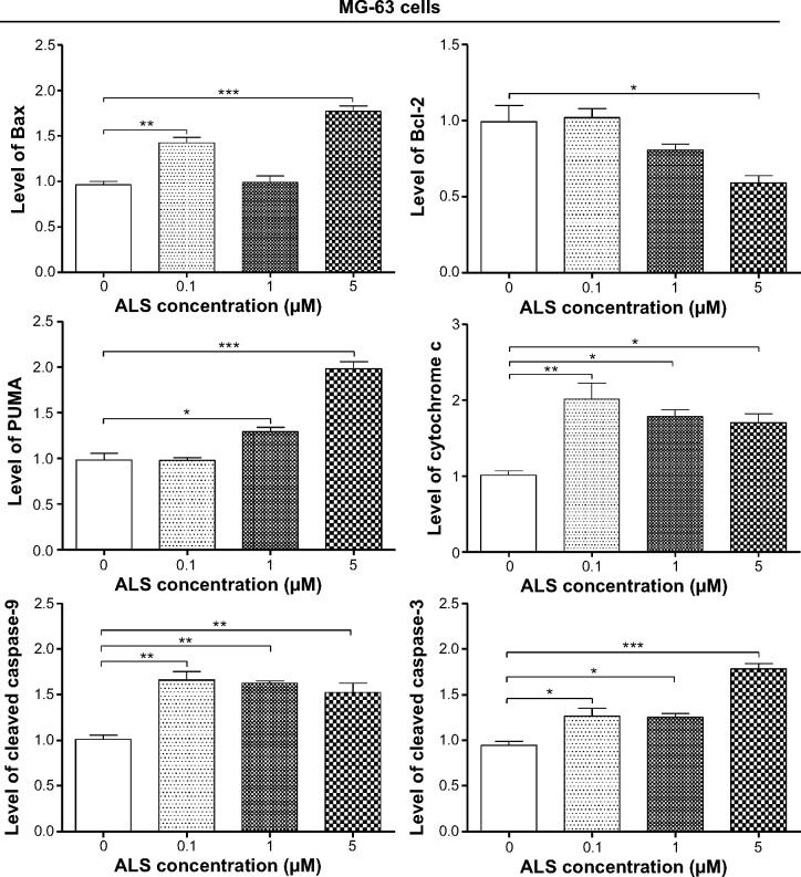
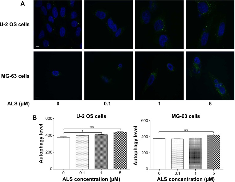
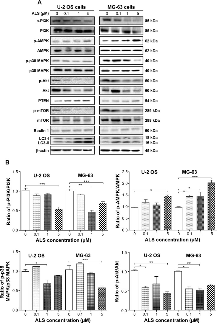
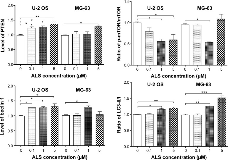
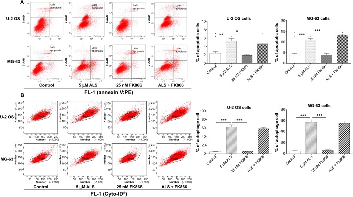
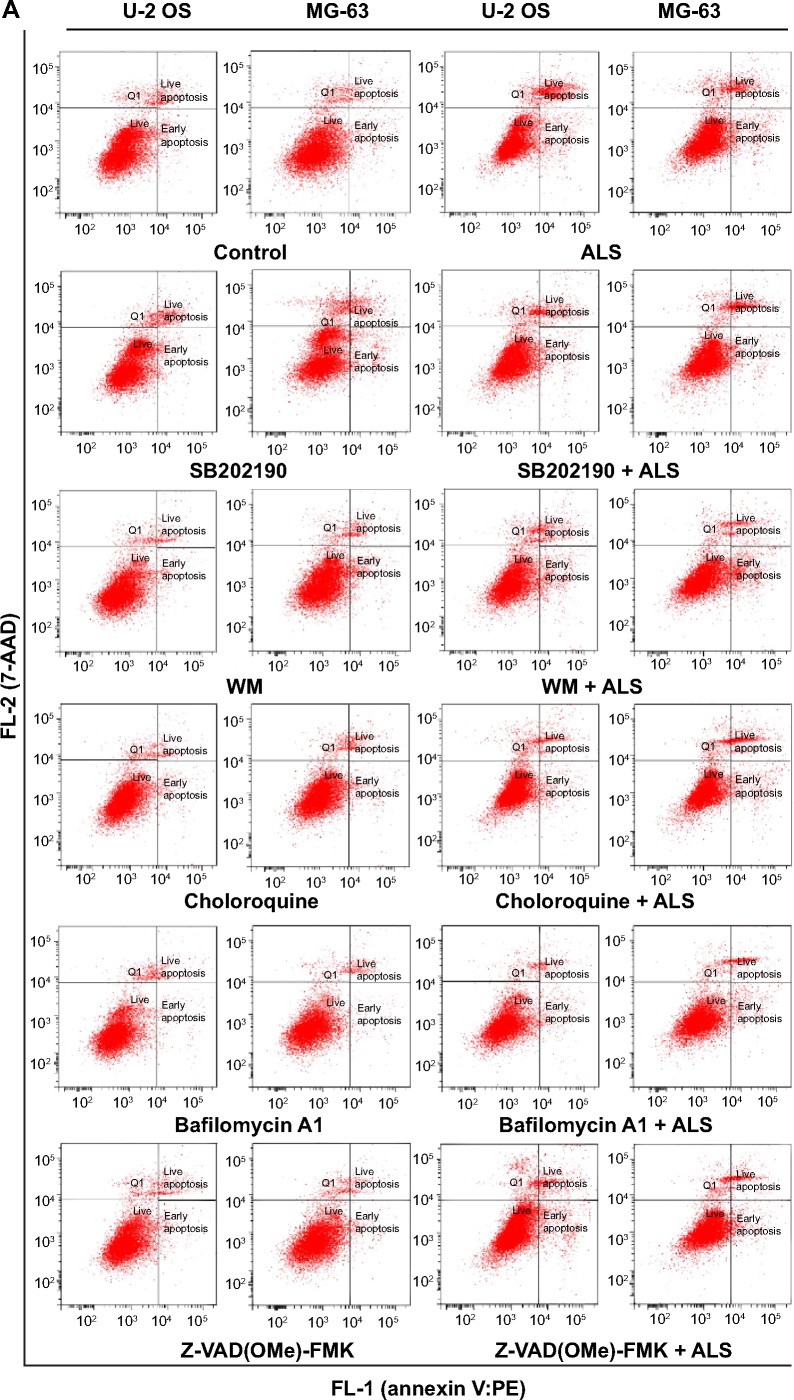
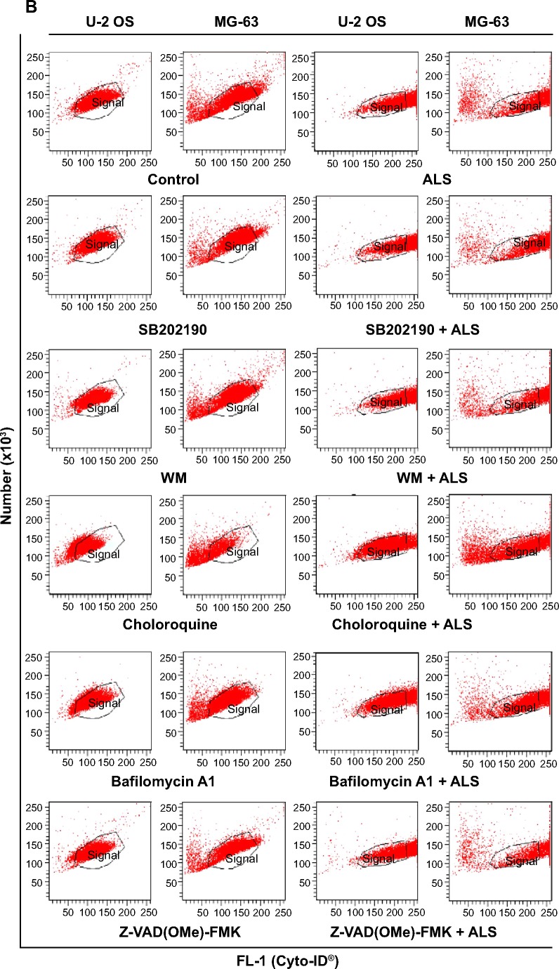
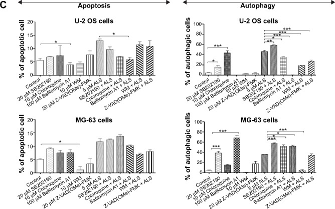
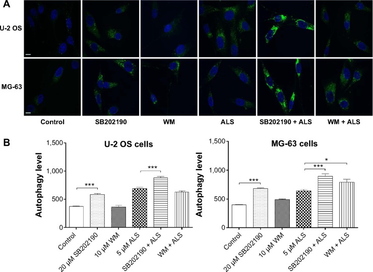
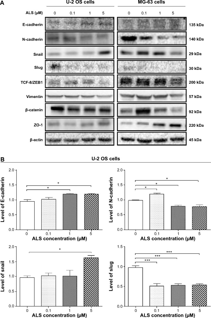
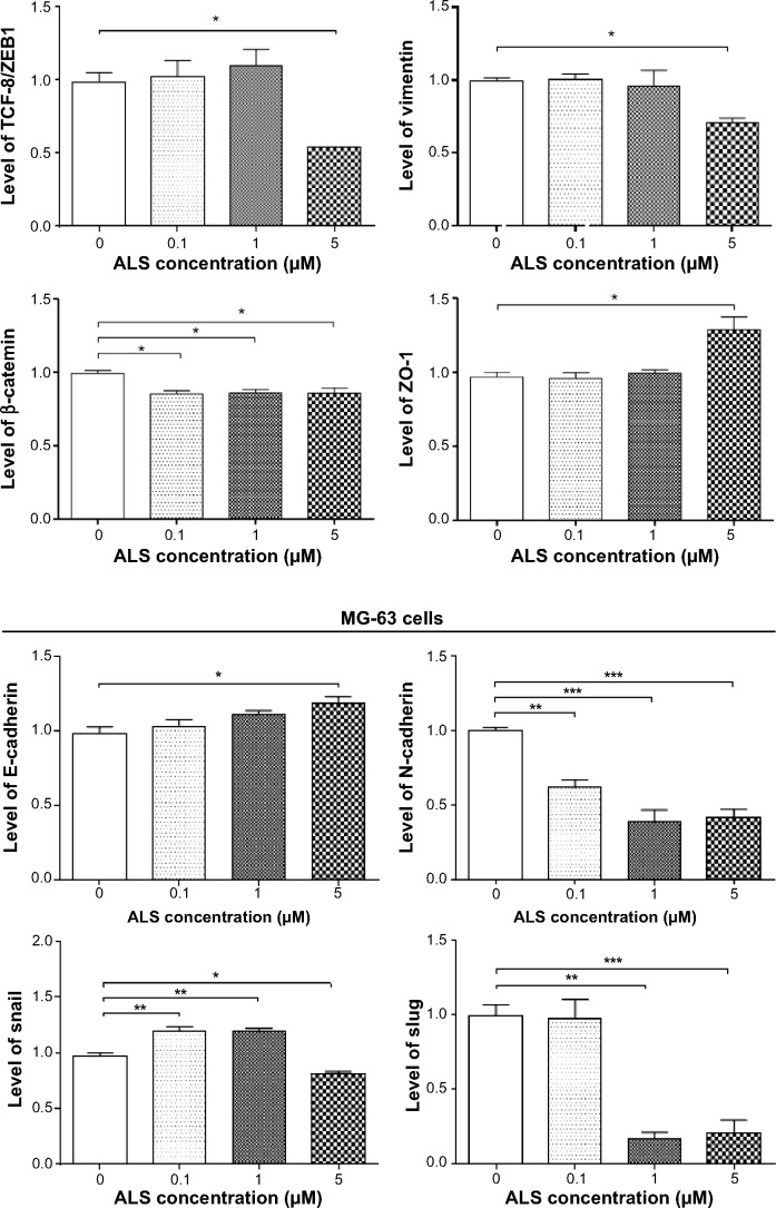
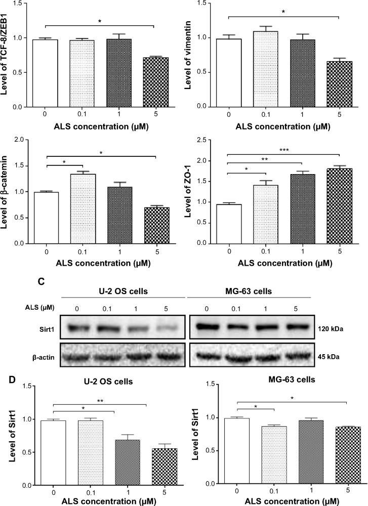
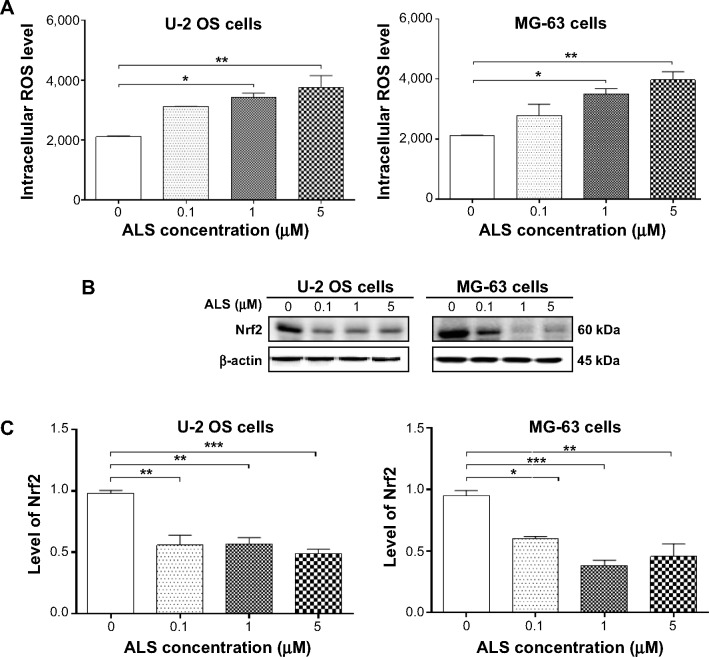
Osteosarcoma (OS) is the most common malignant bone tumor occurring mostly in children and adolescents between 10 and 20 years of age with poor response to current therapeutics. Alisertib (ALS, MLN8237) is a selective Aurora kinase A inhibitor that displays anticancer effects on several types of cancer. However, the role of ALS in the treatment of OS remains unknown. This study aimed to investigate the effects of ALS on the cell growth, apoptosis, autophagy, and epithelial to mesenchymal transition (EMT) and the underlying mechanisms in two human OS cell lines U-2 OS and MG-63. The results showed that ALS had potent growth inhibitory, pro-apoptotic, pro-autophagic, and EMT inhibitory effects on U-2 OS and MG-63 cells. ALS remarkably induced G2/M arrest and down-regulated the expression levels of cyclin-dependent kinases 1 and 2 and cyclin B1 in both U-2 OS and MG-63 cells. ALS markedly induced mitochondria-mediated apoptosis with a significant increase in the expression of key pro-apoptotic proteins and a decrease in main anti-apoptotic proteins. Furthermore, ALS promoted autophagic cell death via the inhibition of phosphatidylinositol 3-kinase (PI3K)/protein kinase B (Akt)/mammalian target of rapamycin (mTOR) and p38 mitogen-activated protein kinase (p38 MAPK) signaling pathways, and activation of 5'-AMP-dependent kinase (AMPK) signaling pathway. Inducers or inhibitors of apoptosis or autophagy simultaneously altered ALS-induced apoptotic and autophagic death in both U-2 OS and MG-63 cells, suggesting a crosstalk between these two primary modes of programmed cell death. Moreover, ALS suppressed EMT-like phenotypes with a marked increase in the expression of E-cadherin but a decrease in N-cadherin in U-2 OS and MG-63 cells. ALS treatment also induced reactive oxygen species (ROS) generation but inhibited the expression levels of sirtuin 1 and nuclear factor-erythroid-2-related factor 2 (Nrf2) in both cell lines. Taken together, these findings show that ALS promotes apoptosis and autophagy but inhibits EMT via PI3K/Akt/mTOR, p38 MAPK, and AMPK signaling pathways with involvement of ROS- and sirtuin 1-associated pathways in U-2 OS and MG-63 cells. ALS is a promising anticancer agent in OS treatment and further studies are needed to confirm its efficacy and safety in OS chemotherapy.

Keywords: ALS; EMT; PI3K/Akt/mTOR pathway; apoptosis; autophagy; osteosarcoma.


Figures

Figure 1
Figure 1
(A) The chemical structure of ALS and (B) the cytotoxicity of ALS toward U-2 OS and MG-63 cells. Notes: The cytotoxic effects of ALS on U-2 OS and MG-63 were determined by the MTT assay. The cells were treated with ALS at 0.01 to 50 μM for 24 hours. Data are the mean ± SD of three independent experiments. Abbreviations: ALS, alisertib; MTT, thiazolyl blue tetrazolium bromide; SD, standard deviation; IC50, half maximal inhibitory concentration; OS, osteosarcoma.
Figure 2
Figure 2
ALS inhibits the proliferation of U-2 OS and MG-63 cells and induces G2/M arrest in both cell lines. Notes: U-2 OS and MG-63 cells were treated with ALS and then subjected to flow cytometric analysis using PI as the DNA stain. (A) Representative DNA fluorescence histograms showing the effect of ALS treatment on cell cycle distribution of U-2 OS and MG-63 cells. Cells were treated with ALS at 0.1 to 5 μM for 24 hours and then analyzed by flow cytometry and (B) representative DNA fluorescence histograms showing the effect of 5 μM ALS treatment on cell cycle distribution over a 72-hour period. The bar graphs show the percentage of U-2 OS and MG-63 cells in G1, S, and G2/M phases. Cells in sub G1 are those undergoing apoptosis. Data are the mean ± SD of three independent experiments. Abbreviations: ALS, alisertib; SD, standard deviation; hr, hour; DMSO, dimethyl sulfoxide; OS, osteosarcoma; PI, propidium iodide; Dip, diploid.
Figure 3
Figure 3
ALS modulates the expression of CDK2, CDC2, cyclin B1, p21 Waf1/Cip1, and p53 in U-2 OS and MG-63 cells. Notes: U-2 OS and MG-63 cells were treated with ALS at 0.1, 1, and 5 μM for 24 hours and then subjected to Western blotting assay. (A) Representative blots of CDK2, CDC2, cyclin B1, p21 Waf1/Cip1, and p53 and (B) bar graphs showing the relative expression levels of CDK2, CDC2, cyclin B1, p21 Waf1/Cip1, and p53 in U-2 OS and MG-63 cells. β-actin was used as the internal control. Data are the mean ± SD of three independent experiments. *P<0.05; **P<0.01; and ***P<0.001 by one-way ANOVA. Abbreviations: ALS, alisertib; SD, standard deviation; ANOVA, analysis of variance; CDK, cyclin-dependent kinase; OS, osteosarcoma.
Figure 3
Figure 3
ALS modulates the expression of CDK2, CDC2, cyclin B1, p21 Waf1/Cip1, and p53 in U-2 OS and MG-63 cells. Notes: U-2 OS and MG-63 cells were treated with ALS at 0.1, 1, and 5 μM for 24 hours and then subjected to Western blotting assay. (A) Representative blots of CDK2, CDC2, cyclin B1, p21 Waf1/Cip1, and p53 and (B) bar graphs showing the relative expression levels of CDK2, CDC2, cyclin B1, p21 Waf1/Cip1, and p53 in U-2 OS and MG-63 cells. β-actin was used as the internal control. Data are the mean ± SD of three independent experiments. *P<0.05; **P<0.01; and ***P<0.001 by one-way ANOVA. Abbreviations: ALS, alisertib; SD, standard deviation; ANOVA, analysis of variance; CDK, cyclin-dependent kinase; OS, osteosarcoma.
Figure 4
Figure 4
ALS induces apoptotic cell death in U-2 OS and MG-63 cells. Notes: U-2 OS and MG-63 cells were treated with ALS at 0.1, 1, and 5 μM for 24 hours and then subjected to flow cytometric analysis. (A) Flow cytometric plots showing specific cell populations (live, early apoptosis, and late apoptosis) in U-2 OS and MG-63 cells and (B) bar graphs showing percentage of total apoptotic cells in U-2 OS and MG-63 cells. Data are the mean ± SD of three independent experiments. *P<0.05 and **P<0.01 by one-way ANOVA. Abbreviations: 7-AAD, 7-amino-actinomycin D; ALS, alisertib; SD, standard deviation; ANOVA, analysis of variance; OS, osteosarcoma; Q1, debris.
Figure 5
Figure 5
ALS modulates the expression of key pro- and anti-apoptotic proteins in U-2 OS and MG-63 cells. Notes: U-2 OS and MG-63 cells were treated with ALS at 0.1, 1, and 5 μM for 24 hours and then subjected to Western blotting assay. (A) Representative blots of Bax, Bcl-2, PUMA, cytochrome c, cleaved caspase-3, and cleaved caspase-9 in U-2 OS and MG-63 cells and (B) bar graphs showing the expression levels of Bax, Bcl-2, PUMA, cytochrome c, cleaved caspase-3, and cleaved caspase-9 in U-2 OS and MG-63 cells. β-actin was used as the internal control. Data are the mean ± SD of three independent experiments. *P<0.05; **P<0.01; and ***P<0.001 by one-way ANOVA. Abbreviations: ALS, alisertib; SD, standard deviation; ANOVA, analysis of variance; Bax, Bcl-associated X protein; PUMA, p53 up-regulated modulator of apoptosis; OS, osteosarcoma.
Figure 5
Figure 5
ALS modulates the expression of key pro- and anti-apoptotic proteins in U-2 OS and MG-63 cells. Notes: U-2 OS and MG-63 cells were treated with ALS at 0.1, 1, and 5 μM for 24 hours and then subjected to Western blotting assay. (A) Representative blots of Bax, Bcl-2, PUMA, cytochrome c, cleaved caspase-3, and cleaved caspase-9 in U-2 OS and MG-63 cells and (B) bar graphs showing the expression levels of Bax, Bcl-2, PUMA, cytochrome c, cleaved caspase-3, and cleaved caspase-9 in U-2 OS and MG-63 cells. β-actin was used as the internal control. Data are the mean ± SD of three independent experiments. *P<0.05; **P<0.01; and ***P<0.001 by one-way ANOVA. Abbreviations: ALS, alisertib; SD, standard deviation; ANOVA, analysis of variance; Bax, Bcl-associated X protein; PUMA, p53 up-regulated modulator of apoptosis; OS, osteosarcoma.
Figure 6
Figure 6
ALS promotes autophagic cell death in U-2 OS and MG-63 cells determined by flow cytometry. Notes: U-2 OS and MG-63 cells were treated with ALS at 0.1, 1, and 5 μM for 24 hours and then subjected to flow cytometric analysis using the Cyto-ID® green stain. (A) Flow cytometric dot plots and (B) bar graphs showing the percentage of autophagy in U-2 OS and MG-63 cells. Data are the mean ± SD of three independent experiments. **P<0.01 and ***P<0.001 by one-way ANOVA. Abbreviations: ALS, alisertib; SD, standard deviation; ANOVA, analysis of variance; OS, osteosarcoma.
Figure 7
Figure 7
ALS induces autophagic cell death in U-2 OS and MG-63 cells determined by confocal microscopy. Notes: U-2 OS and MG-63 cells were treated with ALS at 0.1, 1, and 5 μM for 24 hours and then subjected to confocal microscopic examination. The level of autophagy was evaluated using a lysosome-specific fluorescence dye and the images were analyzed. (A) Confocal microscopic images. Blue: nucleus, green: intracellular autophagic level and (B) quantitative autophagy level in U-2 OS and MG-63 cells. Data are the mean ± SD of three independent experiments. Scale bar, 3 μm; magnification, ×60. *P<0.05 and **P<0.01 by one-way ANOVA. Abbreviations: ALS, alisertib; SD, standard deviation; ANOVA, analysis of variance; OS, osteosarcoma.
Figure 8
Figure 8
ALS regulates the expression of key pro- and anti-autophagic proteins in U-2 OS and MG-63 cells. Notes: U-2 OS and MG-63 cells were treated with ALS at 0.1, 1, and 5 μM for 24 hours and then subjected to Western blotting assay. (A) Representative blots of the phosphorylated PI3K, AMPK, p38 MAPK, Akt, and mTOR and the total protein levels of PI3K, AMPK, p38 MAPK, Akt, mTOR, PTEN, beclin 1, LC3-I, and LC3-II in U-2 OS and MG-63 cells and (B) the ratio of p-PI3K/PI3K, p-AMPK/AMPK, p-p38 MAPK/p38 MAPK, p-Akt/Akt, p-mTOR/mTOR, and LC3-II/I and the expression levels of PTEN and beclin 1 in U-2 OS and MG-63 cells. β-actin was used as the internal control. Data are the mean ± SD of three independent experiments. *P<0.05; **P<0.01; and ***P<0.001 by one-way ANOVA. Abbreviations: ALS, alisertib; SD, standard deviation; ANOVA, analysis of variance; PI3K, phosphatidylinositol 3-kinase; AMPK, AMP-activated protein kinase; MAPK, mitogen-activated protein kinase; Akt, protein kinase B; mTOR, mammalian target of rapamycin; PTEN, phosphatase and tensin homolog; LC3, microtubule-associated protein 1A/1B-light chain 3; OS, osteosarcoma.
Figure 8
Figure 8
ALS regulates the expression of key pro- and anti-autophagic proteins in U-2 OS and MG-63 cells. Notes: U-2 OS and MG-63 cells were treated with ALS at 0.1, 1, and 5 μM for 24 hours and then subjected to Western blotting assay. (A) Representative blots of the phosphorylated PI3K, AMPK, p38 MAPK, Akt, and mTOR and the total protein levels of PI3K, AMPK, p38 MAPK, Akt, mTOR, PTEN, beclin 1, LC3-I, and LC3-II in U-2 OS and MG-63 cells and (B) the ratio of p-PI3K/PI3K, p-AMPK/AMPK, p-p38 MAPK/p38 MAPK, p-Akt/Akt, p-mTOR/mTOR, and LC3-II/I and the expression levels of PTEN and beclin 1 in U-2 OS and MG-63 cells. β-actin was used as the internal control. Data are the mean ± SD of three independent experiments. *P<0.05; **P<0.01; and ***P<0.001 by one-way ANOVA. Abbreviations: ALS, alisertib; SD, standard deviation; ANOVA, analysis of variance; PI3K, phosphatidylinositol 3-kinase; AMPK, AMP-activated protein kinase; MAPK, mitogen-activated protein kinase; Akt, protein kinase B; mTOR, mammalian target of rapamycin; PTEN, phosphatase and tensin homolog; LC3, microtubule-associated protein 1A/1B-light chain 3; OS, osteosarcoma.
Figure 9
Figure 9
Effects of FK866 on ALS-induced apoptosis and autophagy in U-2 OS and MG-63 cells. Notes: U-2 OS and MG-63 cells were pretreated with FK866 for 1 hour and then incubated with or without 5 μM ALS for 24 hours. The apoptotic and autophagic cells were examined by flow cytometry. (A) Effects of FK866 on the basal and ALS-induced apoptosis in U-2 OS and MG-63 cells detected using the double stain assay and (B) effects of the FK866 on the basal and ALS-induced autophagy in U-2 OS and MG-63 cells using Cyto-ID® to detect autophagy. The bar graphs show the levels of the total apoptosis and autophagy of U-2 OS and MG-63 cells. Data are the mean ± SD of three independent experiments. *P<0.05; **P<0.01; and ***P<0.001 by one-way ANOVA. Abbreviations: ALS, alisertib; SD, standard deviation; ANOVA, analysis of variance; FK866, (E)-N-(4-(1-benzoylpiperidin-4-yl)butyl)-3-(pyridin-3-yl)acrylamide; OS, osteosarcoma; 7-AAD, 7-aminoactinomycin D; PE, phycoerythrin.
Figure 10
Figure 10
Effects of a series of inducers and inhibitors on the apoptosis and autophagy induced by ALS in U-2 OS and MG-63 cells. Notes: The effects of the compounds on the basal and ALS-induced apoptosis (A) and autophagy (B) of U-2 OS and MG-63 cells; and (C) the bar graphs showing the levels of total apoptosis and autophagy of U-2 OS and MG-63 cells. The apoptosis was determined using the double stain (7-AAD plus annexin V:PE) assay with flow cytometry. The autophagy was determined using the Cyto-ID® green stain with flow cytometry. The flow cytometer collected 10,000 events for apoptosis and autophagy analysis. Data are the mean ± SD of three independent experiments. *P<0.05; **P<0.01; and ***P<0.001 by one-way ANOVA. Abbreviations: ALS, alisertib; SD, standard deviation; ANOVA, analysis of variance; WM, wortmannin; OS, osteosarcoma; 7-AAD, 7-aminoactinomycin D; PE, phycoerythrin; Q1, debris.
Figure 10
Figure 10
Effects of a series of inducers and inhibitors on the apoptosis and autophagy induced by ALS in U-2 OS and MG-63 cells. Notes: The effects of the compounds on the basal and ALS-induced apoptosis (A) and autophagy (B) of U-2 OS and MG-63 cells; and (C) the bar graphs showing the levels of total apoptosis and autophagy of U-2 OS and MG-63 cells. The apoptosis was determined using the double stain (7-AAD plus annexin V:PE) assay with flow cytometry. The autophagy was determined using the Cyto-ID® green stain with flow cytometry. The flow cytometer collected 10,000 events for apoptosis and autophagy analysis. Data are the mean ± SD of three independent experiments. *P<0.05; **P<0.01; and ***P<0.001 by one-way ANOVA. Abbreviations: ALS, alisertib; SD, standard deviation; ANOVA, analysis of variance; WM, wortmannin; OS, osteosarcoma; 7-AAD, 7-aminoactinomycin D; PE, phycoerythrin; Q1, debris.
Figure 11
Figure 11
Effects of SB202190 and WM on ALS-induced autophagy in U-2 OS and MG-63 cells by confocal microscopy. Notes: Cells were treated with 5 μM alone or in combination with 20 μM SB202190 or 10 μM WM for 24 hours. (A) Fluorescent images of autophagic U-2 OS and MG-63 cells. Blue: nucleus, green: intracellular autophagy level and (B) quantitative fluorescent level showing the effects of SB202190 and WM on ALS-induced autophagy in U-2 OS and MG-63 cells. Data are the mean ± SD of three independent experiments. Scale bar, 3 μm; magnification, ×60. *P<0.05 and ***P<0.001 by one-way ANOVA. Abbreviations: ALS, alisertib; SD, standard deviation; ANOVA, analysis of variance; WM, wortmannin; OS, osteosarcoma.
Figure 12
Figure 12
ALS modulates the expression levels of the key EMT markers and inhibits Sirt1 expression in U-2 OS and MG-63 cells. Notes: U-2 OS and MG-63 cells were treated with ALS at 0.1, 1, and 5 μM for 24 hours and then subjected to Western blotting assay. (A) Representative blots of E-cadherin, N-cadherin, snail, slug, TCF-8/ZEB1, vimentin, β-catenin, and ZO-1 in U-2 OS and MG-63 cells, (B) quantitative expression levels of E-cadherin, N-cadherin, snail, slug, TCF-8/ZEB1, vimentin, β-catenin, and ZO-1 in U-2 OS and MG-63 cells, (C) representative blots of Sirt1 in U-2 OS and MG-63 cells and (D) quantitative expression levels of Sirt1 in U-2 OS and MG-63 cells. β-actin was used as the internal control. Data represent the mean ± SD of three independent experiments. *P<0.05; **P<0.01; and ***P<0.001 by one-way ANOVA. Abbreviations: ALS, alisertib; SD, standard deviation; ANOVA, analysis of variance; EMT, epithelial to mesenchymal transition; Sirt1, sirtuin 1; ZO-1, zona occludens protein 1; TCF-8/ZEB1, zinc finger E-box-binding homeobox 1; OS, osteosarcoma.
Figure 12
Figure 12
ALS modulates the expression levels of the key EMT markers and inhibits Sirt1 expression in U-2 OS and MG-63 cells. Notes: U-2 OS and MG-63 cells were treated with ALS at 0.1, 1, and 5 μM for 24 hours and then subjected to Western blotting assay. (A) Representative blots of E-cadherin, N-cadherin, snail, slug, TCF-8/ZEB1, vimentin, β-catenin, and ZO-1 in U-2 OS and MG-63 cells, (B) quantitative expression levels of E-cadherin, N-cadherin, snail, slug, TCF-8/ZEB1, vimentin, β-catenin, and ZO-1 in U-2 OS and MG-63 cells, (C) representative blots of Sirt1 in U-2 OS and MG-63 cells and (D) quantitative expression levels of Sirt1 in U-2 OS and MG-63 cells. β-actin was used as the internal control. Data represent the mean ± SD of three independent experiments. *P<0.05; **P<0.01; and ***P<0.001 by one-way ANOVA. Abbreviations: ALS, alisertib; SD, standard deviation; ANOVA, analysis of variance; EMT, epithelial to mesenchymal transition; Sirt1, sirtuin 1; ZO-1, zona occludens protein 1; TCF-8/ZEB1, zinc finger E-box-binding homeobox 1; OS, osteosarcoma.
Figure 13
Figure 13
ALS induces ROS generation and down-regulates the expression of Nrf2 in U-2 OS and MG-63 cells. Notes: U-2 OS and MG-63 cells were treated with ALS at 0.1, 1, and 5 μM for 24 hours and then subjected to intracellular ROS measurement and Western blotting assay. (A) ALS induces ROS generation in U-2 OS and MG-63 cells, (B) representative blots of the expression level of Nrf2 in U-2 OS and MG-63cells and (C) bar graphs showing the relative expression level of Nrf2 in U-2 OS and MG-63 cells. β-actin was used as the internal control. Data represent the mean ± SD of three independent experiments. *P<0.05; **P<0.01; and ***P<0.001 by one-way ANOVA. Abbreviations: ALS, alisertib; SD, standard deviation; ANOVA, analysis of variance; ROS, reactive oxygen species; Nrf2, nuclear factor erythroid 2-related factor 2; OS, osteosarcoma.

Similar articles

Cited by

References

    1. Marulanda GA, Henderson ER, Johnson DA, Letson GD, Cheong D. Orthopedic surgery options for the treatment of primary osteosarcoma. Cancer Control. 2008;15(1):13–20. - PubMed
    1. Mirabello L, Troisi RJ, Savage SA. Osteosarcoma incidence and survival rates from 1973 to 2004: data from the Surveillance, Epidemiology, and End Results Program. Cancer. 2009;115(7):1531–1543. - PMC - PubMed
    1. Ritter J, Bielack SS. Osteosarcoma. Ann Oncol. 2010;21(Suppl 7):vii320–5. - PubMed
    1. Ottaviani G, Jaffe N. The epidemiology of osteosarcoma. In: Jaffe, Bruland OS, Bielack S, editors. Pediatric and Adolescent Osteosarcoma. Springer; 2010. pp. 3–13.
    1. Ta HT, Dass CR, Choong PF, Dunstan DE. Osteosarcoma treatment: state of the art. Cancer Metastasis Rev. 2009;28(1–2):247–263. - PubMed

MeSH terms