. 2015 Jul;22(7):884-903.
doi: 10.1177/1933719115576727. Epub 2015 Mar 16.

Mouse Ovarian Very Small Embryonic-Like Stem Cells Resist Chemotherapy and Retain Ability to Initiate Oocyte-Specific Differentiation

Affiliations

Mouse Ovarian Very Small Embryonic-Like Stem Cells Resist Chemotherapy and Retain Ability to Initiate Oocyte-Specific Differentiation

Kalpana Sriraman et al. Reprod Sci. 2015 Jul.

Abstract

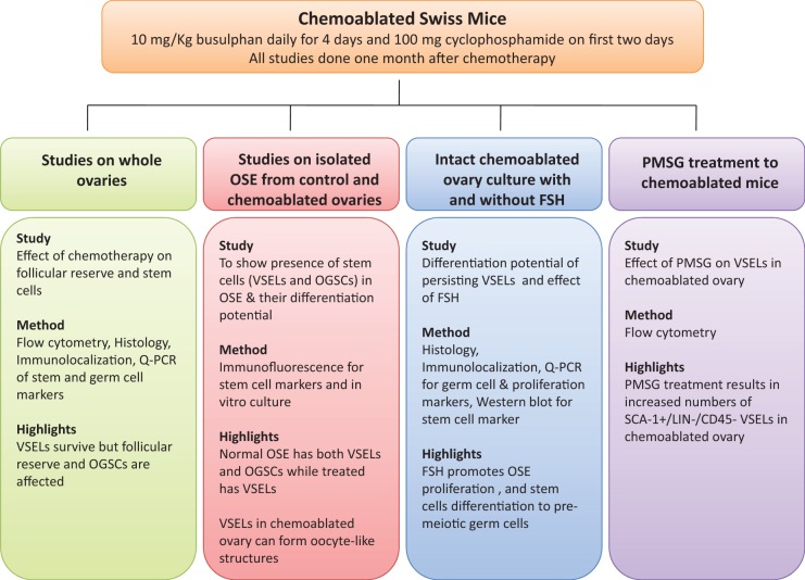
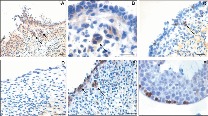
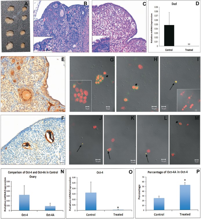
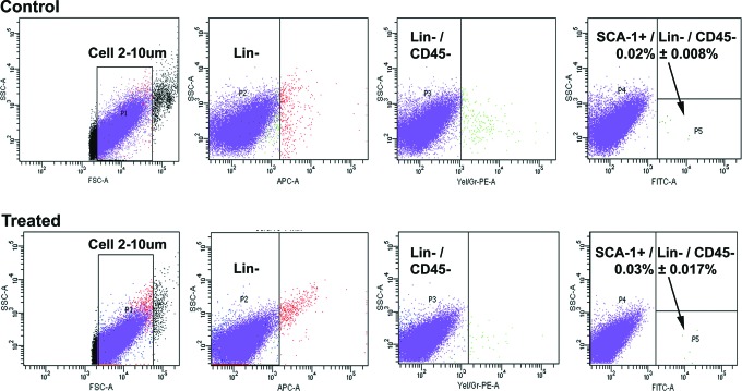
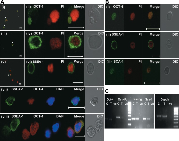
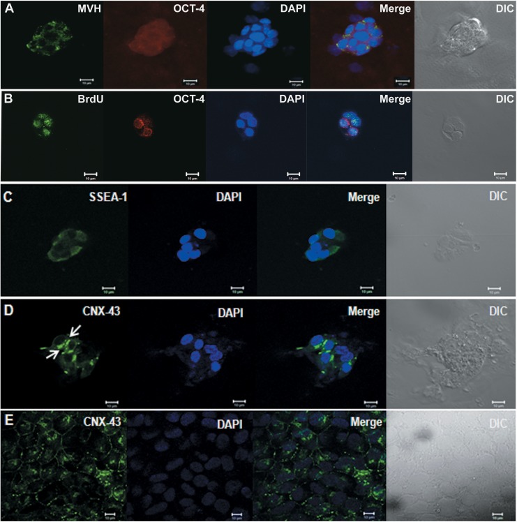
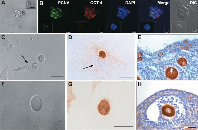
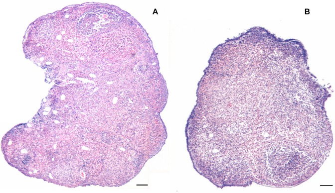
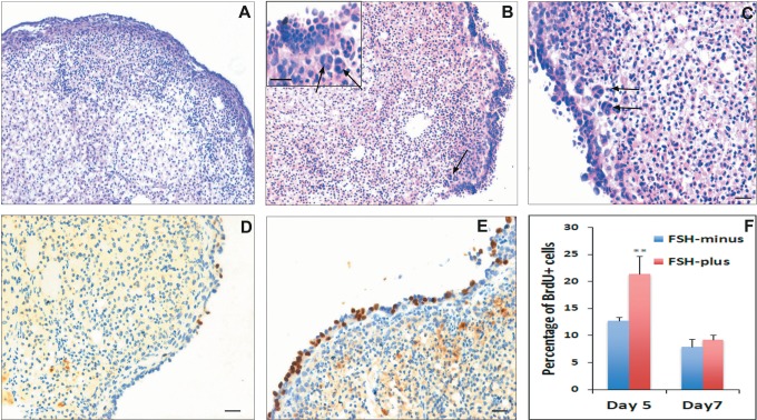
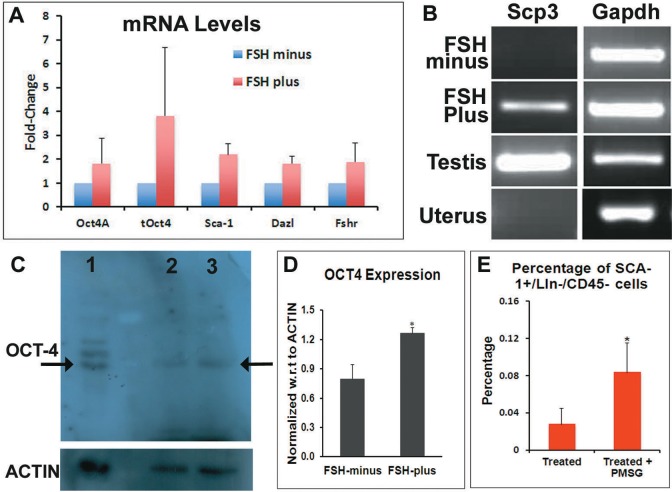
This study was undertaken to investigate stem cells in adult mouse ovary, the effect of chemotherapy on them and their potential to differentiate into germ cells. Very small embryonic-like stem cells (VSELs) that were SCA-1+/Lin-/CD45-, positive for nuclear octamer-binding transforming factor 4 (OCT-4), Nanog, and cell surface stage-specific embryonic antigen 1, were identified in adult mouse ovary. Chemotherapy resulted in complete loss of follicular reserve and cytoplasmic OCT-4 positive progenitors (ovarian germ stem cells) but VSELs survived. In ovarian surface epithelial (OSE) cell cultures from chemoablated ovary, proliferating germ cell clusters and mouse vasa homolog/growth differentiation factor 9-positive oocyte-like structure were observed by day 6, probably arising as a result of differentiation of the surviving VSELs. Follicle-stimulating hormone (FSH) exerted a direct stimulatory action on the OSE and induced stem cells proliferation and differentiation into premeiotic germ cell clusters during intact chemoablated ovaries culture. The FSH analog pregnant mare serum gonadotropin treatment to chemoablated mice increased the percentage of surviving VSELs in ovary. The results of this study provide evidence for the presence of potential VSELs in mouse ovaries and show that they survive chemotherapy, are modulated by FSH, and retain the ability to undergo oocyte-specific differentiation. These results show relevance to women who undergo premature ovarian failure because of oncotherapy.

Keywords: FSH; OCT-4; VSELs; ovary; stem cells.


Conflict of interest statement

Declaration of Conflicting Interests: The author(s) declared no potential conflicts of interest with respect to the research, authorship, and/or publication of this article.

Figures

Figure 1.
Figure 1.
An overview of the study.
Figure 2.
Figure 2.
Effect of chemotherapy (busulfan and cyclophosphamide) on mouse ovary and ovarian stem cells. A, Chemoablated ovaries are smaller in size compared to control. B, Hematoxylin and eosin (H&E)-stained control ovarian sections with multiple follicles including (B inset) primordial follicles and single layer of surface epithelial cells (OSE). C, Chemoablated ovary is completely devoid of follicles; however, OSE appeared multilayered (inset). Effect of chemotherapy on oocytes was studied using a germ cell marker DAZL. D, Note significant reduction (P < .01) in Dazl transcripts in chemoablated ovary compared to control with respect to Gapdh by Q-PCR analysis. Immunolocalization using anti-DAZL antibody revealed several oocytes in control ovarian sections (E) compared to no oocytes in chemoablated ovary (F). Effect of chemotherapy on ovarian stem cells was studied using OCT-4 as a marker. Two population of stem cells stained positive for OCT-4 in control ovarian smears (G-I) including small stem cells (VSELs) with nuclear OCT-4 (arrows) and slightly larger cells (OGSCs) with cytoplasmic OCT-4 (asterisks). Occasionally, the OGSCs appeared as clusters (G) and inset represents cluster negative for OCT-4. Negative control for anti-OCT-4 antibody (inset I). In contrast to control, only small nuclear OCT-4 positive VSELs were observed in chemoablated ovarian smears (J-L, arrows). Cytoplasmic OCT-4 positive cells were rarely observed and had relatively low intensity of staining (M, asterisks). The images in (G-I) and (J-M) are merge of green (OCT-4), red (PI), and DIC channels. The Q-PCR results for Oct-4A transcript (representative of nuclear OCT-4 positive VSELs) and total Oct-4 (representative of all Oct-4 isoforms including cytoplasmic OCT-4 positive OGSCs) correlated well with immunolocalization results. Control ovaries revealed lesser expression of Oct-4A compared to total Oct-4 (N), implying the rarity of VSELs compared to OGSCs. In chemoablated ovaries, total Oct-4 transcripts were significantly lower (P < .05; O), however percentage of Oct-4A within the total Oct-4 was significantly higher (P < .05; P) compared to control, and implying predominance of VSELs. Oct-4 and Oct-4A levels represented here were normalized using Gapdh as housekeeping gene. Bar in (B), (C), (E), and (F) equals 20 μm. DAZL indicates deleted in azoospermia like; OCT-4, octamer-binding transforming factor 4; OGSC, ovary germ stem cell; OSE, ovarian surface epithelial; Q-PCR, quantitative polymerase chain reaction; VSELs, very small embryonic-like stem cells. (The color version of this figure is available in the online version at http://rs.sagepub.com/.)
Figure 3.
Figure 3.
Flow cytometry analysis of SCA-1+/Lin−/CD45− VSELs in ovaries from control and chemotreated mice. Cells between 2 and 10 µm were gated using size calibration beads (1) followed by sequential selection for LIN negative population (2), CD45 negative population (3), and then SCA-1 positive population (4) in both control and treated mice. The average percentage of SCA-1+/Lin−/CD45− VSELs with standard deviation from minimum of 4 animals is reported. VSELs indicates very small embryonic-like stem cells.
Figure 4.
Figure 4.
Immunofluorescence characterization of stem cells in OSE smears and their differentiation potential. A, Characterization of stem cells in OSE from control mice. (i-iv) Immunofluorescence using anti-OCT-4 antibody (green) with propidium iodide (red) as counterstain. Note the presence of 2 types of OCT-4 positive cells similar to those observed in whole ovarian smears (Figure 2). The smaller VSELs are positive for nuclear OCT-4 (i-arrows; ii) and slightly larger OGSCs are positive for cytoplasmic OCT-4 (i and iii-asterisks; iv). v-vi, Immunofluorescence using anti-SSEA-1 antibody (green) and PI (red) as counterstain on control OSE smears. The size of SSEA-1 positive cells ranged from 5 to 10 μm probably representing both VSELs and OGSCs. vi, Represents zoomed image of potential VSEL. vii-viii, Dual labeling with anti-OCT-4 (red) and anti-SSEA-1 (green) antibody counterstained with 4′,6-diamidino-2-phenylindole (DAPI; blue). Both nuclear OCT-4 positive VSELs (vii) and cytoplasmic OCT-4 positive OGSCs (viii) were positive for SSEA-1. B, Characterization of stem cells in OSE from treated mice: Immunofluorescence analysis of OSE preparation from chemoablated ovary for OCT-4 (i), SSEA-1 (ii), and SCA-1 (iii) counterstained with propidium iodide (PI; red). Note that OCT-4 shows nuclear localization. C, Real-time polymerase chain reaction (RT-PCR) analysis of RNA isolated from OSE. Image shows presence of Oct-4A, Oct-4, Sca-1, and Nanog in both control and chemoabalated mice. Gapdh was used as housekeeping gene. In the image, C represents control, T represents OSE from chemoablated ovaries, and −ve represents PCR negative control. OCT-4 indicates octamer-binding transforming factor 4; OGSC, ovary germ stem cell; OSE, ovarian surface epithelial; SSEA-1, stage-specific embryonic antigen 1; VSELs, very small embryonic-like stem cells. (The color version of this figure is available in the online version at http://rs.sagepub.com/.)
Figure 5.
Figure 5.
In vitro culture of OSE from control mouse ovaries. When OSEs from control mouse were cultured for 48 hours in the presence of follicle-stimulating hormone (FSH), clusters of cells that were positive for both MVH (green) and cytoplasmic OCT-4 (red; A) were observed. When cultured with BrdU, the OCT-4 positive clusters showed incorporation of BrdU as seen by colocalization of cytoplasmic OCT-4 (red) and BrdU (green; B) suggesting that the ovarian stem cells show the differentiation potential into proliferating germ cells cyst-like structures. Further characterization of the clusters showed expression of germ cell cyst markers stage-specific embryonic antigen 1 (SSEA-1; green; C) and Connexin-43 (CNX-43, green; D). Expression of CNX-43 (green) on human embryonic stem cell line KIND1 (E) was taken as positive control. 4′,6-Diamidino-2-phenylindole (DAPI; blue) was used as nuclear stain. All bars equals 10 μm. BrdU indicates bromodeoxyuridine; MVH, mouse vasa homolog; OCT-4, octamer-binding transforming factor 4; OSE, ovarian surface epithelial. (The color version of this figure is available in the online version at http://rs.sagepub.com/.)
Figure 6.
Figure 6.
In vitro culture of ovarian surface epithelial (OSE) isolated from chemoablated mouse ovaries. A, Direct microscopic observation of culture on day 2. Clusters of cells (arrowhead) containing fewer cells per cluster with ovoid shape and large nucleus overlying epithelial/fibroblastic cells were observed. Inset represents a different cell cluster with small cells. B, Immunofluorescence analysis shows the cell clusters with ovoid cells were positive for PCNA (green) in nucleus and OCT-4 (red) in cytoplasm suggesting they are proliferating stem cell clusters. Blue represents the nuclear counterstain 4′,6-diamidino-2-phenylindole (DAPI). Insets in respective channels represent nongerm cell cluster showing no staining for OCT-4 and PCNA. C and F, Oocyte-like cells observed on day 6 of culture. They were found in close association with underlying fibroblast cell (arrow). D and G, Immunocytochemical staining of cultured cells harvested on day 6 shows that the oocyte-like cells were positive for germ cell marker MVH (D) and oocyte specific marker GDF-9 (G). In D, arrow represents the nucleus of underlying fibroblast cell. E and H, represents the positive control (mouse ovarian sections) for MVH (E) and GDF-9 (H) that was used to check specificity of the antibodies. Bar equals 20 μm (A and C-H) and 10 μm (B). GDF-9 indicates growth differentiation factor 9; MVH, mouse vasa homolog; OCT-4, octamer-binding transforming factor 4; PCNA, proliferating cell nuclear antigen. (The color version of this figure is available in the online version at http://rs.sagepub.com/.)
Figure 7.
Figure 7.
Low magnification comparison of hematoxylin and eosin (H&E)-stained ovarian sections from chemoablated ovaries cultured with and without FSH. The chemoablated ovaries are completely devoid of follicles (similar to that shown in Figure 2C). FSH treatment in vitro for 7 days results in extensive proliferation of ovarian surface epithelial (OSE), which appears thick and multilayered (B) compared to FSH-minus group (A). Bar equals 100 μm. FSH indicates follicle-stimulating hormone.
Figure 8.
Figure 8.
Effect of FSH on in vitro cultured chemoablated ovaries: higher magnification of hematoxylin and eosin (H&E)-stained ovarian sections showed presence of multilayered and highly proliferative OSE in FSH-plus group (B and C) compared to FSH-minus group (A). Frequently, clusters of cells (presumably differentiating germ cell clusters, refer Figure 9) close to OSE were observed in FSH-plus group (B and C; arrows). BrdU incorporation studies to investigate the proliferation status of cells showed a higher number of cells incorporate BrdU in the FSH-plus group on day 5 (E) compared to FSH-minus group (D) indicating proliferation of OSE in response to FSH. The percentage of BrdU+ cells was significantly (P < .01) higher on day 5 in FSH-plus group confirming increase in proliferation of OSE (F). However, on day 7, there was no difference between the 2 groups and the levels were comparable to that of percentage in FSH-minus group on day 5 suggesting reduced proliferation with time during culture. Bar equals 20 μm. BrdU indicates bromodeoxyuridine; FSH, follicle-stimulating hormone; OSE, ovarian surface epithelial. (The color version of this figure is available in the online version at http://rs.sagepub.com/.)
Figure 9.
Figure 9.
Immunolocalization of MVH, STRA-8, and BrdU on in vitro cultured chemoablated ovaries. A and B, Immunolocalization of MVH on ovaries from the FSH-plus group. Note the cluster of cells (arrows) close to OSE is positive for MVH. C, Immunolocalization of BrdU on ovaries from the FSH-plus group. The clusters of cells are also positive for BrdU (arrows) indicating their proliferative state. Immunolocalization of STRA-8 on ovaries from (D) the FSH-minus group and (E) the FSH-plus group. Cell clusters positive (arrows) for Stra8 are observed close to OSE and also on OSE. Mouse testicular section was used as positive control for STRA-8 in which spermatogonia along the basement membrane express STRA-8 (F). Bar equals 20 μm. BrdU indicates bromodeoxyuridine; FSH, follicle-stimulating hormone; MVH, mouse vasa homolog. OSE, ovarian surface epithelial. (The color version of this figure is available in the online version at http://rs.sagepub.com/.)
Figure 10.
Figure 10.
Analysis of stem/germ cell markers by Q-PCR/Western blot and flowcytometry analysis of SCA-1+/Lin−/CD45− VSELs in ovaries of PMSG-treated chemoablated mice. A, Q-PCR analysis for stem (Oct-4A, Sca-1), germ cell (Oct-4, Dazl) markers, and FSH receptors showed increase in FSH-plus group compared to FSH-minus group. 18s was used for normalization. B, Real-time polymerase chain reaction (RT-PCR) analysis for meiotic marker Scp3 showed the presence of Scp3 mRNA in FSH-plus group and positive control mouse testicular sample, while Scp3 was absent in FSH-minus group and negative control uterine sample. Gapdh was used as housekeeping gene. C, Western blot analysis using anti-OCT-4 antibody on total cell protein extracts from whole ovary showed a band of OCT-4 both in FSH-minus (lane 2) and FSH-plus group (lane 3). Human embryonic stem cells extract was used as positive control (lane 1). Actin was used as housekeeping protein (C lower panel). D, Densitometry analysis of the OCT-4 band with respect to Actin showed the amount of OCT-4 in FSH-plus group is significantly higher (P < .05) compared to FSH-minus group. E, Comparison of SCA-1+/Lin−/CD45− VSELs in ovaries of chemoablated mice (treated) and chemoablated mice treated with PMSG (treated + PMSG) by flowcytometry. The percentage of SCA-1+/Lin−/CD45− was significantly more (P < .1) in treated + PMSG than just treated mice. BrdU indicates bromodeoxyuridine; FSH, follicle-stimulating hormone; PMSG, pregnant mare serum gonadotropin; OCT-4, octamer-binding transforming factor 4; Q-PCR, quantitative polymerase chain reaction; VSELs, very small embryonic-like stem cells. (The color version of this figure is available in the online version at http://rs.sagepub.com/.)

Similar articles

Cited by

References

    1. Johnson J, Canning J, Kaneko T, Pru JK, Tilly JL. Germline stem cells and follicular renewal in the postnatal mammalian ovary. Nature. 2004;428 (6979):145–150. - PubMed
    1. Parte S, Bhartiya D, Telang J, et al. Detection, characterization, and spontaneous differentiation in vitro of very small embryonic-like putative stem cells in adult mammalian ovary. Stem Cells Dev. 2011;20 (8):1451–1464. - PMC - PubMed
    1. Pacchiarotti J, Maki C, Ramos T, et al. Differentiation potential of germ line stem cells derived from the postnatal mouse ovary. Differentiation 2010;79 (3):159–170. - PubMed
    1. Zou K, Yuan Z, Yang Z, et al. Production of offspring from a germline stem cell line derived from neonatal ovaries. Nat Cell Biol. 2009;11 (5):631–636. - PubMed
    1. Zhang D, Fouad H, Zoma WD, Salama SA, Wentz MJ, Al-Hendy A. Expression of stem and germ cell markers within nonfollicle structures in adult mouse ovary. Reprod Sci. 2008;15 (2):139–146. - PubMed

Publication types

Substances