

Peak inflammation in atherosclerosis, primary biliary cirrhosis and autoimmune arthritis is counter-intuitively associated with regulatory T cell enrichment
- PMID: 25770018
- PMCID: PMC4457006
- DOI: 10.1016/j.imbio.2015.02.006
Peak inflammation in atherosclerosis, primary biliary cirrhosis and autoimmune arthritis is counter-intuitively associated with regulatory T cell enrichment
Abstract
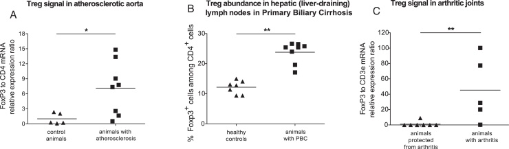
Regulatory T cells (Treg) influence the development of autoimmunity and their use is increasingly proposed for clinical applications. The well-characterized suppressive potential of Treg frequently leads to the assumption that Treg presence in prevailing numbers is indicative of immunosuppression. We hypothesized that this assumption may be false. We examined models of three different diseases caused by organ-specific autoimmune responses: primary biliary cirrhosis, atherosclerosis and rheumatoid arthritis (RA). We examined indicators of relative abundance of Treg compared to pro-inflammatory T cells, during peak inflammation. In all cases, the results were compatible with a relative enrichment of Treg at the site of inflammation or its most proximal draining lymph node. Conversely, in healthy mice or mice successfully protected from disease via a Treg-mediated mechanism, the data did not suggest that any Treg accumulation was occurring. This counter-intuitive finding may appear to be at odds with the immunosuppressive nature of Treg. Yet extensive previous studies in RA show that an accumulation of Treg occurs at peak inflammation, albeit without resulting in suppression, as the Treg suppressive function is overcome by the cytokine-rich environment. We suggest that this is a ubiquitous feature of autoimmune inflammation. Treg abundance in patient samples is increasingly used as an indicator of a state of immunosuppression. We conclude that this strategy should be revisited as it may potentially be a source of misinterpretation.
Keywords: Atherosclerosis; Autoimmunity; Inflammation; Primary biliary cirrhosis; Regulatory T cells; Rheumatoid arthritis.
Copyright © 2015 The Authors. Published by Elsevier GmbH.. All rights reserved.
Figures
Similar articles
-
The CII-specific autoimmune T-cell response develops in the presence of FTY720 but is regulated by enhanced Treg cells that inhibit the development of autoimmune arthritis.Arthritis Res Ther. 2016 Jan 12;18:8. doi: 10.1186/s13075-015-0909-6. Arthritis Res Ther. 2016. PMID: 26757712 Free PMC article.
-
Th17 and regulatory T lymphocytes in primary biliary cirrhosis and systemic sclerosis as models of autoimmune fibrotic diseases.Autoimmun Rev. 2012 Dec;12(2):300-4. doi: 10.1016/j.autrev.2012.05.004. Epub 2012 May 23. Autoimmun Rev. 2012. PMID: 22634708 Review.
-
The role of FOXP3+ regulatory T cells in human autoimmune and inflammatory diseases.Clin Exp Immunol. 2019 Jul;197(1):24-35. doi: 10.1111/cei.13288. Epub 2019 Mar 24. Clin Exp Immunol. 2019. PMID: 30830965 Free PMC article. Review.
-
STAT4 Regulates the CD8+ Regulatory T Cell/T Follicular Helper Cell Axis and Promotes Atherogenesis in Insulin-Resistant Ldlr-/- Mice.J Immunol. 2017 Nov 15;199(10):3453-3465. doi: 10.4049/jimmunol.1601429. Epub 2017 Oct 20. J Immunol. 2017. PMID: 29055004 Free PMC article.
-
TNF receptor 2 signaling prevents DNA methylation at the Foxp3 promoter and prevents pathogenic conversion of regulatory T cells.Proc Natl Acad Sci U S A. 2019 Oct 22;116(43):21666-21672. doi: 10.1073/pnas.1909687116. Epub 2019 Oct 9. Proc Natl Acad Sci U S A. 2019. PMID: 31597740 Free PMC article.
Cited by
-
T cell costimulation blockade blunts pressure overload-induced heart failure.Nat Commun. 2017 Mar 6;8:14680. doi: 10.1038/ncomms14680. Nat Commun. 2017. PMID: 28262700 Free PMC article.
-
IL10 Secretion Endows Intestinal Human iNKT Cells with Regulatory Functions Towards Pathogenic T Lymphocytes.J Crohns Colitis. 2022 Sep 8;16(9):1461-1474. doi: 10.1093/ecco-jcc/jjac049. J Crohns Colitis. 2022. PMID: 35358301 Free PMC article.
-
Regulatory T Cells Beyond Autoimmunity: From Pregnancy to Cancer and Cardiovascular Disease.Front Immunol. 2020 Mar 31;11:509. doi: 10.3389/fimmu.2020.00509. eCollection 2020. Front Immunol. 2020. PMID: 32296427 Free PMC article. Review.
-
Bile acid receptor agonists in primary biliary cholangitis: Regulation of the cholangiocyte secretome and downstream T cell differentiation.FASEB Bioadv. 2019 Apr 22;1(5):332-343. doi: 10.1096/fba.2018-00046. eCollection 2019 May. FASEB Bioadv. 2019. PMID: 32123836 Free PMC article.
-
Immune checkpoints in cardiac physiology and pathology: therapeutic targets for heart failure.Nat Rev Cardiol. 2024 Jul;21(7):443-462. doi: 10.1038/s41569-023-00986-9. Epub 2024 Jan 26. Nat Rev Cardiol. 2024. PMID: 38279046 Review.
References
-
- Ait-Oufella H., Salomon B.L., Potteaux S., Robertson A.K., Gourdy P., Zoll J., Merval R., Esposito B., Cohen J.L., Fisson S., Flavell R.A., Hansson G.K., Klatzmann D., Tedgui A., Mallat Z. Natural regulatory T cells control the development of atherosclerosis in mice. Nat. Med. 2006;12:178–180. - PubMed
-
- Allan S.E., Broady R., Gregori S., Himmel M.E., Locke N., Roncarolo M.G., Bacchetta R., Levings M.K. CD4+ T-regulatory cells: toward therapy for human diseases. Immunol. Rev. 2008;223:391–421. - PubMed
-
- Ammirati E., Cianflone D., Vecchio V., Banfi M., Vermi A.C., De Metrio M., Grigore L., Pellegatta F., Pirillo A., Garlaschelli K., Manfredi A.A., Catapano A.L., Maseri A., Palini A.G., Norata G.D. Effector memory T cells are associated with atherosclerosis in humans and animal models. J. Am. Heart Assoc. 2012;1:27–41. - PMC - PubMed
-
- Benito-Miguel M., Garcia-Carmona Y., Balsa A., Perez de Ayala C., Cobo-Ibanez T., Martin-Mola E., Miranda-Carus M.E. A dual action of rheumatoid arthritis synovial fibroblast IL-15 expression on the equilibrium between CD4+CD25+ regulatory T cells and CD4+CD25− responder T cells. J. Immunol. 2009;183:8268–8279. - PubMed
Publication types
MeSH terms
Substances
Grants and funding
LinkOut - more resources
Full Text Sources
Other Literature Sources
Medical
