

Free ISG15 triggers an antitumor immune response against breast cancer: a new perspective
- PMID: 25749047
- PMCID: PMC4466680
- DOI: 10.18632/oncotarget.3372
Free ISG15 triggers an antitumor immune response against breast cancer: a new perspective
Abstract
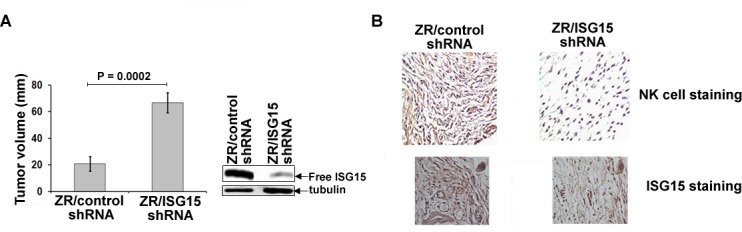
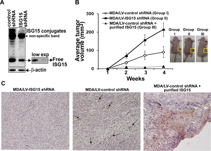
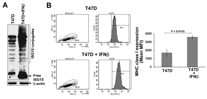
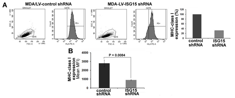
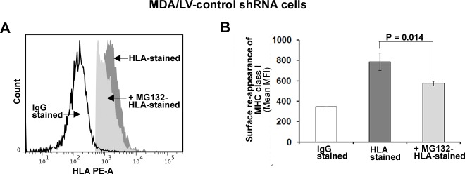
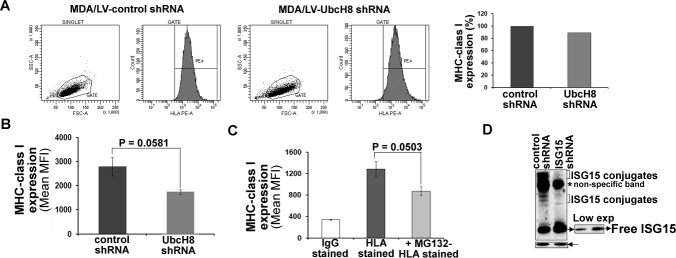
Interferon-Stimulated Gene 15 (ISG15), an antagonist of the canonical ubiquitin pathway, is frequently overexpressed in various cancers. In cancer cells, ISG15 is detected as free (intracellular) and conjugated to cellular proteins (ISGylation). Free ISG15 is also secreted into the extracellular milieu. ISGylation has protumor functions and extracellular free ISG15 has immunomodulatory properties in vitro. Therefore, whether ISG15 is a tumor suppressor or tumor promoter in vivo remains controversial. The current study aimed to clarify the role of free ISG15 in tumorigenesis. Breast cancer cells stably expressing control, ISG15, and UbcH8 (ISG15-specific E2 ligase) shRNAs were used to assess the immunoregulatory and antitumor function of free ISG15 in cell culture (in vitro) and in nude mice (in vivo). We show that extracellular free ISG15 suppresses breast tumor growth and increases NK cell infiltration into xenografted breast tumors in nude mice, and intracellular free ISG15 enhances major histocompatibility complex (MHC) class I surface expression in breast cancer cells. We conclude that free ISG15 may have antitumor and immunoregulatory function in vivo. These findings provides the basis for developing strategies to increase systemic levels of free ISG15 to treat cancer patients overexpressing the ISG15 pathway.
Keywords: ISG15; antitumor; breast cancer; immune system; ubiquitin/proteasome.
Conflict of interest statement
We declare no conflict of interest.
Figures
Similar articles
-
The Functional Roles of ISG15/ISGylation in Cancer.Molecules. 2023 Jan 31;28(3):1337. doi: 10.3390/molecules28031337. Molecules. 2023. PMID: 36771004 Free PMC article. Review.
-
ISGylation governs the oncogenic function of Ki-Ras in breast cancer.Oncogene. 2014 Feb 6;33(6):794-803. doi: 10.1038/onc.2012.633. Epub 2013 Jan 14. Oncogene. 2014. PMID: 23318454
-
ISG15 disrupts cytoskeletal architecture and promotes motility in human breast cancer cells.Exp Biol Med (Maywood). 2012 Jan;237(1):38-49. doi: 10.1258/ebm.2011.011236. Epub 2011 Dec 20. Exp Biol Med (Maywood). 2012. PMID: 22185919
-
Elevated expression of ISG15 in tumor cells interferes with the ubiquitin/26S proteasome pathway.Cancer Res. 2006 Jan 15;66(2):921-8. doi: 10.1158/0008-5472.CAN-05-1123. Cancer Res. 2006. PMID: 16424026
-
Regulation and action of interferon-stimulated gene 15 in breast cancer cells.Hum Cell. 2020 Oct;33(4):954-962. doi: 10.1007/s13577-020-00414-x. Epub 2020 Aug 19. Hum Cell. 2020. PMID: 32813218 Review.
Cited by
-
The Role of EBV-Encoded LMP1 in the NPC Tumor Microenvironment: From Function to Therapy.Front Oncol. 2021 Feb 25;11:640207. doi: 10.3389/fonc.2021.640207. eCollection 2021. Front Oncol. 2021. PMID: 33718235 Free PMC article. Review.
-
ISGylation in Innate Antiviral Immunity and Pathogen Defense Responses: A Review.Front Cell Dev Biol. 2021 Nov 25;9:788410. doi: 10.3389/fcell.2021.788410. eCollection 2021. Front Cell Dev Biol. 2021. PMID: 34901029 Free PMC article. Review.
-
WBSCR22 and TRMT112 synergistically suppress cell proliferation, invasion and tumorigenesis in pancreatic cancer via transcriptional regulation of ISG15.Int J Oncol. 2022 Mar;60(3):24. doi: 10.3892/ijo.2022.5314. Epub 2022 Jan 28. Int J Oncol. 2022. PMID: 35088887 Free PMC article.
-
The Functional Roles of ISG15/ISGylation in Cancer.Molecules. 2023 Jan 31;28(3):1337. doi: 10.3390/molecules28031337. Molecules. 2023. PMID: 36771004 Free PMC article. Review.
-
ISGylation is induced in neurons by demyelination driving ISG15-dependent microglial activation.J Neuroinflammation. 2022 Oct 20;19(1):258. doi: 10.1186/s12974-022-02618-4. J Neuroinflammation. 2022. PMID: 36261842 Free PMC article.
References
-
- Sgorbissa A, Brancolini C. IFNs, ISGylation and cancer: Cui prodest? Cytokine Growth Factor Rev. 2012;23:307–314. - PubMed
-
- Andersen JB, Hassel BA. The interferon regulated ubiquitin-like protein, ISG15, in tumorigenesis: friend or foe? Cytokine Growth Factor Rev. 2006;17:411–421. - PubMed
-
- Bektas N, Noetzel E, Veeck J, Press MF, Kristiansen G, Naami A, Hartmann A, Dimmler A, Beckmann MW, Knüchel R, Fasching PA, Dahl E. The ubiquitin-like molecule interferon-stimulated gene 15 (ISG15) is a potential prognostic marker in human breast cancer. Breast Cancer Res. 2008;10:R58. - PMC - PubMed
Publication types
MeSH terms
Substances
Grants and funding
LinkOut - more resources
Full Text Sources
Other Literature Sources
Medical
Research Materials
Miscellaneous
