Activation of the Wnt/β-catenin signaling cascade after traumatic nerve injury
- PMID: 25743255
- PMCID: PMC5384639
- DOI: 10.1016/j.neuroscience.2015.02.049
Activation of the Wnt/β-catenin signaling cascade after traumatic nerve injury
Abstract
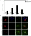
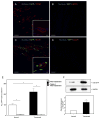
Recent data have shown that preservation of the neuromuscular junction (NMJ) after traumatic nerve injury helps to improve functional recovery with surgical repair via matrix metalloproteinase-3 (MMP3) blockade. As such, we sought to explore additional pathways that may augment this response. Wnt3a has been shown to inhibit acetylcholine receptor (AChR) clustering via β-catenin-dependent signaling in the development of the NMJ. Therefore, we hypothesized that Wnt3a and β-catenin are associated with NMJ destabilization following traumatic denervation. A critical size nerve defect was created by excising a 10-mm segment of the sciatic nerve in mice. Denervated muscles were then harvested at multiple time points for immunofluorescence staining, quantitative real-time PCR, and western blot analysis for Wnt3a and β-catenin levels. Moreover, a novel Wnt/β-catenin transgenic reporter mouse line was utilized to support our hypothesis of Wnt activation after traumatic nerve injury. The expression of Wnt3a mRNA was significantly increased by 2 weeks post-injury and remained upregulated for 2 months. Additionally, β-catenin was activated at 2 months post-injury relative to controls. Correspondingly, immunohistochemical analysis of denervated transgenic mouse line TCF/Lef:H2B-GFP muscles demonstrated that the number of GFP-positive cells was increased at the motor endplate band. These collective data support that post-synaptic AChRs destabilize after denervation by a process that involves the Wnt/β-catenin pathway. As such, this pathway serves as a potential therapeutic target to prevent the motor endplate degeneration that occurs following traumatic nerve injury.
Keywords: Wnt signaling; neuromuscular junction; peripheral nerve; traumatic nerve injury.
Copyright © 2015 IBRO. Published by Elsevier Ltd. All rights reserved.
Figures


Similar articles
-
Wnt/beta-catenin signaling suppresses Rapsyn expression and inhibits acetylcholine receptor clustering at the neuromuscular junction.J Biol Chem. 2008 Aug 1;283(31):21668-75. doi: 10.1074/jbc.M709939200. Epub 2008 Jun 9. J Biol Chem. 2008. PMID: 18541538
-
Muscle Yap Is a Regulator of Neuromuscular Junction Formation and Regeneration.J Neurosci. 2017 Mar 29;37(13):3465-3477. doi: 10.1523/JNEUROSCI.2934-16.2017. Epub 2017 Feb 17. J Neurosci. 2017. PMID: 28213440 Free PMC article.
-
Matrix metalloproteinase 3 deletion preserves denervated motor endplates after traumatic nerve injury.Ann Neurol. 2013 Feb;73(2):210-23. doi: 10.1002/ana.23781. Epub 2012 Dec 31. Ann Neurol. 2013. PMID: 23281061
-
Dual roles for Wnt signalling during the formation of the vertebrate neuromuscular junction.Acta Physiol (Oxf). 2012 Jan;204(1):128-36. doi: 10.1111/j.1748-1716.2011.02295.x. Epub 2011 May 7. Acta Physiol (Oxf). 2012. PMID: 21554559 Review.
-
Crosstalk between Agrin and Wnt signaling pathways in development of vertebrate neuromuscular junction.Dev Neurobiol. 2014 Aug;74(8):828-38. doi: 10.1002/dneu.22190. Epub 2014 Jun 4. Dev Neurobiol. 2014. PMID: 24838312 Review.
Cited by
-
Exuberant circumferential fibroproliferative neuromas in lipomatosis of nerve: a unifying theory. Illustrative case.J Neurosurg Case Lessons. 2024 Jan 15;7(3):CASE23661. doi: 10.3171/CASE23661. Print 2024 Jan 15. J Neurosurg Case Lessons. 2024. PMID: 38224588 Free PMC article.
-
Denervation-Related Neuromuscular Junction Changes: From Degeneration to Regeneration.Front Mol Neurosci. 2022 Feb 24;14:810919. doi: 10.3389/fnmol.2021.810919. eCollection 2021. Front Mol Neurosci. 2022. PMID: 35282655 Free PMC article. Review.
-
A Subtle Network Mediating Axon Guidance: Intrinsic Dynamic Structure of Growth Cone, Attractive and Repulsive Molecular Cues, and the Intermediate Role of Signaling Pathways.Neural Plast. 2019 Apr 14;2019:1719829. doi: 10.1155/2019/1719829. eCollection 2019. Neural Plast. 2019. PMID: 31097955 Free PMC article. Review.
-
Inhibition of Wnt/β-catenin signaling ameliorates osteoarthritis in a murine model of experimental osteoarthritis.JCI Insight. 2018 Feb 8;3(3):e96308. doi: 10.1172/jci.insight.96308. eCollection 2018 Feb 8. JCI Insight. 2018. PMID: 29415892 Free PMC article.
-
Neuromuscular junction degeneration in muscle wasting.Curr Opin Clin Nutr Metab Care. 2016 May;19(3):177-81. doi: 10.1097/MCO.0000000000000267. Curr Opin Clin Nutr Metab Care. 2016. PMID: 26870889 Free PMC article. Review.
References
-
- Aguilera O, Fraga MF, Ballestar E, Paz MF, Herranz M, Espada J, García JM, Muñoz A, Esteller M, González-Sancho JM. Epigenetic inactivation of the Wnt antagonist DICKKOPF-1 (DKK-1) gene in human colorectal cancer. Oncogene. 2006;25:4116–4121. - PubMed
-
- Bafico A, Liu G, Goldin L, Harris V, Aaronson SA. An autocrine mechanism for constitutive Wnt pathway activation in human cancer cells. Cancer Cell. 2004;6:497–506. - PubMed
-
- Bentolila V, Nizard R, Bizot P, Sedel L. Complete traumatic brachial plexus palsy. Treatment and outcome after repair. J Bone Joint Surg Am. 1999;81:20–28. - PubMed
-
- Chao T, Frump D, Lin M, Caiozzo VJ, Mozaffar T, Steward O, Gupta R. Matrix metalloproteinase 3 deletion preserves denervated motor endplates after traumatic nerve injury. Ann Neurol. 2013;73:210–223. - PubMed
Publication types
MeSH terms
Substances
Grants and funding
LinkOut - more resources
Full Text Sources
Other Literature Sources
Molecular Biology Databases
Research Materials
Miscellaneous

