. 2015 Mar 20;6(8):6160-78.
doi: 10.18632/oncotarget.3349.

Elevated NIBP/TRAPPC9 mediates tumorigenesis of cancer cells through NFκB signaling

Affiliations

Elevated NIBP/TRAPPC9 mediates tumorigenesis of cancer cells through NFκB signaling

Yonggang Zhang et al. Oncotarget. .

Abstract

Regulatory mechanisms underlying constitutive and inducible NFκB activation in cancer remain largely unknown. Here we investigated whether a novel NIK- and IKK2-binding protein (NIBP) is required for maintaining malignancy of cancer cells in an NFκB-dependent manner. Real-time polymerase chain reaction analysis of a human cancer survey tissue-scan cDNA array, immunostaining of a human frozen tumor tissue array and immunoblotting of a high-density reverse-phase cancer protein lysate array showed that NIBP is extensively expressed in most tumor tissues, particularly in breast and colon cancer. Lentivirus-mediated NIBP shRNA knockdown significantly inhibited the growth/proliferation, invasion/migration, colony formation and xenograft tumorigenesis of breast (MDA-MB-231) or colon (HCT116) cancer cells. NIBP overexpression in HCT116 cells promoted cell proliferation, migration and colony formation. Mechanistically, NIBP knockdown in cancer cells inhibited cytokine-induced activation of NFκB luciferase reporter, thus sensitizing the cells to TNFα-induced apoptosis. Endogenous NIBP bound specifically to the phosphorylated IKK2 in a TNFα-dependent manner. NIBP knockdown transiently attenuated TNFα-stimulated phosphorylation of IKK2/p65 and degradation of IκBα. In contrast, NIBP overexpression enhanced TNFα-induced NFκB activation, thus inhibiting constitutive and TNFα-induced apoptosis. Collectively, our data identified important roles of NIBP in promoting tumorigenesis via NFκΒ signaling, spotlighting NIBP as a promising target in cancer therapeutic intervention.

Keywords: NFκB; TRAPPC9; cancer cells; trans-Golgi network; tumorigenesis.


Conflict of interest statement

Disclosure of Potential Conflicts of Interest

No potential conflicts of interest were disclosed.

Figures
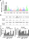
Figure 1
Figure 1. NIBP expression is upregulated in most tumor tissues
(A) A TissueScan cancer survey qPCR analysis identified the increased expression of NIBP mRNA in most tumor tissues. The data represent fold changes of NIBP mRNA expression in a tumor sample to a mean value of the corresponding normal tissues after β-actin normalization. (B) Semi-quantitative evaluation of NIBP-like immunoreactivity in the frozen tissue microarray from indicated cancer patients showed dramatic increases in NIBP protein expression in most tumor tissues as compared to corresponding normal tissues. A 5-grade scoring method was employed on the basis of the area and positivity of immunofluorescent staining. (C) A high-density reverse-phase cancer protein lysate array was evaluated for NIBP protein expression in 11 tumor tissues at various concentrations (250 and 500 μg/ml) of loaded protein lysates. * p<0.05 and ** P<0.01 indicates significant difference between tumor samples and corresponding normal tissues using Student's t test.
Figure 2
Figure 2. NIBP knockdown by lentivirus-mediated shRNAs inhibits cancer cell growth/proliferation
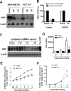
(A-C) The efficacy of NIBP knockdown in cancer cells was validated in cancer cells. The MDA-MB-231 (A) or HCT116 (A-C) cells were transduced with indicated lentiviral vectors encoding shRNA targeting 5′-coding region (NR), 3′-coding region (CR) and 3′-untranslated (UTR) regions of human NIBP. After cell sorting with an internal GFP marker and passaging four times, the levels of NIBP mRNA (A, B) and protein (C) were determined by Northern blot (A), RT-qPCR (B) and immunoblotting analyses (C). The β-actin or GAPDH was used for loading control. The pRK-Flag-NIBP transfected cells were used as a positive control for immunoblotting. (D-F) Hemocytometry (D) and Cell-Titer Glo luminescence viability assays (E, F) showed significant inhibition of cell growth in MDA-MB-231(D, E) and HCT116 (F) cells at passage 4. ** P<0.01 indicates a significant decrease in time-dependent viability/proliferation of NIBP-CR shRNA knockdown cells as compared with corresponding empty vector controls.
Figure 3
Figure 3. Lentivirus-mediated shRNA knockdown of NIBP inhibits the colony formation of cancer cells in vitro
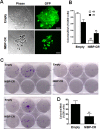
(A, B) Anchorage-dependent colony formation was significantly inhibited in NIBP-CR shRNA transduced HCT116 cells (passage 5). An equal number of exponentially growing cancer cells with or without stable NIBP shRNA knockdown were seeded in a 6-well tissue culture plate and incubated for 2 weeks. All colonies with at least 10 cells were counted under a standard inverted fluorescent microscope. The values (B) represent mean ± SD of three independent experiments each with triplicate wells. (C, D) Anchorage-independent growth was significantly reduced in NIBP-CR shRNA transduced MDA-MB-231 cells (passage 4). Equal numbers of cancer cells were resuspended in 1 ml of 0.3% top agar and plated on 2 ml of 0.8% bottom agar in each well of a 6-well plate. After 3 weeks, cell colonies were visualized by crystal violet staining (C) and quantified by counting the colony number (D). ** p<0.01 indicates a significant decrease in NIBP-CR shRNA knockdown as compared with corresponding empty vector control. Scale bar = 50 μm (A) or 10 mm (C).
Figure 4
Figure 4. NIBP promotes cancer cell invasion/migration
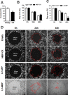
(A) Lentivirus-mediated NIBP knockdown significantly inhibited the invasion of HCT116 cells as determined by the Boyden chamber assay. Equal numbers of cells (300,000 per well) were seeded into polycarbonate membrane inserts in a 24-transwell culture plate. After 24 h incubation, cells having invaded the membrane were extracted for spectrophotometric measurement (560 nm). Values represent percentage change over 100% arbitrarily set for the empty vector control. (B-D) NIBP knockdown inhibited (B) while NIBP overexpression promoted (C) the migration of HCT116 cells as determined by a gap closure assay. Equal number of cells (10,000 per well) were seeded at 10 wells each group in a 384-well plate. A circular gap was generated by removing a biocompatible hydrogel in each well. At 24 and 48 h, the gap area (%) was measured by micrograph and image analysis. Representative micrographs were shown in D. * p<0.05 and ** p<0.01 indicates significant changes in the NIBP knockdown or overexpression group as compared with the corresponding empty control group.
Figure 5
Figure 5. Lentivirus-mediated shRNA knockdown of NIBP inhibits the xenograft tumor formation of HCT116 (A) and MDA-MB-231 (B-D) cancer cells in nude mice
Equal numbers of exponentially growing cells (1×10⁷ cells per site) stably expressing indicated shRNAs were mixed in matrigel and injected subcutaneously into the left and right flanks of female nu/J mice (n=5). Each tumor was measured twice a week for 9 weeks. A, B, Representative pictures taken at 8 weeks after injection showing tumor/injection sites (arrow). C, Tumor weight at the end of experiments showing the tumors growing at the left (L) and right (R) flanks of animals injected with NIBP-UTR lentivirus. D, The tumor growth curve for the ineffective NIBP-UTR group, the effective NIBP-CR group and the IKK2-shRNA group.
Figure 6
Figure 6. NIBP enhances NFκB activation in cancer cells
(A, B) Stable NIBP knockdown significantly inhibited cytokine-stimulated NFκB activation in HCT116 (A) and MDA-MB-231 (B) cells. The cancer cells at passage 5 were infected with adenovirus carrying an NFκB-firefly-luciferase reporter. After 24 h, cells were serum-starved overnight and treated with indicated cytokines for 24 h before GloOne luminescence assay. (C, D) Lentivirus (LV)-mediated overexpression of NIBP induced constitutive and TNFα-stimulated NFκB activation in HCT116 (C) and MDA-MB231 (D) cells. Cells were infected with indicated lentiviruses followed by an adenovirus-mediated NFκB firefly-luciferase reporter assay. The Flag-tagged NIBP expression was validated by immunoblotting with anti-Flag antibody and anti-GAPDH antibody used as loading control. The data represent mean ± SD of 3-5 independent experiments. * p<0.05 and ** p<0.01 indicates significant changes in the NIBP knockdown or overexpression group as compared with empty control lentiviral vectors.
Figure 7
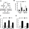
Figure 7. NIBP interacts with phosphorylated IKK2 and modulates TNFα-induced activation of the classical IKK2/IκBα/p65 pathway in cancer cells
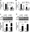
(A, B) Endogenous NIBP was coimmunoprecipated with phosphorylated IKK2 in HCT116 cells. Cells were treated with TNFα for 5-30 min and whole cell lysates were immunoprecipitated with anti-NIBP antibody or control IgG followed by immunoblotting with antibodies against NIBP (A), IKK1/2 (A, B) or phosphorylated IKK1/2 (A). Input accounts for 1% of used lysates. (C) TNFα-induced activation of the classical IKK2/IκBα/p65 pathway in MDA-MB-231 cells was inhibited by stable NIBP knockdown at 5-30 min. Cells were infected with lentiviruses carrying empty vector or NIBP-CR shRNA and sorted by FACS. After 5 passages, equal numbers of cells in 6-well plates were treated with TNFα for the indicated time intervals and the expression of indicated proteins was analyzed by immunoblotting using the Odyssey CLx Infrared Fluorescent Immunoblotting system. The signal intensities were determined using the LI-COR imaging software and the numbers below the blot indicate relative fold changes compared to empty vector vehicle control after normalization by total IKK1/2, p65 or β-actin respectively.
Figure 8
Figure 8. NIBP maintains cancer cell survival
(A) Schematic representation of the cancer cell NIBP/NFκB-dependent regulation of chemoresistance. (B) TNFα-induced cell death in HCT116 cells with NIBP knockdown was examined. An equal number of cells (10,000 cells/well) were seeded in 96-well plates. After 24 h, cells were treated with or without TNFα (10 ng/ml) for 1 and 3 d and the viable cell number was determined by trypan blue staining and hemocytometry. The data represent fold change related to the corresponding vehicle control at 1 d. (C) NIBP shRNA knockdown sensitized TNFα-induced apoptosis in HCT116 cells. Equal numbers of cells stably engineered with indicated shRNA were cultured in a 96-well plate (5,000 cells per well in quadruplicate) and treated with TNFα (10 ng/ml) for 24 h before Caspase-Glo® 3/7 assay was performed. (D) NIBP overexpression inhibited constitutive and TNFα-induced apoptosis in HCT116 cells. Cells were infected with indicated lentivirus (LV) and the third passage cells (5,000 cells per well in quadruplicate) were treated with TNF (10 ng/ml) for 24 h before Caspase-Glo® 3/7 assay was performed. * P<0.05 indicates a significant difference compared with the corresponding empty lentiviral vector.

Similar articles

Cited by

References

    1. Bonizzi G, Karin M. The two NF-kappaB activation pathways and their role in innate and adaptive immunity. Trends Immunol. 2004;25(6):280–288. - PubMed
    1. Mattson MP, Meffert MK. Roles for NF-kappaB in nerve cell survival, plasticity, and disease. Cell Death Differ. 2006;13(5):852–860. - PubMed
    1. O'Sullivan NC, Croydon L, McGettigan PA, Pickering M, Murphy KJ. Hippocampal region-specific regulation of NF-kappaB may contribute to learning-associated synaptic reorganisation. Brain Res Bull. 2010;81(4-5):385–390. - PubMed
    1. Karin M. Nuclear factor-kappaB in cancer development and progression. Nature. 2006;441(7092):431–436. - PubMed
    1. Mantovani A, Balkwill F. RalB signaling: a bridge between inflammation and cancer. Cell. 2006;127(1):42–44. - PubMed

Publication types

MeSH terms

LinkOut - more resources