. 2015 Feb 18;35(7):3155-73.
doi: 10.1523/JNEUROSCI.0586-14.2015.

Regulation of postsynaptic function by the dementia-related ESCRT-III subunit CHMP2B

Affiliations

Regulation of postsynaptic function by the dementia-related ESCRT-III subunit CHMP2B

Romain Chassefeyre et al. J Neurosci. .

Erratum in

  • J Neurosci. 2015 May 20;35(20):8035-7

Abstract

The charged multivesicular body proteins (Chmp1-7) are an evolutionarily conserved family of cytosolic proteins that transiently assembles into helical polymers that change the curvature of cellular membrane domains. Mutations in human CHMP2B cause frontotemporal dementia, suggesting that this protein may normally control some neuron-specific process. Here, we examined the function, localization, and interactions of neuronal Chmp2b. The protein was highly expressed in mouse brain and could be readily detected in neuronal dendrites and spines. Depletion of endogenous Chmp2b reduced dendritic branching of cultured hippocampal neurons, decreased excitatory synapse density in vitro and in vivo, and abolished activity-induced spine enlargement and synaptic potentiation. To understand the synaptic effects of Chmp2b, we determined its ultrastructural distribution by quantitative immuno-electron microscopy and its biochemical interactions by coimmunoprecipitation and mass spectrometry. In the hippocampus in situ, a subset of neuronal Chmp2b was shown to concentrate beneath the perisynaptic membrane of dendritic spines. In synaptoneurosome lysates, Chmp2b was stably bound to a large complex containing other members of the Chmp family, as well as postsynaptic scaffolds. The supramolecular Chmp assembly detected here corresponds to a stable form of the endosomal sorting complex required for transport-III (ESCRT-III), a ubiquitous cytoplasmic protein complex known to play a central role in remodeling of lipid membranes. We conclude that Chmp2b-containing ESCRT-III complexes are also present at dendritic spines, where they regulate synaptic plasticity. We propose that synaptic ESCRT-III filaments may function as a novel element of the submembrane cytoskeleton of spines.

Keywords: ESCRT filaments; frontotemporal dementia; postsynaptic scaffold; spinoskeleton; structural plasticity.


Figures

Figure 1.
Figure 1.
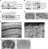
Chmp2b expression in the mouse brain. A, An adult male mouse was dissected, and equal amounts of total protein from each tissue were analyzed by SDS-PAGE and immunoblotting with the indicated antibodies. The arrowhead indicates the Chmp4b-specific band. α-Tubulin was used as the loading control. B, Mouse brains at increasing developmental ages were analyzed by immunoblotting with the indicated antibodies. PSD-95 was used as a marker of synaptogenesis. P, Postnatal age (days); 1 M, 1 month. C, Equal protein amounts from the indicated regions of an adult mouse brain were analyzed by immunoblotting as above. D, Frontal (left) or sagittal (right) sections from adult mouse brain were stained by immunoperoxidase with anti-Chmp2b antibody. Cx, Cortex; cc, corpus callosum; Hpc, hippocampus; Hb, habenula; Ce, central amygdala; OB, olfactory bulb; CB, cerebellum. E, Left, Immunostained CA1 layer in a frontal section of the hippocampus. Note the strong staining of pyramidal cell bodies and the weaker but definite immunoreactivity of the neuropil. SO, Stratum oriens; Pyr, stratum pyramidale; SR, stratum radiatum. Right, Higher magnification of the boxed region. Immunoreactivity is observed in apical dendrites (arrowheads) and interneurons (arrows). F, Specificity of Chmp2b immunostaining. As a control, we used brain sections from Chmp2b KO mice (Ghazi-Noori et al., 2012), here called Chmp2b KD because of the presence of residual Chmp2b protein in these mice (Fig. 7G,H). Chmp2b immunoreactivity was revealed by immunoperoxidase in a frontal section of the brain of a 5-week-old wild-type (Wt) mouse (left) or in an equivalent section of a sibling KD mouse (right). Note that development of DAB staining was performed identically for the two sections, but for a longer time than in D, which therefore appears lighter than F (Wt).
Figure 2.
Figure 2.
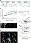
Chmp2b forms clusters in dendrites. A, Cultured mouse cortical neurons were lysed at various stages of maturation, and equal amounts of lysate protein were analyzed by immunoblotting with the indicated antibodies. PSD-95 was used as a marker for synaptogenesis. B, Hippocampal neurons were transfected with empty pSuper-mCherry vector, fixed at 21 DIV, and stained by dual immunofluorescence with anti-Chmp2b and anti-PSD-95 antibodies. Left, Punctate staining of a dendritic segment in a transfected neuron (single confocal plane). Staining of nontransfected cells is also visible. Merge, Overlap (yellow) between Chmp2b and PSD-95 puncta, in dendritic shafts and spines, visualized with mCherry (white contour). The numbered arrowheads indicate spines with colocalized puncta. Right, Enlarged views of the same spines. Scale bar, 20 μm. C, Top, Dendritic segment double stained by anti-Chmp2b and anti-PSD-95. Bottom, Same after randomization of Chmp2b staining pattern. The correlation coefficient (r) between actual Chmp2b and PSD-95 staining intensities is significantly higher than the mean r value generated by randomized Chmp2b patterns (p < 0.0001, z test). D, Specificity of Chmp2b immunofluorescence. Hippocampal neurons were transfected with pSuper-mCherry expressing Chmp2b-specific shRNA, stained by immunofluorescence with anti-Chmp2b, and imaged by confocal microscopy. Left, Representative transfected neuron, visualized by mCherry. Middle, Chmp2b immunostaining of the same field. Arrowheads indicate the position of the transfected cell dendrites and soma. N, Neighboring, nontransfected neuron. Right, Immunostaining (red hot pseudocolor) is mostly absent from the transfected neuron (outlined in white), though clearly detectable in the nontransfected cell (N). Scale bar, 10 μm.
Figure 3.
Figure 3.
Chmp2b regulates dendritic morphology in an ESCRT-III-dependent manner. A, Hippocampal neurons were cotransfected at 10 DIV with the indicated mixtures of pSuper-based and rescue (pBI-based) plasmids. pSuper vectors expressed mCherry together with the indicated shRNA [GFP shRNA (shGFP); Chmp2b shRNA (shChmp2b or sh2b)]. Rescue plasmids expressed ZsGreen, alone or together with the indicated version of RNAi-resistant Chmp2b (2B*). Images of doubly transfected neurons at 14 DIV were acquired by confocal microscopy. Depicted is the morphology of representative neurons in each condition, as visualized by mCherry fluorescence (negative contrast, maximum intensity projections). B, Sholl analysis of dendritic arborization in neurons transfected as in A. For each plasmid mix, the graph shows mean numbers of dendritic branches as a function of radial distance (n = 40 neurons per condition). Chmp2b knockdown strongly reduced the distance-dependent increase in branch number (***p = 0.00014 compared with control curve, Kolmogorov–Smirnov test with Bonferroni's correction). Expression of wild-type Chmp2b* restored the control phenotype (p = 1). Chmp4b-binding-defective (Chmp2b*R19/22/26A) and membrane-binding-defective (Chmp2b* L4D/F5D) mutants of Chmp2b were unable to fully restore the control phenotype (**p = 0.004 and p = 0.006, respectively). C, Hippocampal neurons were triply cotransfected at 10 DIV with HA-tagged Chmp2bWT, Flag-tagged Chmp4b, and mCherry plasmids (1 μg of each plasmid). The cells were fixed 24 h later and stained by dual immunofluorescence with anti-HA and anti-Flag antibodies. Confocal microscopy images (z-projections) of dendritic fluorescence are shown for a representative neuron. Chmp2b was recruited to Chmp4b puncta, some of which were found at dendritic branch points (arrowheads) or at the base of spine-looking protrusions (asterisk). Scale bar, 10 μm. D, N2A cells were transfected with the indicated combinations of RNAi-inducing plasmids and rescue plasmids. The cells were lysed and analyzed by immunoblotting with anti-Chmp2b antibody. E, HeLa cells were cotransfected with HA-tagged Chmp2bWT or Chmp2bR19/22/26A together with Chmp4b-Flag. Cells were stained by dual immunofluorescence with anti-HA and anti-Flag antibodies and imaged by confocal microscopy. Insets, High magnification of the boxed regions. Scale bars, 20 μm. Colocalization of Chmp2b and Chmp4b was 80% for Chmp2bWT and 12% for Chmp2bR19/22/26A (quantified as the percentage of Chmp2b-HA-positive pixels overlapping Chmp4b-Flag puncta). F, HEK-293T cells were transfected with Flag-tagged Chmp4b plasmid, alone or together with HA-tagged Chmp2bWT or Chmp2bR19/22/26A. Anti-HA immunoprecipitates prepared from the transfected cells were analyzed by immunoblotting with anti-Flag or anti-HA antibodies. G, HEK-293T cells were transfected with Flag-tagged Chmp2bWT or Chmp2bL4D/F5D. The transfected cells were homogenized, the membrane (P) and cytosolic (S) fractions were separated by centrifugation, and equivalent amounts of each fraction were analyzed by immunoblotting with anti-Flag antibody.
Figure 4.
Figure 4.
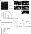
Chmp2b is involved in the maintenance of excitatory synapses in cultured neurons. A, Hippocampal neurons were transfected at 16 DIV with pSuper-mCherry encoding control (GFP) shRNA. The neurons were fixed at 18 DIV, stained by dual immunofluorescence with anti-PSD-95 and anti-synapsin I antibodies, and dendrites were imaged by confocal microscopy. Postsynaptic spines (arrowheads) were identified as dendritic protrusions (top) containing PSD-95 puncta (middle). Nearly all of the spines colocalized with the presynaptic marker Synapsin (bottom). mCherry fluorescence was used as a mask to facilitate visualization of puncta belonging to the transfected neuron. Scale bar, 15 μm. B, Neurons were transfected at 16 DIV with the same plasmid combinations as in Figure 3A. Images of dendritic morphology were acquired, and postsynaptic spines were identified as in A and counted. A representative field is shown for each condition. Scale bar, 5 μm. C, Mean density of postsynaptic spines along the dendrites of neurons transfected as in B. Compared with the control (GFP) shRNA, the Chmp2b-depleting shRNA induced a large drop in spine density (***p = 2.4 × 10⁻¹¹, two-sided Wilcoxon test with Holm correction). Coexpressing wild-type Chmp2b* completely restored density (p = 1 vs control, ***p = 6.3 × 10⁻¹⁴ vs Chmp2b shRNA alone). Coexpressing the Chmp4b binding-defective mutant Chmp2b*R19/22/26A did not rescue spine density at all (***p = 3.8 × 10⁻⁶ vs Chmp2b*WT, p = 0.14 vs Chmp2b shRNA alone); neither did the membrane binding-defective mutant Chmp2b* L4D/F5D (***p = 2.9 × 10⁻⁷ vs Chmp2bWT, p = 1 vs Chmp2b shRNA alone). Neurons per condition: control, n = 34; Chmp2b shRNA, n = 33; Chmp2b*WT, n = 30; Chmp2b*R19/22/26A, n = 16; Chmp2b*L4D/F5D, n = 14. D–F, Hippocampal neurons were transfected at 18 DIV with the indicated plasmids. Transfected neurons were identified by fluorescence at 21 DIV, and their miniature EPSCs were measured by patch-clamp recordings in whole-cell configuration. D, Sample traces from neurons transfected as indicated. E, Histogram of mean mEPSC amplitudes in neurons transfected as indicated (n = 5 cells, >100 minis per cell in each condition). The amplitude of mEPSCs was markedly reduced in Chmp2b-deficient neurons (**p = 0.005, two-sided t test). F, Histogram of mean mEPSC frequencies in neurons transfected as indicated. Frequencies were lower in Chmp2b-deprived neurons (*p = 0.012, two-sided t test). G, Neurons transfected as above were stimulated at 21 DIV by bath-applied NMDA (control, n = 6 cells; Chmp2b shRNA, n = 5 cells). The density of NMDA-evoked currents (calculated as current normalized by capacitance) was strongly decreased in Chmp2b-deprived neurons (*p = 0.031, two-sided t test).
Figure 5.
Figure 5.
Chmp2b is involved in the maintenance of excitatory synapses in vivo. A, Brain sections from 5-week-old Chmp2b knockdown mice (KD) or sibling controls (Wild type) were stained by the Golgi impregnation technique. Stained dendritic segments were visualized by bright-field microscopy of the stratum radiatum. Shown are representative images of dendritic shafts and spines in both genotypes. B, Spines were classified into thin, stubby, or mushroom types, and their density per unit dendritic length was measured for each type (n = 4 animals, 36 dendrites per genotype). Left, Histogram showing the mean density of each spine type in KD and WT mice. The total spine density was significantly lower in KD mice than in controls (*p = 0.039, two-sided t test). Right, Histogram showing the mean proportion of the three spine types. The percentage of mushroom spines was significantly reduced in the KD mice (*p = 0.02). C, Representative electron micrographs of the CA1 stratum radiatum from Wt and KD siblings. Presynaptic profiles are highlighted in blue, and postsynaptic spines (s) are in brown. D, Quantification of synapse density in the electron micrographs. WT: n = 3 animals, 851 axo-spinous synapses from 30 micrographs; KD: n = 3 animals, 627 axo-spinous synapses from 30 micrographs. The density of postsynaptic spines per unit surface was significantly lower in the KD (***p = 0.0008).
Figure 6.
Figure 6.
Chmp2b is required for stable increase in spine volume and in synaptic efficacy during chemical LTP. A, B, Hippocampal neurons were transfected with pSuper-mCherry and rescue plasmids. Confocal image stacks of dendritic segments (visualized by mCherry fluorescence) were acquired before and after induction of cLTP. 3D models of dendritic morphology were built from the stacks with NeuronStudio and used to measure changes in spine volume. A, Maximal intensity projection of confocal images (top) and 3D models (bottom) of a representative dendritic segment before and after cLTP in a neuron transfected with control plasmid. Arrows indicate spines that have expanded. B, The percentage increase in spine volume was measured for each stable spine of a segment 5 min before and 45 min after cLTP induction, and median increases were calculated per transfected neuron. The box plot shows the distribution of median increases in neurons transfected as indicated (n = 8–9 neurons per condition, 50–100 spines per neuron). vec, Vector expressing GFP alone. The spines of control neurons were significantly expanded after cLTP induction (p = 0.0075, Wilcoxon one-sample test). The increase was abolished by Chmp2b knockdown (*p = 0.011 compared with control, Wilcoxon test with Holm correction) and reinstated by expression of Chmp2b* (p = 0.336 compared with control), but not by GFP alone (p = 0.041 vs control). C, D, Hippocampal neurons were transfected at 18 DIV with the indicated plasmids and cLTP was induced as in A. mEPSCs were recorded before and after cLTP induction by patch-clamp recording in the whole-cell configuration. C, Sample traces obtained before and after cLTP in neurons transfected as indicated. D, The histogram shows the relative potentiation of mEPSCs (mean percentage increase per neuron) compared with the mean amplitude before cLTP. The dotted line indicates 100% of basal amplitude (mean mEPSC amplitude before LTP). LTP was abolished in Chmp2b-deprived neurons (*p = 0.033 compared with control, Holm-corrected t test). Expression of Chmp2b* rescued LTP (p = 0.046 compared with basal amplitude in the same cells, paired t test; **p = 0.0062 vs nonrescued cells; p = 0.34 vs control cells, two-sided t tests). Control, n = 5 cells; Chmp2b shRNA plus vector, n = 11 cells; Chmp2b shRNA plus Chmp2b*WT, n = 9 cells.
Figure 7.
Figure 7.
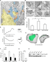
Ultrastructural localization of Chmp2b in dendritic spines. A, Electron micrograph of Chmp2b immunoreactivity in the mouse stratum radiatum, detected by pre-embedding immunogold labeling. D, Dendrites; S, spines; at, axon terminals. B, High-magnification views of typical dendritic spines. Chmp2b usually appears on the periphery of the PSD or on the neck of the spine, beneath the plasma membrane. Bottom right, Labeled MVB in a dendritic shaft. C, Quantitative analysis of immunogold labeling in the stratum radiatum. The graph shows mean density of gold particles in three neuronal compartments. The ultrastructural localization of 2100 gold particles was analyzed in 30 fields similar to that shown in A. Chmp2b density was significantly higher in spines than in shafts or axonal boutons (p = 0.0498, n = 3 animals, 3 compartments and 10 fields per animal, Friedman test). D, Distribution of gold particle positions inside dendritic spines (n = 335 particles, 3 animals). Top, Histogram of normalized positions along the spine radius. Inset, Coordinate 0 corresponds to the spine center, and 1.0 corresponds to the plasma membrane. Particles concentrate in the subplasmalemmal cortex. Bottom, Histogram of normalized particle distances from the PSD. Inset, Coordinate 0 corresponds to the PSD edge, and 1.0 corresponds to the point equidistant from both edges of the PSD. Particles concentrate in the proximity of the PSD and become scarcer further away, with a slight rebound in frequency at the usual position of the neck. E, 3D reconstruction of a typical spine using immuno-electron micrographs of serial sections. The plasma membrane is in gray, PSD in green, and particles are marked by dots. Particles appear beneath the perisynaptic membrane around the PSD and also in the neck. Left, View from above the PSD; right, lateral view. An animated version of the 3D model appears in Movie 1 (see Notes). F, Quantitation of immunogold labeling in the stratum radiatum of WT and KD siblings. The density of gold particles in the three neuronal compartments is much lower in the KD animals (p < 10−5, n = 3 animals per genotype, Kruskal–Wallis test, density vs genotype). G, Comparison of Chmp2b amounts in WT and KD brains. Total brain extract was obtained from three animals of each genotype and analyzed by SDS-PAGE and Western immunoblotting with anti-Chmp2b antibody. Actin was used as a loading control. Quantitation of the Chmp2b band in each lane was obtained by densitometry and normalized to the respective actin control. KD brain still expressed 30% of the wild-type amount. H, Western blot used for the quantitation in G.
Figure 8.
Figure 8.
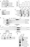
Chmp2b is part of a stable ESCRT-III-type complex. A, Rat forebrain homogenates were fractionated as described in Materials and Methods. Equivalent amounts of each fraction were analyzed by Western immunoblotting with the indicated antibodies. Both Chmp2b and Chmp4b were enriched in the PSD fraction. B, PSDI was re-extracted with either 0.5% Triton X-100 or 3% Sarkosyl to yield insoluble fractions PSDII or PSDIII, respectively. Equivalent amounts of each fraction were analyzed by Western immunoblotting. C, Synaptoneurosomes were solubilized with the indicated detergents and centrifuged at high speed. The resulting supernatants and pellets were analyzed by immunoblotting with the indicated antibodies. D, Gradient-purified synaptoneurosomes were lysed in 1% DOC, insoluble material was removed, and the extract was fractionated by size-exclusion chromatography on a Sephacryl S-300 column. Column fractions were analyzed by immunoblotting with the indicated antibodies. Apparent molecular weights (MW) were calibrated with protein markers. A subset of Chmp2b displayed the same high MW as the PSD-95 complex. E, Monomeric recombinant His-tagged Chmp2b was incubated with either 1% Triton X-100 or 1% DOC and fractionated by size-exclusion chromatography on a Sephacryl S-300 column. Fractions were analyzed by immunoblotting with an anti-His tag antibody. DOC did not cause detectable aggregation of monomeric Chmp2b. The elution peaks of synaptic Chmp2b-containing fractions resolved on the same column are indicated for reference. F, Fractions corresponding to HMW and LMW Chmp2b populations were separately pooled and immunoprecipitated with anti-Chmp2b antibody or control IgG. The immunoprecipitates were analyzed by immunoblotting with anti-Chmp2b or anti-Chmp4b antibody. Chmp4b coprecipitated with Chmp2b in the HMW peak. G, Synaptoneurosome extracts were prepared as in D and immunoprecipitated with anti-Chmp2b antibody or control IgG. The immunoprecipitates were analyzed by immunoblotting with the indicated antibodies.

Similar articles

Cited by

References

    1. Alvarez VA, Ridenour DA, Sabatini BL. Retraction of synapses and dendritic spines induced by off-target effects of RNA interference. J Neurosci. 2006;26:7820–7825. doi: 10.1523/JNEUROSCI.1957-06.2006. - DOI - PMC - PubMed
    1. Bajorek M, Schubert HL, McCullough J, Langelier C, Eckert DM, Stubblefield WM, Uter NT, Myszka DG, Hill CP, Sundquist WI. Structural basis for ESCRT-III protein autoinhibition. Nat Struct Mol Biol. 2009;16:754–762. doi: 10.1038/nsmb.1621. - DOI - PMC - PubMed
    1. Baldys A, Raymond JR. Critical role of ESCRT machinery in EGFR recycling. Biochemistry. 2009;48:9321–9323. doi: 10.1021/bi900865u. - DOI - PubMed
    1. Bartlett WP, Banker GA. An electron microscopic study of the development of axons and dendrites by hippocampal neurons in culture. J Neurosci. 1984;4:1944–1953. - PMC - PubMed
    1. Belly A, Bodon G, Blot B, Bouron A, Sadoul R, Goldberg Y. CHMP2B mutants linked to frontotemporal dementia impair maturation of dendritic spines. J Cell Sci. 2010;123:2943–2954. doi: 10.1242/jcs.068817. - DOI - PMC - PubMed

Publication types

MeSH terms

Substances

LinkOut - more resources