. 2015 Mar 11;43(5):2874-87.
doi: 10.1093/nar/gkv048. Epub 2015 Feb 19.

Alu RNA regulates the cellular pool of active ribosomes by targeted delivery of SRP9/14 to 40S subunits

Affiliations

Alu RNA regulates the cellular pool of active ribosomes by targeted delivery of SRP9/14 to 40S subunits

Elena Ivanova et al. Nucleic Acids Res. .

Abstract

The human genome contains about 1.5 million Alu elements, which are transcribed into Alu RNAs by RNA polymerase III. Their expression is upregulated following stress and viral infection, and they associate with the SRP9/14 protein dimer in the cytoplasm forming Alu RNPs. Using cell-free translation, we have previously shown that Alu RNPs inhibit polysome formation. Here, we describe the mechanism of Alu RNP-mediated inhibition of translation initiation and demonstrate its effect on translation of cellular and viral RNAs. Both cap-dependent and IRES-mediated initiation is inhibited. Inhibition involves direct binding of SRP9/14 to 40S ribosomal subunits and requires Alu RNA as an assembly factor but its continuous association with 40S subunits is not required for inhibition. Binding of SRP9/14 to 40S prevents 48S complex formation by interfering with the recruitment of mRNA to 40S subunits. In cells, overexpression of Alu RNA decreases translation of reporter mRNAs and this effect is alleviated with a mutation that reduces its affinity for SRP9/14. Alu RNPs also inhibit the translation of cellular mRNAs resuming translation after stress and of viral mRNAs suggesting a role of Alu RNPs in adapting the translational output in response to stress and viral infection.


Figures

Figure 1.
Figure 1.
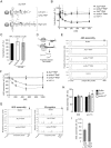
Alu RNPs repress translation initiation by inhibiting 48S complex formation. (A) Secondary structure model of AluYA RNA. Binding sites of h9/14 are indicated by analogy to SRP RNA. Preparation of Alu RNPs is described in Supplementary Figure S1. (B) Translation efficiencies of uncapped pPL mRNA in RRL supplemented with increasing concentrations of Alu RNPs or the h9/14 protein or the AluYA/scAluYA RNAs alone. [35S]-labeled proteins were resolved by SDS-PAGE and quantified by phosphorimager (see Supplementary Figure S2A and B for SDS-PAGE autoradiographs). Translation efficiency was calculated as a percentage of the value obtained in the absence of RNPs. Error bars are shown as SD (n ≥ 3). (C) Translation efficiencies of uncapped pPL mRNA in RRL translation reaction synchronized by 0.5 μM edeine in the presence of 100 nM AluYA/scAluYA RNP. Translation reactions were incubated 3 min at 30°C before the addition of 0.5 μM edeine. Translation mixtures were incubated another 1 min and then supplemented with the RNPs. See also Supplementary Figure S2C. (D) Schematic representation of the toeprint analysis. Reverse transcription by avian myeloblastosis virus reverse transcriptase (AMV) stalls upon encounter with ribosomal complexes. Fluorescently labeled cDNAs were analyzed by capillary electrophoresis. (E) Toeprinting analysis of 48S complexes reconstituted in the presence of 500 nM Alu RNPs, h9/14 or scAluYA RNA. See also Supplementary Figure S2D for toeprint analysis of MVHL-stop mRNA in the absence of 40S and translation factors. (F) Relative efficiency of 48S complex formation in the presence of increasing concentrations of Alu RNPs, h9/14 or scAluYA RNA. To calculate the efficiency of 48S complex assembly, the intensity of the 48S toeprint signal was quantified as described in ‘Materials and Methods’ section. The relative efficiency of 48S complex assembly was expressed as a percentage of the value obtained in the absence of RNPs (buffer). Error bars are shown as SD (n ≥ 3). (G) Toeprint analysis of 80S complexes (left panel) and preTC (right panel) reconstituted in the presence of 500 nM AluYA RNP or 1 μM scAluYA RNP. Quantification is shown in (H). Error bars are shown as SD (n ≥ 3). (I) Relative efficiency of 48S complex assembly in the presence of 500 nM scAluYA RNP added either to the preassembled 43S complexes (43S) or to preassembled 48S complexes (48S). Error bars are shown as SD (n ≥ 3).
Figure 2.
Figure 2.
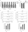
The inhibitory activity of the Alu RNP depends on the identity of its RNA component and the presence of the two positively charged patches in SRP9/14. (A) Translation efficiency of uncapped pPL mRNA in RRL supplemented with increasing concentrations of the SA110 or the scAluYL RNP. Error bars are shown as SD (n ≥ 3). See also Supplementary Figure S3B. (B) Translation efficiency of uncapped pPL mRNA in RRL supplemented with 100 nM SA151, SA86, scALuYa5, scAluYf2 or scAluYj4 RNPs. Error bars are shown as SD (n ≥ 3). See also Supplementary Figure S3D. (C) Relative efficiency of 48S complex formation in the presence of increasing concentrations of SA110 or scAluYL RNPs. Error bars are shown as SD (n ≥ 3). (D) Mutations introduced in the h14 and h9 proteins. Numbers correspond to the human sequence. Amino acid residues of interest are shown in black. Mutated residues are shown in grey. (E) Translation efficiency of uncapped pPL mRNA in RRL supplemented with increasing concentrations of scAluYA, scAluYA 14A5 or scAluYA 9-3A RNPs. Error bars are shown as SD (n ≥ 3). See also Supplementary Figure S4B. (F) Relative efficiency of 48S complex formation in the presence of increasing concentrations of scAluYA, scAluYA 14A5 or scAluYA 9-3A RNPs. Error bars are shown as SD (n ≥ 3).
Figure 3.
Figure 3.
Alu RNPs do not affect 43S complex formation but prevent its recruitment to the mRNA. (A, B) 43S and 48S complexes were reconstituted in the presence of 0.5 μM AluYA, scAluYA or SA110 RNPs and either [35S]-Met-tRNAiMet (A) or [32P]-labeled MVHL-stop mRNA (B). Reconstitution reactions were fractionated on 5–20% sucrose gradients. Results are expressed as percentage of total cpm across all fractions. All experiments were repeated twice.
Figure 4.
Figure 4.
Alu RNPs inhibit cap-dependent and IRES-dependent translation. Translation efficiencies in RRL of the indicated reporter mRNAs in the presence of (A) 100 nM AluYA or scAluYA RNP; (B) 100 nM scAluYA 14A5 RNP or scAluYA 9–3A RNP. The translation efficiency of each mRNA in the absence of the RNP (buffer) was set to 100%. Error bars are shown as SD (n ≥ 3). See also Supplementary Figure S5A. (C) Toeprint analysis of 48S complex assembly on HCV-NS’ mRNA (23) in the presence of 1 μM Alu RNPs as indicated. Quantification is shown in lower panel. Dashed line indicates 48S complex assembly in the absence of RNPs, which was set to 100%. Error bars are shown as SD (n ≥ 3). (D) Toeprint analysis of 48S complex assembly on CrPV-VHLM mRNA in the presence of 0.5 μM Alu RNPs. Quantification is shown in lower panel. Dashed line indicates 48S complex assembly in the absence of RNPs, which was set to 100%. Error bars are shown as SD (n ≥ 3). See also Supplementary Figure S5B.
Figure 5.
Figure 5.
Alu RNA is required for the specific delivery of SRP9/14 to an inhibitory site in the 40S subunit. Binding of scAluYA RNP (A) or h9/14 (B) to the 40S (top panel) or 60S (bottom panel) subunits. Equimolar amounts of RNP were incubated with ribosomal subunits and the binding reactions were fractionated on 5–20% sucrose gradients. Fractions 13 and 14 contain 40S dimers. (C) Binding of scAluYA RNP (upper panel) or h9/14 (lower panel) to the reassembled 80S ribosomes. To form 80S ribosomes, equimolar amounts of 40S and 60S were incubated at 37°C for 10 min. Alu RNP was added and the binding reaction was incubated for another 10 min and fractionated on 10–30% sucrose gradients. 40S- and 60S-containing fractions were identified by probing for S15 and L9, respectively. (D) Binding of scAluYA RNP (left panel) or SA110 RNP (right panel) to the 43S complex. SRP9/14 in ribosomal fractions was quantified from Western blots against h14. The signal intensities in 40S-containing fractions (9 and 10) were expressed as a percentage of the total signal intensity across all fractions. Data are shown as mean ± SD (n ≥ 3). (E) Binding of scAluYA 14A5 RNP (top panel) or scAluYA 9-3A RNP (bottom panel) to 40S subunits. (F) Translation efficiency of pPL mRNA in RRL supplemented with 100 or 200 nM h9/14 (left panel), scAluYA RNA (middle panel) or h9/14A5 (right panel). scAluYA RNP were added to 100 nM. Error bars are shown as SD (n ≥ 3). *P < 0.05, **P < 0.01 by Student's t-test. See also Supplementary Figure S7.
Figure 6.
Figure 6.
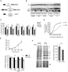
Expression of scAlu RNA reduces the translational capacity of HEK 293T cells. (A) Constructs used for luciferase reporter assay. Sequences of ncRNAs were cloned downstream of the 7SL gene enhancer element (Enh). (B) Northern blot analysis of transiently transfected HEK 293T cells. Membranes containing 0.5 μg of total RNA per lane were hybridized with [32P]-labeled oligonucleotides complementary to 4.5S RNA, 7SL RNA, scAluYNF1 RNA and 18S RNA (loading control). (C) Translation efficiency of reporter mRNAs in HEK 293T cells expressing different ncRNAs. Luciferase activity was measured 24 h post-transfection, normalized to μg of protein in the cell lysate and expressed as a percentage of the value obtained in control cells (ctrl) transfected with the empty vector pDL7Enh. Error bars are shown as SEM (n ≥ 5). *P < 0.05, **P < 0.01 by Student's t-test. See Supplementary Figure S8A for northern blot analysis of reporter mRNAs. (D) Bioluminescence recordings of HEK 293T cells expressing the estradiol-induced luciferase reporter gene along with pDLscAlu (scAlu) or pDL7enh (Ctrl) following stimulation with 100 nM estradiol. (E) Relative total protein synthesis in HEK 293T cells after arsenite treatment (top panel). Control cells were treated with 500 μM sodium arsenite for 30 min at 48 h post-transfection, allowed to recover for the indicated times and pulse labeled with [35S]-methionine/cysteine for 15 min. 100 μg of total protein was precipitated in 10% TCA, [35S]-incorporation was determined by scintillation counting and expressed as a percentage of that in untreated cells. Error bars are shown as SEM (n ≥ 5). Bottom panel shows total protein synthesis in untreated cells (-Ars) and cells expressing scAluYNF1 or 4.5S RNA treated for 30 minutes with 500 μM sodium arsenite and allowed to recover for 60, 90 and 120 min (+Ars). Total protein synthesis is expressed as a percentage of that in control cells measured in parallel. Error bars are shown as SEM (n ≥ 5). *P < 0.05, **P < 0.01 by Student's t-test. Expression of scAluYNF1 RNA is shown in Supplementary Figure S8D. (F) Representative autoradiograph following SDS-PAGE of VSV-infected cell lysates. HEK 293T cells expressing scAluYNF1, 4.5S RNA or control cells were infected with VSV for 6 h, pulse labeled with [35S]-methionine/cysteine for 15 min and 100 μg of total protein was displayed by 5–20% gradient SDS-PAGE. Bands corresponding to viral proteins are indicated. Coomassie-blue staining (bottom panel) was used to check uniform loading. Quantification of the viral protein synthesis is shown on the right. Viral protein synthesis in cells expressing scAluYNF1 RNA or 4.5S RNA was calculated as the sum of the intensities of the viral protein bands and expressed as a percentage of the control, which was set to 100%. Error bars are shown as SD (n ≥ 3). **P < 0.01 by Student's t-test. Expression of scAluYNF1 RNA is shown in Supplementary Figure S8E.

Similar articles

Cited by

References

    1. Sonenberg N., Hinnebusch A.G. Regulation of translation initiation in eukaryotes: mechanisms and biological targets. Cell. 2009;136:731–745. - PMC - PubMed
    1. Hershey J.W., Sonenberg N., Mathews M.B. Principles of translational control: an overview. Cold Spring Harb. Perspect. Biol. 2012;4 doi:10.1101/cshperspect.a011528. - PMC - PubMed
    1. Aitken C.E., Lorsch J.R. A mechanistic overview of translation initiation in eukaryotes. Nat. Struct. Mol. Biol. 2012;19:568–576. - PubMed
    1. Hasler J., Strub K. Alu RNP and Alu RNA regulate translation initiation in vitro. Nucleic Acids Res. 2006;34:2374–2385. - PMC - PubMed
    1. Quentin Y. Origin of the Alu family: a family of Alu-like monomers gave birth to the left and the right arms of the Alu elements. Nucleic Acids Res. 1992;20:3397–3401. - PMC - PubMed

Publication types

MeSH terms