Effect of GuiXiong Xiaoyi Wan in Treatment of Endometriosis on Rats
- PMID: 25691906
- PMCID: PMC4322821
- DOI: 10.1155/2015/208514
Effect of GuiXiong Xiaoyi Wan in Treatment of Endometriosis on Rats
Abstract
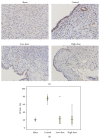
Objective. To evaluate the effect of GuiXiong Xiaoyi Wan (GXXYW) on the development of endometriosis in a rat model. Methods. Sprague-Dawley rats with surgically induced endometriosis were randomly treated with low-dose GXXYW, high-dose GXXYW, or vehicle (negative control) for 28 days. Immunohistochemistry was used to assess cell proliferation in the lesions. The terminal deoxynucleotidyl transferase- (TdT-) mediated dUTP biotin nick end labelling (TUNEL) method was performed to analyse the apoptosis induced by GuiXiong Xiaoyi Wan. The percentages of CD3+ lymphocytes, CD4+ lymphocytes, and CD8+ lymphocytes in the spleens of the rats were evaluated using flow cytometric analysis. Results. Treatment with GXXYW significantly decreased the lesion size, inhibited cell proliferation, and induced apoptosis in endometriotic tissue. The spleens of GXXYW-treated rats also demonstrated a significant increase in the percentage of CD4+ lymphocytes and a significant decrease in the percentage of CD8+ lymphocytes. Conclusions. These results suggest that, in a rat model, GXXYW may be effective in the suppression of the growth of endometriosis, possibly through the inhibition of cell proliferation, the induction of apoptosis of endometriotic cells, and the regulation of the immune system.
Figures


Similar articles
-
Shaofu Zhuyu Decoction Regresses Endometriotic Lesions in a Rat Model.Evid Based Complement Alternat Med. 2018 Jan 30;2018:3927096. doi: 10.1155/2018/3927096. eCollection 2018. Evid Based Complement Alternat Med. 2018. PMID: 29636775 Free PMC article.
-
Natural therapies assessment for the treatment of endometriosis.Hum Reprod. 2013 Jan;28(1):178-88. doi: 10.1093/humrep/des369. Epub 2012 Oct 18. Hum Reprod. 2013. PMID: 23081870
-
ENMD-1068, a protease-activated receptor 2 antagonist, inhibits the development of endometriosis in a mouse model.Am J Obstet Gynecol. 2014 Jun;210(6):531.e1-8. doi: 10.1016/j.ajog.2014.01.040. Epub 2014 Feb 1. Am J Obstet Gynecol. 2014. PMID: 24495669
-
Guizhi fuling capsule, an ancient Chinese formula, attenuates endometriosis in rats via induction of apoptosis.Climacteric. 2014 Aug;17(4):410-6. doi: 10.3109/13697137.2013.876618. Epub 2014 Feb 23. Climacteric. 2014. PMID: 24559203
-
Protective effect of Honokiol against endometriosis in Rats via attenuating Survivin and Bcl-2: A mechanistic study.Cell Mol Biol (Noisy-le-grand). 2016 Jan 11;62(1):1-5. Cell Mol Biol (Noisy-le-grand). 2016. PMID: 26828978
Cited by
-
Celecoxib, a selective COX-2 inhibitor, markedly reduced the severity of tamoxifen-induced adenomyosis in a murine model.Exp Ther Med. 2020 May;19(5):3289-3299. doi: 10.3892/etm.2020.8580. Epub 2020 Mar 6. Exp Ther Med. 2020. PMID: 32266025 Free PMC article.
-
Effect of Protoberberine-Rich Fraction of Chelidonium majus L. on Endometriosis Regression.Pharmaceutics. 2021 Jun 23;13(7):931. doi: 10.3390/pharmaceutics13070931. Pharmaceutics. 2021. PMID: 34201532 Free PMC article.
-
Baicalein reduces endometriosis by suppressing the viability of human endometrial stromal cells through the nuclear factor-κB pathway in vitro.Exp Ther Med. 2017 Oct;14(4):2992-2998. doi: 10.3892/etm.2017.4860. Epub 2017 Aug 1. Exp Ther Med. 2017. PMID: 28912852 Free PMC article.
-
Assessing Pain Behavioral Responses and Neurotrophic Factors in the Dorsal Root Ganglion, Serum and Peritoneal Fluid in Rat Models of Endometriosis.J Family Reprod Health. 2020 Dec;14(4):259-268. doi: 10.18502/jfrh.v14i4.5210. J Family Reprod Health. 2020. PMID: 34054998 Free PMC article.
-
Possible therapeutic effect of royal jelly on endometriotic lesion size, pain sensitivity, and neurotrophic factors in a rat model of endometriosis.Physiol Rep. 2021 Nov;9(22):e15117. doi: 10.14814/phy2.15117. Physiol Rep. 2021. PMID: 34806344 Free PMC article.
References
LinkOut - more resources
Full Text Sources
Other Literature Sources
Research Materials

