Mitochondria of a human multidrug-resistant hepatocellular carcinoma cell line constitutively express inducible nitric oxide synthase in the inner membrane
- PMID: 25691007
- PMCID: PMC4459854
- DOI: 10.1111/jcmm.12528
Mitochondria of a human multidrug-resistant hepatocellular carcinoma cell line constitutively express inducible nitric oxide synthase in the inner membrane
Abstract
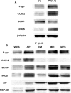
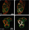
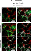
Mitochondria play a crucial role in pathways of stress conditions. They can be transported from one cell to another, bringing their features to the cell where they are transported. It has been shown in cancer cells overexpressing multidrug resistance (MDR) that mitochondria express proteins involved in drug resistance such as P-glycoprotein (P-gp), breast cancer resistant protein and multiple resistance protein-1. The MDR phenotype is associated with the constitutive expression of COX-2 and iNOS, whereas celecoxib, a specific inhibitor of COX-2 activity, reverses drug resistance of MDR cells by releasing cytochrome c from mitochondria. It is possible that COX-2 and iNOS are also expressed in mitochondria of cancer cells overexpressing the MDR phenotype. This study involved experiments using the human HCC PLC/PRF/5 cell line with and without MDR phenotype and melanoma A375 cells that do not express the MDR1 phenotype but they do iNOS. Western blot analysis, confocal immunofluorescence and immune electron microscopy showed that iNOS is localized in mitochondria of MDR1-positive cells, whereas COX-2 is not. Low and moderate concentrations of celecoxib modulate the expression of iNOS and P-gp in mitochondria of MDR cancer cells independently from inhibition of COX-2 activity. However, A375 cells that express iNOS also in mitochondria, were not MDR1 positive. In conclusion, iNOS can be localized in mitochondria of HCC cells overexpressing MDR1 phenotype, however this phenomenon appears independent from the MDR1 phenotype occurrence. The presence of iNOS in mitochondria of human HCC cells phenotype probably concurs to a more aggressive behaviour of cancer cells.
Keywords: COX-2; MDR; coxib; iNOS; mitochondrion.
© 2015 The Authors. Journal of Cellular and Molecular Medicine published by John Wiley & Sons Ltd and Foundation for Cellular and Molecular Medicine.
Figures


Similar articles
-
The MDR phenotype is associated with the expression of COX-2 and iNOS in a human hepatocellular carcinoma cell line.Hepatology. 2002 Apr;35(4):843-52. doi: 10.1053/jhep.2002.32469. Hepatology. 2002. PMID: 11915030
-
P-glycoprotein mediates celecoxib-induced apoptosis in multiple drug-resistant cell lines.Cancer Res. 2007 May 15;67(10):4915-23. doi: 10.1158/0008-5472.CAN-06-3952. Cancer Res. 2007. PMID: 17510421
-
P-gp localization in mitochondria and its functional characterization in multiple drug-resistant cell lines.Exp Cell Res. 2006 Dec 10;312(20):4070-8. doi: 10.1016/j.yexcr.2006.09.005. Epub 2006 Sep 16. Exp Cell Res. 2006. PMID: 17027968
-
Novel approaches to reversing anti-cancer drug resistance using gene-specific therapeutics.Hum Cell. 2001 Sep;14(3):172-84. Hum Cell. 2001. PMID: 11774737 Review.
-
Cyclooxygenase-2: potential role in regulation of drug efflux and multidrug resistance phenotype.Curr Pharm Des. 2004;10(6):647-57. doi: 10.2174/1381612043453117. Curr Pharm Des. 2004. PMID: 14965327 Review.
Cited by
-
Mitochondrial fission factor promotes cisplatin resistancein hepatocellular carcinoma.Acta Biochim Biophys Sin (Shanghai). 2022 Mar 25;54(3):301-310. doi: 10.3724/abbs.2022007. Acta Biochim Biophys Sin (Shanghai). 2022. PMID: 35538029 Free PMC article.
-
Reversal of multidrug resistance of hepatocellular carcinoma cells by metformin through inhibiting NF-κB gene transcription.World J Hepatol. 2016 Aug 18;8(23):985-93. doi: 10.4254/wjh.v8.i23.985. World J Hepatol. 2016. PMID: 27621764 Free PMC article.
-
Solanine reverses multidrug resistance in human myelogenous leukemia K562/ADM cells by downregulating MRP1 expression.Oncol Lett. 2018 Jun;15(6):10070-10076. doi: 10.3892/ol.2018.8563. Epub 2018 Apr 25. Oncol Lett. 2018. PMID: 29928376 Free PMC article.
-
Clinical characterization and therapeutic targets of vitamin A in patients with hepatocholangiocarcinoma and coronavirus disease.Aging (Albany NY). 2021 Jun 27;13(12):15785-15800. doi: 10.18632/aging.203220. Epub 2021 Jun 27. Aging (Albany NY). 2021. PMID: 34176789 Free PMC article.
-
Models for Understanding Resistance to Chemotherapy in Liver Cancer.Cancers (Basel). 2019 Oct 29;11(11):1677. doi: 10.3390/cancers11111677. Cancers (Basel). 2019. PMID: 31671735 Free PMC article. Review.
References
-
- Staud F, Ceckova M, Micuda S, et al. Expression and function of p-glycoprotein in normal tissues: effect on pharmacokinetics. Methods Mol Biol. 2010;596:199–222. - PubMed
-
- Sharom FJ. ABC multidrug transporters: structure, function and role in chemoresistance. Pharmacogenomics. 2008;9:105–27. - PubMed
-
- Solazzo M, Fantappie O, Lasagna N, et al. P-gp localization in mitochondria and its functional characterization in multiple drug-resistant cell lines. Exp Cell Res. 2006;312:4070–8. - PubMed
-
- Shen Y, Chu Y, Yang Y, et al. Mitochondrial localization of P-glycoprotein in the human breast cancer cell line MCF-7/ADM and its functional characterization. Oncol Rep. 2012;27:1535–40. - PubMed
Publication types
MeSH terms
Substances
LinkOut - more resources
Full Text Sources
Other Literature Sources
Research Materials
Miscellaneous

