. 2015 Feb 28;6(6):3874-86.
doi: 10.18632/oncotarget.2885.

Shed syndecan-2 enhances tumorigenic activities of colon cancer cells

Affiliations

Shed syndecan-2 enhances tumorigenic activities of colon cancer cells

Sojoong Choi et al. Oncotarget. .

Abstract

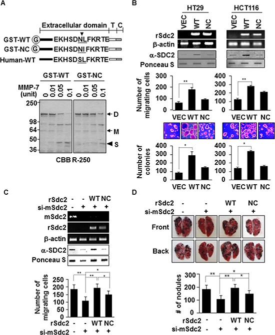
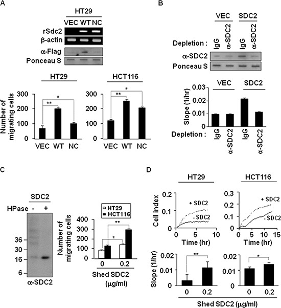
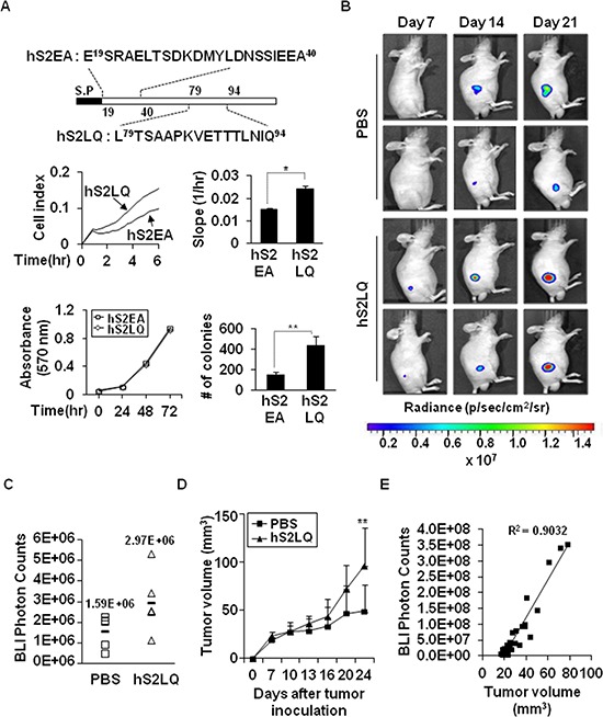
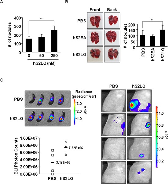
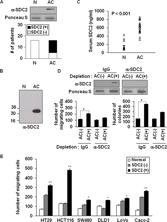
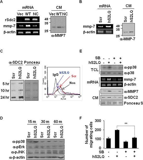
Because earlier studies showed the cell surface heparan sulfate proteoglycan, syndecan-2, sheds from colon cancer cells in culture, the functional roles of shed syndecan-2 were assessed. A non-cleavable mutant of syndecan-2 in which the Asn148-Leu149 residues were replaced with Asn148-Ile149, had decreased shedding, less cancer-associated activities of syndecan-2 in vitro, and less syndecan-2-mediated metastasis of mouse melanoma cells in vivo, suggesting the importance of shedding on syndecan-2-mediated pro-tumorigenic functions. Indeed, shed syndecan-2 from cancer-conditioned media and recombinant shed syndecan-2 enhanced cancer-associated activities, and depletion of shed syndecan-2 abolished these effects. Similarly, shed syndecan-2 was detected from sera of patients from advanced carcinoma (625.9 ng/ml) and promoted cancer-associated activities. Furthermore, a series of syndecan-2 deletion mutants showed that the tumorigenic activity of shed syndecan-2 resided in the C-terminus of the extracellular domain and a shed syndecan-2 synthetic peptide (16 residues) was sufficient to establish subcutaneous primary growth of HT29 colon cancer cells, pulmonary metastases (B16F10 cells), and primary intrasplenic tumor growth and liver metastases (4T1 cells). Taken together, these results demonstrate that shed syndecan-2 directly enhances colon cancer progression and may be a promising therapeutic target for controlling colon cancer development.


Figures

Figure 1
Figure 1. Extracellular domain shedding is necessary for syndecan-2 functions in colon cancer
(A) Equivalent amounts of GST-fused rat syndecan-2 wild-type (WT) or the Leu149® Ile non-cleavable mutant (NC) were incubated with pro-MMP-7, separated by SDS-PAGE, and stained with Coomassie Brilliant Blue. The arrows indicate the dimeric (D) and monomeric (M) forms of syndecan-2. The arrowhead indicates the shed fragment of syndecan-2 (S). (B) Colon cancer cells were transfected with 1 μg of VEC, WT- or NC-syndecan-2. Syndecan-2 mRNA expression was evaluated by RT-PCR using the indicated primer. Conditioned medium was subjected to slot blotting with the anti-syndecan-2 antibody (top). Transfected cells were allowed to migrate on gelatin-coated (10 μg/ml) Transwell plates, and migrated cells were stained with hematoxylin and eosin, and counted (middle). n = 3; **p < 0.01. Transfected cells were added to bottom agar and colonies were counted after 2 weeks (bottom). n = 5; *p < 0.05. (C) B16F10 cells were transfected with siRNA targeting mouse syndecan-2 alone or with 1 μg of the indicated rat syndecan-2 constructs. Syndecan-2 mRNA expression, shed syndecan-2 in the conditioned media and cell migration were analyzed as described in Figure 1B. n = 5; **p < 0.01, *p < 0.05. (D) BALB/c mice were injected with the indicated B16F10 melanoma cells. After 2 weeks, lungs were removed and examined. Two independent experiments were performed (n = 5/each group). Representative photographs of the front and back sides of each lung are shown (top). The bar graph indicates the numbers of metastatic lung nodules (bottom). The columns represent the mean (± s.d.) number of lung metastatic nodules (n = 3). *p < 0.05, **p < 0.01.
Figure 2
Figure 2. Shedding of syndecan-2 plays a critical role in colon cancer cell migration
(A) HT29 cells were transfected with indicated cDNAs, and syndecan-2 mRNA expression was evaluated by RT-PCR. Conditioned media were subjected to slot blotting with the anti-Flag antibody (top). HT29 and HCT116 cells were treated with HT29 conditioned media (final 10% v/v) from VEC, WT-or NC-syndecan-2 mutant transfected cells and allowed to migrate on Transwell apparatus (bottom). n = 5; *p < 0.05, **p < 0.01. (B) Conditioned media were immunodepleted with control IgG- or anti-syndecan-2 antibody-conjugated protein G beads. The supernatants were subjected to slot blotting with anti-syndecan-2 antibody (top). Mixture of HCT116 cells with each supernatant (final 10% v/v) were added to the upper chambers of CIM-plates and migration curves were monitored using the xCELLigence system. The rates of cell migration over 24 hr were analyzed using the RTCA software to each RTCA CIM-Plate wells (bottom). (C) Shed syndecan-2 in HT29 cell conditioned media was isolated by DEAE-Sepharose column chromatography. Final elution fractions were digested by heparinase and analyzed by immunoblotting using anti-syndecan-2 antibody (left). Cells were treated with 0.2 μg/ml of purified shed syndecan-2, and Transwell migration assay was performed (right). n = 5; *p = 0.05, **p < 0.01. (D) Cells were treated with 0.2 μg/ml of purified shed syndecan-2, and a real-time migration assay was analyzed by xCELLigence system. n = 5; *p = 0.05, **p < 0.01.
Figure 3
Figure 3. The syndecan-2 synthetic peptide significantly increases subcutaneous tumor growths in vivo
(A) Schematic diagram depicting the synthetic peptides of human syndecan-2. Peptides corresponding to human syndecan-2 extracellular domain regions consisting of residues 79–94 (hS2LQ: LTSAAPKVETTTLNIQ) and residues 19–40 (hS2EA: ESRAELTSDKDMYLDNSSIEEA) were synthesized (top). HCT116 cells were mixed with synthetic peptides (final 5 nM). Cells migration was analyzed using the provided RTCA software (middle). n = 5; *p < 0.05. Cell viability was determined by MTT assay (bottom left). Colony formation was analyzed as described above (bottom right). N-terminal human syndecan-2 synthetic peptide (hS2EA) was used as a control peptide. n = 4; **p < 0.01. (B) HT29-luc cells (5 × 10⁶ cells/mouse) preincubated with synthetic peptides were injected subcutaneously into the right flanks of BALB/c nude mice (n = 5/group). Representative images of in vivo tumor development at the injection sites, which received HT29-luc cells incubated with human syndecan-2 synthetic peptide or PBS from the same mouse and taken weekly after implantation, are shown. (C) Tumor growth was quantified (as photon/s) weekly by IVIS at 7 days after injection. PBS, mean = 1.59E+06 photon/s, 95% CI = 1.35E+06 to 1.83E+06 photon/s; hS2LQ, mean = 2.97E+06 photon/s, 95% CI = 2.50E+06 to 3.44E+06 photon/s; p < 0.05. (D) Tumor volume (mm3) represented as average at 3–4 days. Average tumor volumes for 21 days were 42.75 mm3 with syndecan-2 peptide, 95% CI = 34.52 mm3 to 51.00 mm3 than the 29.21 mm3 with PBS, 95% CI = 24.94 mm3 to 33.48 mm3. **p < 0.01. (E) Correlation between tumor volume measurement and photon imaging (R2 = 0.9032).
Figure 4
Figure 4. The syndecan-2 synthetic peptide significantly increases the metastasis in mouse models
(A) BALB/c mice (n = 5/group) were injected with B16F10 melanoma cells (1 × 10⁵ cells/mice) incubated with the synthetic peptides into the tail vein. Mice were sacrificed after 2 weeks, and the number of metastatic tumor nodules was counted in the lungs. The bar graph indicates the numbers of metastatic lung nodules. The columns represent the mean ± s.d. of number of lung metastatic nodules, **p < 0.01. (B) B16F10 melanoma cells incubated with the indicated peptide (final 250 nM) were injected into BALB/c mice (n = 7/group) via the tail vein. Representative photographs of the front and back sides of each lung are shown. The columns represent the mean ± s.d. of number of lung metastatic nodules, *p < 0.05. (C) Mouse mammary cancer 4T1-luc cells (1 × 10⁵ cells/mice) were incubated with syndecan-2 synthetic peptide (final 250 nM) or PBS, and injected into spleens of BALB/c mice. Bioluminescence images of spleen (left, top; PBS, mean = 3.17E+06 photon/s, 95% CI = 2.10E+06 to 4.24E+06 photon/s; syndecan-2 peptide, mean = 7.32E+06 photon/s, 95% CI = 2.57E+06 to 12.07E+06 photon/s, p < 0.05) and liver (right) metastatic 4T1-luc cells, taken after 7 days of splenic injection, are shown. The respective photon counts of each mouse are represented by the color scales beside the mouse pictures. The IVIS imaging system acquired pictures were taken 10–20 min after intraperitoneal injection of d-luciferin (150 mg/kg). Quantification of tumor signal is represented as photon counts (left, bottom).
Figure 5
Figure 5. Elevated levels of shed syndecan-2 in serum correlate with increased migratory potential in colon cancer
(A) Serum samples from either a normal donor (N) or advanced stages of colon cancer (AC) patients were subjected to slot blotting with anti-syndecan-2 antibody. The data represent the number of patients found to be serum negative (□) or serum positive (■) for the presence of shed syndecan-2. (B) Serum samples pretreated with HNO2 were resolved by 15% SDS-PAGE followed by blotting with the anti-syndecan-2 antibody. (C) Serum from patients with colon cancer (n = 12) and normal controls (n = 11) was analyzed by ELISA for shed syndecan-2 levels. Median levels are indicated by the horizontal bars. (D) Serum samples from shed syndecan-2 negative (AC−) and positive (AC+) AC patients were incubated with protein A Sepharose, and then the unbound materials were further immunodepleted with either IgG- or anti-syndecan-2 antibody-conjugated protein G Sepharose. The supernatants were subjected to slot blotting with the anti-syndecan-2 antibody (top). The supernatants were then used for Transwell migration assays and anchorage-independent growth assays with HCT116 cells (bottom). n = 4; *p < 0.05. (E) Colon cancer cells were treated with the indicated serum samples and allowed to migrate on gelatin-coated Transwell plates. n = 4; *p < 0.05, **p < 0.01.
Figure 6
Figure 6. Shed syndecan-2 enhances MMP-7 expression via p38 MAP kinase activation in colon cancer cells
(A) HT29 cells were transfected with indicated cDNAs. Expression of SDC2 and MMP-7 was analyzed by RT-PCR (left). Conditioned media (CM) were collected, and proteins were concentrated by TCA precipitation and analyzed by immunoblotting using anti-MMP-7 antibody (right). (B) HT29 cells were treated with either scrambled (Scr) or human (hS2LQ) peptide in SFM for 24 hr, and MMP7 expressions were analyzed by RT-PCR (left). Conditioned media were collected, and proteins were concentrated by TCA precipitation and analyzed by immunoblotting using anti-MMP-7 antibody (right). (C) HT29 cells stably overexpressing syndecan-2 were treated with the indicated peptides and conditioned media were subjected to slot blotting with the anti-syndecan-2 antibody (left). Cells were either untreated (non) or treated with the indicated peptides and cell surface expression of syndecan-2 was analyzed by Flow cytometry (right). (D) HT29 cells treated with the indicated peptides were lysed, and MAPK activation was assessed using an antibody specific for phospho-p38 (pp38). (E) HT29 cells preincubated with SB239063 (1 hr) were treated with the syndecan-2 synthetic peptide. After 15 min, total cell lysates (TCL) were subjected to immunoblotting with the indicated antibodies to determine the phosphorylation of p38 (top). At 6 hr, MMP-7 mRNA expression was analyzed by RT-PCR (middle), and conditioned media were subjected to either immunoblotting with anti-MMP-7 antibody or slot blotting with anti-syndecan-2 antibody (bottom). (F) HT29 cells preincubated with SB239063 (1 hr) were allowed to migrate on Transwell apparatus in the absence or presence of the syndecans-2 peptide. n = 4; **p < 0.01.

Similar articles

Cited by

References

    1. Fakih MM. KRAS mutation screening in colorectal cancer: From paper to practice. Clin Colorectal Cancer. 2010;9:22–30. - PubMed
    1. Shah SN, Hile SE, Eckert KA. Defective mismatch repair, microsatellite mutation bias, and variability in clinical cancer phenotypes. Cancer Res. 2010;70:431–435. - PMC - PubMed
    1. Schvartzman JM, Sotillo R, Benezra R. Mitotic chromosomal instability and cancer: mouse modelling of the human disease. Nat Rev Cancer. 2010;10:102–115. - PMC - PubMed
    1. Suda K, Tomizawa K, Mitsudomi T. Biological and clinical significance of KRAS mutations in lung cancer: an oncogenic driver that contrasts with EGFR mutation. Cancer Metastasis Rev. 2010;29:49–60. - PubMed
    1. Duffy MJ, van Dalen A, Haglund C, Hansson L, Holinski-Feder E, Klapdor R, Lamerz R, Peltomaki P, Sturgeon C, Topolcan O. Tumour markers in colorectal cancer: European Group on Tumour Markers (EGTM) guidelines for clinical use. Eur J Cancer. 2007;43:1348–1360. - PubMed

Publication types

LinkOut - more resources