. 2015 Apr;157(2):175-86.
doi: 10.1016/j.clim.2015.02.001. Epub 2015 Feb 11.

Myeloid-derived suppressor cells are proinflammatory and regulate collagen-induced arthritis through manipulating Th17 cell differentiation

Affiliations

Myeloid-derived suppressor cells are proinflammatory and regulate collagen-induced arthritis through manipulating Th17 cell differentiation

Hui Zhang et al. Clin Immunol. 2015 Apr.

Abstract

Myeloid-derived suppressor cells (MDSC) and Th17 cells were found to expand in collagen-induced arthritis (CIA) significantly. Two subsets of MDSC, polymorphonuclear (PMN) and mononuclear (MO), were detected and their ratios varied during the development of CIA. The depletion of MDSC in vivo resulted in suppression of T-cell proliferation and decreased IL-17A and IL-1β production. The adoptive transfer of MDSC restored the severity of arthritis and Th17 cell differentiation. The depletion of MDSCs on day 35 resulted in arthritis amelioration without reaching a significant difference. Furthermore, MDSCs from CIA mice had higher production of IL-1β and promoted Th17 cell differentiation. The expansion of MDSCs in the peripheral blood of rheumatoid arthritis (RA) patients was in correlation with increased Th17 cells and disease activity DAS28. These results support the hypothesis that MDSC may play a significant proinflammatory role in the pathogenesis of CIA and RA by inducing Th17 development in an IL-1β-dependent manner.

Keywords: Collagen-induced arthritis; IL-1β; Myeloid-derived suppressor cells; Th17 cells.


Conflict of interest statement

Conflicts

All the authors have no interests to declare.

Figures
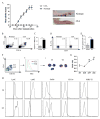
Figure 1
Figure 1
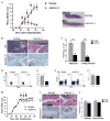
CD11b+Gr-1+ MDSCs consist of two major subsets and were expanded with differentiation of Th17 cells in mice with CIA. (A) Mice were immunized with CII (100 μg) on day 0 and day 21, and clinical arthritis scores were recorded. Photograph on the right show a normal hind limb and one affected by CIA. (B–E) Mice were euthanized on day 35. Spleen and DLN were collected and single-cell suspensions were prepared and analyzed. (B) CD11b+Gr-1+ MDSCs in spleen were measured by flow cytometry and one representative experiment is shown. (C) Percentages of MDSC in the spleens of normal mice and those with CIA. (D) Th17 cells identified as IL-17A+ cells in DLN in normal, and CIA mice were measured by flow cytometry and one representative experiment is shown gating on CD4+ cells. (E) The percentages of Th17 cells in CD4+DNLs in normal mice and mice with CIA. (F) CD11b+Gr-1high and CD11b+Gr-1medium cells were sorted by flow cytometry and spun onto a slide and stained with Giemsa. (G) The ratios of CD11b+Gr-1high (G1) and CD11b+Gr-1medium(G2) cells in the spleen in CIA at different time points of CIA development are shown.(H) Two populations of cells as shown in panel F were stained with anti-Ly6C, anti-Ly6G, anti-F4/80, anti-CD11c, and anti-MHC-II mAbs. Data are summarized from 6 mice in each group and shown as mean ± SD. *p < 0.05, compared to day 28; #p < 0.05, compared to day 35; **p < 0.01.
Figure 2
Figure 2
MDSC depletion at the onset of CIA decreased inflammatory response in CIA mice. Mice were euthanized on day 35. DLNs were collected and single-cell suspensions were prepared. (A) DLN cells were labeled with CFSE and cultured in 96-well round-bottom plate in the presence of CII (50 μg/ml) for 4 days. Anti-CD3/CD28 Abs were used as polyclonal stimulation. Proliferation responses were measured by flow cytometry. (B) Cell proliferation rates in response to anti-CD3/anti-CD28 mAbs and to CII by DLN cells from CII-immunized mice treated with either anti-Gr-1 mAb or isotype control Ab were shown. Data from three experiments were presented. (C) DLN cells from CII immunized mice treated with either anti-Gr-1 mAb or isotype control were cultured in 96-well plate in the presence of CII for 3 days. Supernatants were collected, and the concentration of IL-17A and IL-1β was measured by ELISA. Data from three experiments were presented. (D) Sera were collected when mice were sacrificed, and the concentrations of IL-17A and IL-1β were measured by ELISA. Results are summarized from data of 6 mice in each group and shown as mean ± SD; *p < 0.05 and **p < 0.01.
Figure 3
Figure 3
MDSCs were proinflammatory and regulated Th17 differentiation. (A–G) Anti-Gr-1 Ab treatment was started from day 26 after the initial immunization. (A) Clinical scores and representative images of hind paws in CIA mice treated with anti-Gr-1 mAb or with isotype control Ab. Arrows in red indicate the time when anti-Gr-1 mAb or control Ab was given. (B) For histology assessment, mice were euthanized on day 42. Paraffin-embedded hind paws sections were stained with H&E or Safranin-O/fast green. (C) Pathology scores on hind paws sections. (D–G) Mice were euthanized on day 35. Draining lymph nodes (DLNs) were collected, and single-cell suspension was prepared. (D–E) The frequency of Th17 cells in DLNs was measured by flow cytometry. CD4+ cells were gated. (F–G) Percentages of Treg cells in DLN were measured by flow cytometry. (H–J) Treatments with anti-Gr-1 mAb or isotype control Ab were started on day 35. (H) Clinical scores in CIA mice treated with anti-Gr-1 mAb or isotype control Abs. Arrows in red indicate when anti-Gr-1 mAb or control Ab treatments were given. (I) Histology assessment was carried out on mice euthanized on day 49 after the initial CII immunization. (J) Pathology scores on hind paws sections are shown. Results are summarized from data of 6 mice in each group and shown as mean ± SD. **p < 0.01.
Figure 4
Figure 4
The adoptive transfer of MDSCs into CIA mice treated with anti-Gr1-mAb restored arthritis and Th17 cell response. (A–F) Mice were euthanized on day 49. (A) Clinical scores and representative images of hind paws from CIA mice received anti-Gr-1 Abs or anti-Gr-1 Ab followed by MDSC transfer. Blue arrows indicate the time when anti-Gr-1 treatments were given. Red arrows indicate the time when MDSC transfer was carried out. (B) Paraffin-embedded hind paws sections were stained with H&E or Safranin-O/fast green. (C) Pathology scores on hind paws sections of mice treated with anti-Gr1-mAb with or without MDSC transfer. (D) Cytokines of IL-17A and IL-1β were measured by ELISA on sera obtained from mice sacrificed on day 49. (E) The frequencies of Th17 cells in CD4+DLN from CIA mice received either anti-Gr-1 mAb or anti-Gr-1 mAb followed by MDSC transfer were measured by flow cytometry. (F) Percentages of Th17 cells in DLNs gated in CD4+ cells. Results are summarized from data of 6 mice in each group and shown as mean ± SD. **p < 0.01.
Figure 5
Figure 5
MDSC promoted Th17 cell differentiation from naïve CD4+ T cells in an IL-1β-dependent manner. (A) Mice were euthanized on day 35. Splenic cells stained with anti-CD11b and anti-Gr-1 mAbs were sorted by flow cytometry. Sorted CD11b+Gr-1+ were stained with anti-CD3 mAb. (B) Cell lysates were prepared from CD11b+Gr-1+ cells isolated from mice with CIA or normal mice. IL-1β concentrations in the lysates were determined by ELISA. (C) Naïve CD4+CD25CD62L+ T cells from normal mice and CD11b+Gr-1+MDSCs from CIA or normal mice were sorted by flow cytometry. Naïve CD4+T cells were stimulated with anti-CD3/anti-CD28 mAbs either in the presence or in the absence of CD11b+Gr-1+MDSC. IL-1β concentrations in the culture supernatant were determined by ELISA. (D) MDSCs derived from normal or CIA mice were co-cultured with naïve CD4+ T cells in the presence of anti-CD3/CD28 Ab for 3 days with or without IL-1Ra (300 ng/ml). The concentration of IL-17A in supernatant was measured by ELISA. The results represent three independent experiments. Data are shown as mean ± SD. *p < 0.05 and **p < 0.01.
Figure 6
Figure 6
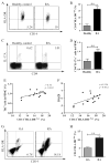
MDSC and Th17 cells were expanded in rheumatoid arthritis (RA). The frequency of CD14+HLA-DR−/low MDSCs in PBMCs from RA patients (n = 15) and healthy individuals (n = 15) was analyzed by flow cytometry. A representative experiment is shown in panel A. The percentages of HLA-DR−/low cells gated in CD14+ cells in PBMCs in these two populations are shown in panel B. The frequency of Th17 cells in PBMCs from RA patients or healthy individuals was analyzed by flow cytometry. A representative experiment is shown in panel C. The percentages of Th17 cells in PBMCs from RA patients and healthy individuals are shown in panel D. The correlation between MDSCs andTh17 cells in PBMCs from RA patients is shown in panel E. The correlation between MDSCs andDAS28 in RA patients is shown in panel F. Single cells were prepared from the synovial tissues from RA (n = 6) and OA (n = 6) patients. The frequency of CD14+HLA-DR−/low MDSC in these synovial cells was analyzed by flow cytometry. A representative experiment is presented in panel G. Percentages of CD14+HLA-DR−/low cells in the synovial tissues of RA and OA patients are presented in (H). **p < 0.01.

Similar articles

Cited by

References

    1. Firestein GS. Evolving concepts of rheumatoid arthritis. Nature. 2003;423(6937):356–361. - PubMed
    1. Sokka T, Kautiainen H, Pincus T, Verstappen SM, Aggarwal A, Alten R, et al. Work disability remains a major problem in rheumatoid arthritis in the 2000s: data from 32 countries in the QUEST-RA study. Arthritis Res Ther. 2010;12(2):R42. - PMC - PubMed
    1. Korn T, Bettelli E, Oukka M, Kuchroo VK. IL-17 and Th17 Cells. Annu Rev Immunol. 2009;27:485–517. - PubMed
    1. McGeachy MJ, Cua DJ. Th17 cell differentiation: the long and winding road. Immunity. 2008;28(4):445–453. - PubMed
    1. Tesmer LA, Lundy SK, Sarkar S, Fox DA. Th17 cells in human disease. Immunol Rev. 2008;223:87–113. - PMC - PubMed

Publication types

MeSH terms