. 2015 Feb 11;35(6):2624-35.
doi: 10.1523/JNEUROSCI.3051-14.2015.

The roles of Cdk5-mediated subcellular localization of FOXO1 in neuronal death

Affiliations

The roles of Cdk5-mediated subcellular localization of FOXO1 in neuronal death

Jiechao Zhou et al. J Neurosci. .

Abstract

Deficiency of cyclin-dependent kinase 5 (Cdk5) has been linked to the death of postmitotic cortical neurons during brain development. We now report that, in mouse cortical neurons, Cdk5 is capable of phosphorylating the transcription factor FOXO1 at Ser249 in vitro and in vivo. Cellular stresses resulting from extracellular stimulation by H2O2 or β-amyloid promote hyperactivation of Cdk5, FOXO1 nuclear export and inhibition of its downstream transcriptional activity. In contrast, a loss of Cdk5 leads to FOXO1 translocation into the nucleus: a shift due to decreased AKT activity but independent of S249 phosphorylation. Nuclear FOXO1 upregulates transcription of the proapoptotic gene, BIM, leading to neuronal death, which can be rescued when endogenous FOXO1 was replaced by the cytoplasmically localized form of FOXO1, FOXO1-S249D. Cytoplasmic, but not nuclear, Cdk5 attenuates neuronal death by inhibiting FOXO1 transcriptional activity and BIM expression. Together, our findings suggest that Cdk5 plays a novel and unexpected role in the degeneration of postmitotic neurons through modulation of the cellular location of FOXO1, which constitutes an alternative pathway through which Cdk5 deficiency leads to neuronal death.

Keywords: Cdk5; FOXO1; neuronal death; phosphorylation.


Figures

Figure 1.
Figure 1.
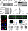
Cdk5 phosphorylates FOXO1 at serine 249. A, B, Lysates of 293T cells transfected with Cdk5/p35, Cdk5/p25, Cdk5-KD/p35, Cdk5-KD/p25 (Cdk5 kinase-dead plasmid), Cdk1/Cyclin B, Cdk2/Cyclin A, or control vector, together with GFP-FOXO1 or the GFP-FOXO1–S249A mutant, were analyzed by immunoblotting with antibodies to pS249-FOXO1, FOXO1, Cdk5, p35/p25, GFP, or GAPGH as shown. C, D, In vitro kinase assays with immunoprecipitated (IP) Cdk5. Recombinant GST-FOXO1 or GST-FOXO1–S249A proteins were subjected to an in vitro kinase assay with IP GFP fusion proteins from 293T cells expressing the indicated constructs. Kinase reaction products were immunoblotted with the pS249-FOXO1 antibody. FOXO1 proteins levels were evaluated by Coomassie Blue staining. E, The interactions among FOXO1, Cdk5, and p35 were investigated in 293T cells. The 293T cells were cotransfected Flag-FOXO1 with GFP-Cdk5 and/or GFP-p35. The antibodies used for IP are indicated in the top of the gel. After immunoprecipitation, the gels were blotted with indicated antibodies. F, Primary cortical neurons were transfected with the GFP-FOXO1-WT or GFP-FOXO1–S249A plasmid together with p25/Cdk5 or p25/Cdk5-KD or control vector in combination with the 3xIRS luciferase reporter system and subjected to luciferase assay. Data are mean ± SEM; n = 5. *p < 0.05. G, Primary cortical neurons were transfected with different Cdk/cyclin pairs together with the 3xIRS-luciferase reporter gene and subjected to luciferase assays. Data are mean ± SEM; n = 5. *p < 0.05.
Figure 2.
Figure 2.
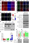
Oxidative stress induces FOXO1 nuclear export by active Cdk5. A, Lysates of 293T cells transfected with GFP-FOXO1 or GFP-FOXO1–S249A, together with Cdk5/p25 or the control vector, were immunoprecipitated with the FOXO1 antibody and immunoblotted with the FOXO1, Cdk5, and 14-3-3β antibodies. B, Primary cortical neurons were treated with 300 μm H2O2 for the indicated time. Whole-cell lysates were then analyzed by immunoblotting with the FOXO1, pS249-FOXO1, p35/p25, and α-tubulin antibodies. C, Quantifications of the relative pS249-FOXO1 intensities. Data are mean ± SEM; n = 3. *p < 0.05. D, E, Primary cortical neurons were treated with 300 μm H2O2 for the indicated time. The cytosolic and nuclear extracts were isolated and immunoblotted with the indicated antibodies. E, Quantifications of the relative nuclear FOXO1 intensities. Data are mean ± SEM; n = 3. *p < 0.05. F, Primary cortical neurons were treated with 50 μm roscovitine or vehicle control (DMSO). Endogenous FOXO1 and Map2 (Microtubule-associated protein 2) were immunostained to investigate the localization of FOXO1 in neurons (Map2-positive cells). G–I, GFP-FOXO1 or GFP-FOXO1–S249D was transfected in primary cortical neurons. G, After 50 μm roscovitine administration, the nuclear translocation time of FOXO1 proteins (GFP-tag) was monitored by live cell imaging. Representative images and quantifications of subcellular localization of GFP-FOXO1 and GFP-FOXO1–S249D are shown in H and I, respectively. J–M, Primary cortical neurons were transfected with control U6 or shCdk5 plasmid together with GFP-FOXO1-WT or GFP-FOXO1–S249A in combination with the 3xIRS luciferase reporter system. On DIV 7, the neurons were treated with 300 μm H2O2 for 3 h or 3 μm β-amyloid for 10 h, and then subjected to luciferase assay. Data are mean ± SEM; n = 5. *p < 0.05.
Figure 3.
Figure 3.
Ser249-FOXO1 is less affected by AKT inhibition. A–F, GFP-FOXO1, GFP-FOXO1–S249A, or GFP-FOXO1–S249D was transfected in primary cortical neurons treated with DMSO or LY294002 for 16 h. Representative images and the quantification of subcellular localization of GFP-FOXO1, GFP-FOXO1–S249A, and GFP-FOXO1–S249D were shown in top and bottom panels, respectively. G–I, GFP-FOXO1-AAA, GFP-FOXO1-AAA-S249A, or GFP-FOXO1-AAA-S249D was transfected in primary cortical neurons. Representative images and the quantification of subcellular localization of GFP-FOXO1-AAA, GFP-FOXO1-AAA-S249A, and GFP-FOXO1-AAA-S249D were shown in top and bottom panels, respectively. J–K, Primary cortical neurons were transfected with GFP-FOXO1-AAA, GFP-FOXO1-AAA-S249A, or GFP-FOXO1-AAA-S249D plasmids together with the 3xIRS-luciferase (J) or Bim-luciferase (K) reporter gene and subjected to luciferase assays. Data are mean ± SEM; n = 5. *p < 0.05.
Figure 4.
Figure 4.
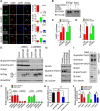
Cdk5 promotes an AKT-independent nuclear export of FOXO1. A, B, Primary cortical neurons were transfected with Flag-FOXO1 together with GFP, GFP-p25/Cdk5, GFP-p35/Cdk5, GFP-p25/Cdk5-KD, or GFP-p35/Cdk5-KD on DIV 3. On DIV 7, the neurons were treated with vehicle control (DMSO) (A) or LY294002 (B) for 16 h. After treatments, the cells were fixed to measure the subcellular localization of FOXO1 by Flag-immunofluorescent staining. Representative images are shown on the left: green represents GFP; red represents Flag; blue represents DAPI. Quantification of subcellular localization of FOXO1 is shown on the right. C, D, Primary cortical neurons were transfected with Flag-FOXO1-AAA or Flag-FOXO1-AAA-S249A together with GFP, GFP-p25/Cdk5, GFP-p35/Cdk5, GFP-p25/Cdk5-KD, or GFP-p35/Cdk5-KD. On DIV 7, the cells were fixed to measure the subcellular localization of FOXO1 by Flag-immunofluorescent staining. Representative images are shown on the left: green represents GFP; red represents Flag; blue represents DAPI. Quantification of subcellular localization of FOXO1 is shown on the right.
Figure 5.
Figure 5.
Cdk5 inhibits FOXO1 transcriptional activity in an AKT-independent way. Primary cortical neurons were transfected with the GFP-FOXO1-WT, GFP-FOXO1–S249A, GFP-FOXO1-AAA, or GFP-FOXO1-AAA-S249A plasmid together with indicated vectors in A, B, C or control vector in combination with the 3xIRS luciferase reporter system. With or without drug treatments, the neurons were subjected to luciferase assay. Data are mean ± SEM; n = 5. *p < 0.05.
Figure 6.
Figure 6.
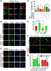
Nuclear accumulation of FOXO1 in Cdk5-deficient neurons leads to the activation of FOXO1. A, Representative images of FOXO1 immunoreactivity in cortical regions of E16.5 Cdk5+/+, Cdk5+/−, and Cdk5−/− mice. B, C, Cdk5+/+ and Cdk5−/− neurons were isolated from E16.5 embryos obtained from crosses of Cdk5+/−mice. B, Subcellular localization of endogenous FOXO1 in Cdk5+/+ and Cdk5−/− neurons was evaluated by FOXO1 immunofluorescence staining in Tuj1-positive neurons. LY294002 treatments induced nuclear accumulation of FOXO1. C, Subcellular localization of exogenous FOXO1 was evaluated following transfection of GFP-FOXO1 in Cdk5+/+ and Cdk5−/− neurons. Representative images are shown on the left, and quantification of subcellular localization of FOXO1 is shown on the right. D, E, The cytoplasmic and nuclear extracts were isolated from cortices of E16.5 Cdk5+/+ and Cdk5−/− mice. Lysates were immunoblotted with the indicated antibodies. E, The relative level of FOXO1 in total/cytoplasm/nucleus was quantified. Data are mean ± SEM; n = 3. *p < 0.05. F, Lysates from primary cortical neurons were transfected with Cdk5-specific siRNA (01, 02, 03) and control siRNA (NC) on DIV 3. On DIV 7, the cytosolic and nuclear extracts were isolated and immunoblotted with the indicated antibodies. G, Cdk5+/+ and Cdk5−/− neurons were transfected with the 3xIRS luciferase reporter system on DIV 3. On DIV 7, luciferase activities for 3xIRS were measured and analyzed. Data are mean ± SEM; n = 5. *p < 0.05. The protein level of Cdk5 in lysates was also provided. H, Primary cortical neurons (DIV 3) were transfected with plasmids or specific siRNA targeting Cdk5 as indicated. On DIV 7, luciferase activities for 3xIRS were measured and analyzed. Data are mean ± SEM; n = 5. *p < 0.05. The protein level of Cdk5 in lysates was also provided.
Figure 7.
Figure 7.
Cdk5 deficiency induces nuclear accumulation of FOXO1 by inactivating AKT. A, Primary cortical neurons were transfected with control U6 or shcdk5 plasmid together with GFP-FOXO1-WT, GFP-FOXO1–S249D, or GFP-FOXO1–S249A on DIV 3, then immunostained with Cdk5 antibody on DIV 7. The subcellular localizations of GFP-FOXO1-WT, GFP-FOXO1–S249D, or GFP-FOXO1–S249A were measured in neurons displaying low Cdk5 expression; arrows indicate shcdk5 effective neurons. Representative images are shown on the left: green represents GFP; red represents Cdk5; blue represents DAPI. Quantification is shown on the right. B, Lysates of 293T cells transfected with Flag-FOXO1 together with control U6 or shcdk5 plasmid were immunoprecipitated with the Flag antibody and immunoblotted with the FOXO1 and 14-3-3β antibodies. C, D, Primary cortical neurons transfected with the GFP-FOXO1-WT or GFP-FOXO1–S249A plasmid together with the control U6 or shcdk5 plasmid plus either the 3xIRS reporter (C) or Bim reporter gene (D). Neurons were subjected to luciferase assays. Data are mean ± SEM; n = 5. *p < 0.05. E, F, N2a cells were transfected with Cdk5/p25, Cdk5/p35, or control Flag vectors. Forty-eight hours after transfection, the cells were treated with DMSO or 50 μm LY294002 for another 12 h. The lysates were immunoblotted with the indicated antibodies. F, The relative levels of pS319-FOXO1 and pS473-Akt1 were quantified. Data are mean ± SEM; n = 3. G, H, N2a cells were transfected with shRNA targeted to either Cdk5 or foxo1. Scrambled shRNA was used as a control. Forty-eight hours after transfection, the lysates were immunoblotted with the indicated antibodies. H, The relative level of pS319-FOXO1 was quantified. Data are mean ± SEM; n = 5. *p < 0.05. I, J, Brain lysates from the cortices of E16.5 Cdk5+/+ and Cdk5−/− mice were prepared, and the levels of pS249-FOXO1 were detected following immunoprecipitation with FOXO1 antibody. The other proteins were measured by directly immunoblotting with the indicated antibodies. J, The relative levels of pS249-FOXO1, pS319-FOXO1, and pS473-Akt1 were quantified. Data are mean ± SEM; n = 3. *p < 0.05.
Figure 8.
Figure 8.
Interaction of Cdk5-dependent cell cycle and cell death activities. A, Cleaved-caspase-3 and BrdU incorporation was double stained on DIV 7 Cdk5−/− neurons. TuJ1 staining serves as neuronal cell marker. B–D, Effects of expressing mutant or wild-type FOXO1 on cortical neuronal apoptosis. B, Primary cortical neurons (DIV 3) were transfected with CMV-mCherry-IRES-U6-shcdk5 or control CMV-mCherry-IRES-U6-scramble vector either alone or together with U6-shfoxo plasmid. Cells were then immunostained with Cdk5 antibody at DIV 7. DAPI was used to counterstain nuclei to measure the neuronal death by nuclei condensation. C, Primary cortical neurons (DIV 3) were transfected with CMV-mCherry-IRES-U6-shcdk5 and shfoxo1 vector together with GFP-FOXO1-WT-Res, GFP-FOXO1–S249A-Res, or GFP-FOXO1–S249D-Res plasmid. The cells were fixed on DIV 7 and stained with DAPI to evaluate neuronal death by nuclei condensation. D, The quantification of neuronal apoptotic rate in B, C is presented in D. Data are mean ± SEM; n = 3. *p < 0.05. F, Primary cortical Cdk5−/− neurons were transfected with GFP-Cdk5-NLS or GFP-Cdk5-NES. Cleaved-caspase-3 and BrdU incorporation was detected by immunostaining. E, G, Primary cortical neurons were transfected with the indicated plasmids together with the 3xIRS-luciferase or Bim-luciferase reporter gene. The neurons were subjected to luciferase assays on DIV 7. Data are mean ± SEM; n = 5. *p < 0.05.

Similar articles

Cited by

References

    1. Ahlijanian MK, Barrezueta NX, Williams RD, Jakowski A, Kowsz KP, McCarthy S, Coskran T, Carlo A, Seymour PA, Burkhardt JE, Nelson RB, McNeish JD. Hyperphosphorylated tau and neurofilament and cytoskeletal disruptions in mice overexpressing human p25, an activator of cdk5. Proc Natl Acad Sci U S A. 2000;97:2910–2915. doi: 10.1073/pnas.040577797. - DOI - PMC - PubMed
    1. Arden KC. FoxO: linking new signaling pathways. Mol Cell. 2004;14:416–418. doi: 10.1016/S1097-2765(04)00213-8. - DOI - PubMed
    1. Burgering BM, Kops GJ. Cell cycle and death control: long live Forkheads. Trends Biochem Sci. 2002;27:352–360. doi: 10.1016/S0968-0004(02)02113-8. - DOI - PubMed
    1. Cheung ZH, Ip NY. Cdk5: mediator of neuronal death and survival. Neurosci Lett. 2004;361:47–51. doi: 10.1016/j.neulet.2003.12.117. - DOI - PubMed
    1. Cicero S, Herrup K. Cyclin-dependent kinase 5 is essential for neuronal cell cycle arrest and differentiation. J Neurosci. 2005;25:9658–9668. doi: 10.1523/JNEUROSCI.1773-05.2005. - DOI - PMC - PubMed

Publication types

MeSH terms