. 2015 Feb 9;32(3):358-372.
doi: 10.1016/j.devcel.2015.01.003.

The ABBA motif binds APC/C activators and is shared by APC/C substrates and regulators

Affiliations

The ABBA motif binds APC/C activators and is shared by APC/C substrates and regulators

Barbara Di Fiore et al. Dev Cell. .

Abstract

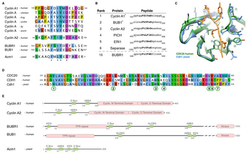
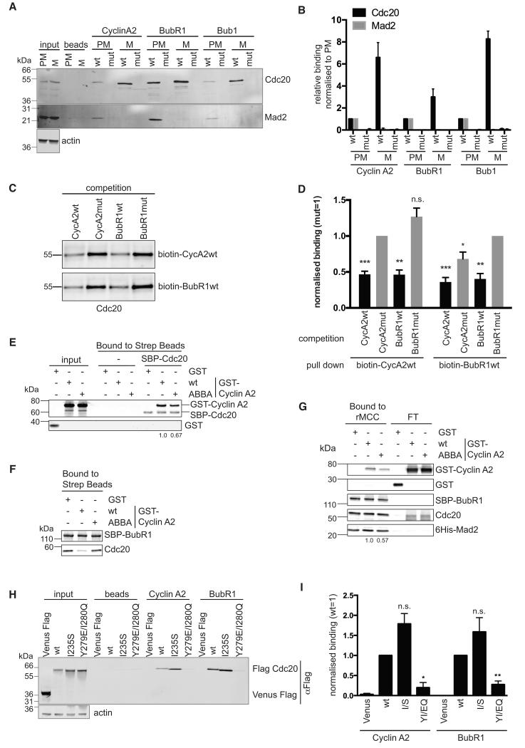
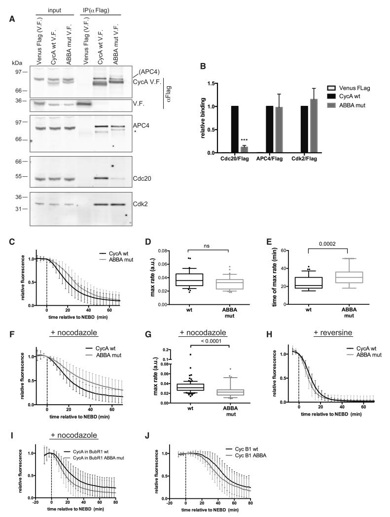
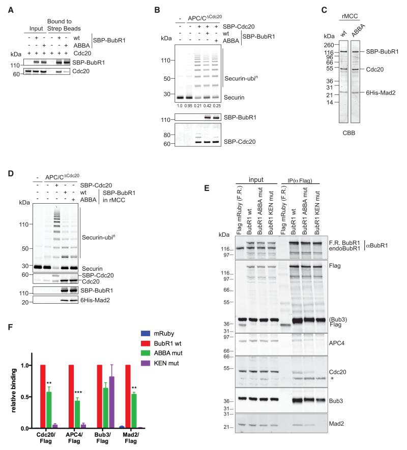
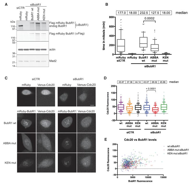
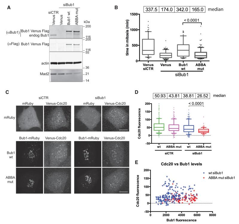
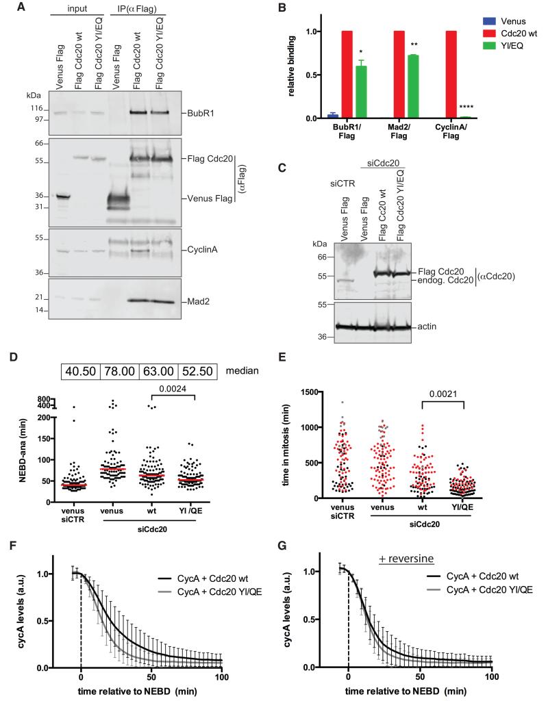
The anaphase-promoting complex or cyclosome (APC/C) is the ubiquitin ligase that regulates mitosis by targeting specific proteins for degradation at specific times under the control of the spindle assembly checkpoint (SAC). How the APC/C recognizes its different substrates is a key problem in the control of cell division. Here, we have identified the ABBA motif in cyclin A, BUBR1, BUB1, and Acm1, and we show that it binds to the APC/C coactivator CDC20. The ABBA motif in cyclin A is required for its proper degradation in prometaphase through competing with BUBR1 for the same site on CDC20. Moreover, the ABBA motifs in BUBR1 and BUB1 are necessary for the SAC to work at full strength and to recruit CDC20 to kinetochores. Thus, we have identified a conserved motif integral to the proper control of mitosis that connects APC/C substrate recognition with the SAC.


Figures

Figure 1
Figure 1. Identification of the ABBA motif and its conservation through evolution
(A) Alignment of the ABBA motif-containing region in Cyclin A1 orthologs, and peptides from human Cyclin A2, BUBR1, BUB1 and yeast Acm1 matching the Fx[ILV][FHY]x[DE] consensus (asterisks). See also supplemental Fig. 1. (B) Top results, ranked by conservation, of a SLiMSearch analysis of the human proteome using the ABBA motif consensus. Five of the top six instances are found in important mitotic proteins (ERI1 has no known cell cycle function). (C) Structural alignment of yeast Cdh1 (blue) bound to the ABBA peptide from Acm1 (orange) (He et al., 2013) and human CDC20 (green) (Tian et al., 2012). The ABBA peptide is numbered to define the position of the six core residues in the consensus. CDC20 is numbered to define the key residues in the ABBA motif-binding pocket. (D) Alignment of the motif-binding pocket in human CDC20, human CDH1, and yeast Cdh1. Numbering refers to the key residues in the ABBA motif-binding pocket defined in (C). (E) Modular architecture of human Cyclin A1, Cyclin A2, BUBR1, BUB1 and yeast Acm1.
Figure 2
Figure 2. The ABBA motif binds to CDC20 between blades 2 and 3 of the WD40 domain
(A) Immobilized peptides containing wild-type (wt) and mutant (mut) ABBA motif from Cyclin A2, BubR1 and Bub1 were incubated with extracts from prometaphase (PM) and metaphase (M) HeLa cells (see Experimental Procedures). Input signals and background binding to beads are shown. For the input, 1/30 of the extract used for the pulldown was loaded. Actin is the loading control for the input. (B) Mean and Standard error of the Mean (SEM) from 4 independent experiments as shown in (A). For each peptide the binding is normalised to the binding of the wt peptide in PM. (C) Sf9 cell extracts expressing Hs CDC20 were incubated with the indicated biotinylated peptides and a 200-fold excess of a competing non-biotinylated peptide followed by purification with streptavidin beads. See also supplemental Fig. 2. (D) Bar charts show mean and SEM from 3 independent experiments normalized to CDC20 binding after competition with the respective Cyclin A or BubR1 mutant peptide. The competition of wild type Cyclin A and BubR1 peptides to the respective mutant peptide was analysed using a two-tailed paired t-test (*, p<0.05; **, p<0.005; ***, p<0.0005; n.s., not significant). (E) Streptavidin beads bound to SBP-CDC20 were incubated with GST, GST-Cyclin A2 wt or ABBA mutant. Proteins retained on the beads were analysed by quantitative immunoblotting and normalized values for Cyclin A2 are shown. Results are representative of 2 experiments. (F) Streptavidin beads were incubated with insect cell lysate expressing SBP-BubR1, unbound proteins washed away, and beads incubated with cell lysate expressing untagged CDC20 with GST, or GST-Cyclin A2 wt or ABBA mutant. Proteins retained on the beads were analysed by quantitative immunoblotting. (G) Streptavidin beads with rMCC bound via SBP-BubR1 were incubated with GST, GST-Cyclin A2 wt or ABBA mutant. Proteins retained on the beads and in the flow-through (FT) were analysed by quantitative immunoblotting and normalized values for Cyclin A2 are shown. Results representative of 2 experiments. (H) Peptide pulldown using immobilised peptides and total extract from siRNA CDC20-depleted prometaphase cells expressing Venus-Flag, or siRNA resistant Flag-CDC20 wild-type or Y279E/I280Q mutant. Actin is a loading control. (I) Mean and SEM from at least 3 independent experiments shown in (H). For each peptide the binding is normalised to the binding of wt CDC20 and values are normalised to input. Paired t-test was used for statistical analysis (*, p<0.05; **, p<0.005; n.s., not significant).
Figure 3
Figure 3. The ABBA motif is required for Cyclin A to be degraded correctly in prometaphase
(A) Ant-Flag immunoprecipitations from nocodazole-treated HeLa FRT cell lines expressing inducible Cyclin A Venus-Flag (V.F.) wt and ABBA mutant. Protein in parenthesis indicates signal from a previous blot. Asterisk marks a weak anti-Flag signal due to cross-reaction of the strong Flag signal in the IP with the 2° antibody. (B) Mean and SEM from 4 independent experiments shown in (A). Paired t-test used for statistical analysis (***, p<0.0005). (C) Mean degradation curves with SD from Hela cells microinjected with wt or ABBA mutant Cyclin A Venus-Flag. 105 cells (n=6) and 34 cells (n=2) were analysed for Cyclin A wt and ABBA mutant, respectively. Time is relative to nuclear envelope breakdown (NEBD). (D) & (E) The maximal degradation rate (D) and the time when maximal degradation rate was reached (E) was measured for wt and ABBA mutant Cyclin A from cells in (C) and plotted as a box and whiskers graph. Unpaired t-test used for statistical analysis (ns, not significant). (F) Cyclin A degradation in cells treated with nocodazole was analysed as in (C). 98 cells (n=5) and 64 cells (n=4) were analysed for Cyclin A wt and ABBA mutant, respectively. (G) The maximal degradation rate was measured for wt and ABBA mutant Cyclin A, for single cells in (F) and plotted as box and whiskers graphs. Unpaired t-test used for statistical analysis. (H) Cyclin A degradation in cells treated with reversine was analysed as in (C). 31 cells and 37 cells (n=3) were analysed for Cyclin A wt and the ABBA mutant, respectively. (I) Cyclin A Venus degradation was analysed as in (C) in Hela FRT cell lines treated with nocodazole expressing wt (n=73) or ABBA mutant mRuby-BUBR1 (n=84) in which the endogenous BUBR1 was depleted by siRNA and Cyclin A Venus-Flag was transiently transfected (n=3). (J) Cyclin B1 Venus degradation was analysed as in (C) in Hela cells microinjected with wt Cyclin B1 (39 cells) or Cyclin B1 with a N-terminal ABBA motif (48 cells) (n=5).
Figure 4
Figure 4. The ABBA motif of BUBR1 contributes to binding to CDC20
(A) Cell lysates from Sf9 cells expressing CDC20 and SBP-BubR1 wt or ABBA mutant were incubated with streptavidin beads and proteins retained on the beads analysed by quantitative immunoblotting. (B) APC/C immunoprecipitated from mitotic siRNA CDC20-depleted HeLa cell extract (APC/CΔCdc20) was incubated with SBP-CDC20, or with SBP-CDC20 preincubated with either BubR1 WT or ABBA mutant. Ubiquitylation reactions were performed with securin as substrate and analysed with a Li-COR Odyssey scanner. Values for unconjugated securin are given below the gel (n=3). SBP-CDC20 and SBP-BubR1 were analysed by quantitative immunoblotting. (C) His6-Mad2, CDC20, and SBP-BubR1 - either wt or ABBA mutant - were co-expressed in insect cells and recombinant MCC (rMCC) purified using Ni-NTA agarose and streptavidin beads. The purified rMCCs were analysed by SDS-PAGE and Coomassie Blue staining (CBB). (D) APC/CΔCdc20 purified as in (B) was incubated with SBP-CDC20 and rMCC generated with either wt BubR1 or the ABBA mutant, and securin ubiquitylation assayed as in (B). (E) Flag-mRuby (F.R.) BubR1 wt and mutants were immunoprecipitated from taxol-arrested HeLa FRT cell lines with an anti-Flag antibody. Asterisk denotes a non-specific band. (F) Mean and SEM of at least 6 independent experiments shown in (E). A paired t-test was used for statistical analysis (**, p<0.005; ***, p<0.0005). See also supplemental Fig. 3.
Figure 5
Figure 5. The ABBA motif of BUBR1 is required to recruit CDC20 to kinetochores and contributes to the strength of the SAC
(A) HeLa FRT cell lines expressing stable inducible siRNA-resistant wt and mutant BubR1 were transfected with control siRNA or siRNA against BubR1. Immunoblotting analysis shows the expression levels compared to endogenous BubR1 and the efficiency of knockdown. Actin is a loading control. (B) Taxol-treated HeLa FRT cell lines stably expressing inducible siRNA-resistant Flag-mRuby BubR1, were transfected with the indicated siRNA. Cells were imaged by DIC time-lapse microscopy. Median values of the time spent in mitosis are shown and analysed using an unpaired t-test. (n=3.) See also supplemental Fig. 4A. (C) RPE1 FRT Venus-CDC20 knock-in cell lines stably expressing inducible siRNA resistant Flag-mRuby BubR1 were treated with control siRNA or siRNA against BubR1 and Taxol added. Images of the first frame after NEBD are shown for each BubR1 protein. Scale bars 10 μm. See also supplemental Figs. 4B & 5. (D) Box and whisker analysis of kinetochore (KT) localised CDC20 in cells transfected with the indicated siRNA and rescued with wt and mutant BubR1, examples shown in (C). KT signal is relative to the background. Median values of the KT intensity are shown. At least 170 KTs were analysed from at least 11 cells. (n=3.) Unpaired t-test used for statistical analysis. (E) Levels of KT localised CDC20 and BubR1, relative to the background, are shown for the same cells analysed in (D).
Figure 6
Figure 6. The ABBA motif of BUB1 helps to recruit CDC20 to kinetochores and contributes to the strength of the SAC
(A) HeLa FRT cell lines expressing inducible siRNA-resistant wt or mutant Bub1 Venus-Flag were transfected with control or Bub1 siRNA. Immunoblot shows expression levels compared to endogenous Bub1. Actin is a loading control. (B) HeLa FRT cell lines expressing inducible siRNA-resistant Bub1 Venus-Flag, were transfected with the indicated siRNA, then treated and analysed as in Fig 5B. Median values of mitotic duration are shown and analysed by an unpaired t-test (n=3). (C) RPE1 FRT Venus-CDC20 knock-in cell lines expressing siRNA resistant mRuby-Flag-tagged Bub1, wild-type or ABBA mutant, were treated with control siRNA or siRNA against Bub1 and analysed as in Fig 5C. Scale bars 10 μm. See also supplemental Figs. 5 & 6. (D) Box and whisker analysis of KT localised CDC20 in cells expressing wt or mutant Bub1 as in (C). Median values of the KT intensity are shown. At least 100 KTs were analysed from at least 7 cell (n=3). Unpaired t-test used for statistical analysis. (E) Levels of KT localised CDC20 and Bub1 are shown for the cells analysed in (D).
Figure 7
Figure 7. A CDC20 mutant that cannot bind the ABBA motif is unable to bind Cyclin A and is partially refractory to the SAC
(A) Flag-tagged CDC20 wt and YI/EQ were immunoprecipitated with Flag mAb from taxol-arrested HeLa FRT cell lines and blotted with the indicated antibodies. (B) Average and SEM of 3 independent experiments shown in (A). Paired t-test used for statistical analysis (*, p<0.05; **, p<0.005; ***, p<0.0005). (C) HeLa FRT cell lines expressing inducible siRNA-resistant wild-type or YI/EQ mutant CDC20 were transfected with control siRNA or siRNA against CDC20. Immunoblot shows expression levels compared to endogenous CDC20 and the efficiency of knockdown. Actin is a loading control. (D) Hela FRT cell lines expressing inducible siRNA-resistant Flag-tagged CDC20, were transfected with the indicated siRNA and analysed as in Fig 5B. Red lines indicate the median value. Unpaired t-test used for statistical analysis (n=3). (E) Rescue experiments as in (D) but in the presence of taxol. Cells that exit mitosis (black), cells that die in mitosis (red), cells arrested in mitosis (grey) are indicated. Unpaired t-test was used for statistical analysis (n=3). (F) Cyclin A-Venus-Flag degradation in cells transfected with siRNA against CDC20 and microinjected with Venus-Flag- wt Cyclin A together with either wt or YI/EQ mutant CDC20. Average degradation curves with SD were obtained from 48 cells (n=3) for wt CDC20, and 63 cells (n=3) for mutant CDC20. Time is relative to NEBD. (G) Average curves were obtained as in (F), but in the presence of reversine. 49 cells (n=3) for wt CDC20, and 45 cells (n=3) for mutant CDC20 were analysed.

Similar articles

Cited by

References

    1. Bolanos-Garcia VM, Blundell TL. BUB1 and BUBR1: multifaceted kinases of the cell cycle. Trends in Biochemical Sciences. 2011;36:141–150. - PMC - PubMed
    1. Burton JL, Solomon MJ. Mad3p, a pseudosubstrate inhibitor of APCCdc20 in the spindle assembly checkpoint. Genes & Development. 2007;21:655–667. - PMC - PubMed
    1. Burton JL, Xiong Y, Solomon MJ. Mechanisms of pseudosubstrate inhibition of the anaphase promoting complex by Acm1. Embo J. 2011;30:1818–1829. - PMC - PubMed
    1. Chang L, Zhang Z, Yang J, McLaughlin SH, Barford D. Molecular architecture and mechanism of the anaphase-promoting complex. Nature. 2014 - PMC - PubMed
    1. Chao WC, Kulkarni K, Zhang Z, Kong EH, Barford D. Structure of the mitotic checkpoint complex. Nature. 2012;484:208–213. - PubMed

Publication types

MeSH terms

LinkOut - more resources