. 2015 Apr;89(8):4296-310.
doi: 10.1128/JVI.03055-14. Epub 2015 Feb 4.

Rice stripe tenuivirus nonstructural protein 3 hijacks the 26S proteasome of the small brown planthopper via direct interaction with regulatory particle non-ATPase subunit 3

Affiliations

Rice stripe tenuivirus nonstructural protein 3 hijacks the 26S proteasome of the small brown planthopper via direct interaction with regulatory particle non-ATPase subunit 3

Yi Xu et al. J Virol. 2015 Apr.

Abstract

The ubiquitin/26S proteasome system plays a vital role in regulating host defenses against pathogens. Previous studies have highlighted different roles for the ubiquitin/26S proteasome in defense during virus infection in both mammals and plants, but their role in the vectors that transmit those viruses is still unclear. In this study, we determined that the 26S proteasome is present in the small brown planthopper (SBPH) (Laodelphax striatellus) and has components similar to those in plants and mammals. There was an increase in the accumulation of Rice stripe virus (RSV) in the transmitting vector SBPH after disrupting the 26S proteasome, indicating that the SBPH 26S proteasome plays a role in defense against RSV infection by regulating RSV accumulation. Yeast two-hybrid analysis determined that a subunit of the 26S proteasome, named RPN3, could interact with RSV NS3. Transient overexpression of RPN3 had no effect on the RNA silencing suppressor activity of RSV NS3. However, NS3 could inhibit the ability of SBPH rpn3 to complement an rpn3 mutation in yeast. Our findings also indicate that the direct interaction between RPN3 and NS3 was responsible for inhibiting the complementation ability of RPN3. In vivo, we found an accumulation of ubiquitinated protein in SBPH tissues where the RSV titer was high, and silencing of rpn3 resulted in malfunction of the SBPH proteasome-mediated proteolysis. Consequently, viruliferous SBPH in which RPN3 was repressed transmitted the virus more effectively as a result of higher accumulation of RSV. Our results suggest that the RSV NS3 protein is able to hijack the 26S proteasome in SBPH via a direct interaction with the RPN3 subunit to attenuate the host defense response.

Importance: We show, for the first time, that the 26S proteasome components are present in the small brown planthopper and play a role in defense against its vectored plant virus (RSV). In turn, RSV encodes a protein that subverts the SBPH 26S proteasome via direct interaction with the 26S proteasome subunit RPN3. Our results imply that the molecular arms race observed in plant hosts can be extended to the insect vector that transmits those viruses.


Figures

FIG 1
FIG 1
Ubiquitin conjugate levels and RSV accumulation increase after RNAi-mediated knockdown of RPN11. Six days after injection of dsRNAs of RPN11, about 20 juvenile planthoppers were used for extraction of total protein. After quantifying the protein, the same amounts of total protein were used for Western blotting and ubiquitin level detection (Fig. 1A). The RSV titer was detected using ELISA with antibody raised against RSV virions (Fig. 1B). Another 30 juvenile planthoppers from the same treatment were used for isolation of RNA for Northern blotting to detect the four RSV genomic RNAs (Fig. 1C) and for qRT-PCR for detection of the RPN11 transcript level (Fig. 1D). Three replicates for each treatment were conducted, and three technical ELISA and qRT–PCR replicates were analyzed for each biological replicate. The error bars represent standard error of the mean (SEM).
FIG 2
FIG 2
SBPH RPN3 and RSV-NS3 interact in a yeast two-hybrid assay. (A) Yeast strain Y2H Gold cotransformed with the indicated plasmids was spotted onto synthetic dextrose dropout medium, SD/−Trp/−Leu/−His/−Ade, in a series of 10-fold dilutions. NS3 and LSRPN3 were cloned as translational fusions with either the Gal4 activation domain (AD) or the Gal4 binding domain (BD). The mouse p53 antitumor protein (P53) and lamin C (Lam) genes were cloned as translational fusions with the Gal4 binding domain and used as positive and negative controls, respectively. (B) Schematic representation of the RPN3-coding sequence. Putative conserved functional domains of RPN3 are indicated and were deduced from InterProScan (http://www.ebi.ac.uk/Tools/InterProScan/).
FIG 3
FIG 3
Identification of domains required for the interaction between RPN3 and NS3. Deletion mutants were constructed based on the conserved domains within the RPN3 protein. The deletion RPN3 (positions 1 to 261) comprises the N-terminal 261 amino acids and lacks the 26S Psome reg C and PCI domains, RPN3 (1 to 425) comprises the N-terminal 425 amino acids and contains the entire 26S Psome reg C domains, RPN3 (426 to 498) contains the entire PCI domain within amino acids 426 through 498, RPN3 (261 to 498) contains both the 26S Psome reg C and PCI domains but lacks the N-terminal 260 amino acids, RPN3 (381 to 498) is the fragment of RPN3 that we isolated as a prey plasmid in the Y2H screen, and RPN3 (M) represents a point mutation (isoleucine-to-threonine) at amino acid 433 of RPN3, within the 26S Psome reg C domain. The various mutants were cloned as translational fusions with the Gal4 activation domain (AD) and used to cotransform yeast strain Y2H Gold with full-length NS3 cloned as a translational fusion to the Gal4 binding domain. Yeast strains were spotted onto synthetic dextrose dropout medium, SD/−Trp/−Leu/−His/−Ade, in a series of 10-fold dilutions. P53 and Lam were used as positive and negative controls, respectively.
FIG 4
FIG 4
Characterization of the expression pattern of SBPH RPN3 mRNA. Graphs represent the expression of RPN3 mRNA in total RNA samples isolated from viruliferous and nonviruliferous planthoppers. Values were calculated by qPCR and represent the means from 3 independent replicates with standard errors of the means. Significance was assessed by one-way ANOVA on the values obtained from the different experiments (*, P < 0.05; **, P < 0.01). (A) Three-instar SBPH nymphs were dissected into head, gut (including foregut, midgut, hindgut, and Malpighian tubule), spermary, and ovarium under a stereomicroscope (Olympus). The expression level measured in the heads of nonviruliferous planthoppers was arbitrarily assigned a value of 1.00. (B) Different stages of SBPH from one- to two-instar to adult were collected, and RNA was isolated. The expression level measured at the one- to two-instar stage of nonviruliferous planthoppers was arbitrarily assigned a value of 1.00.
FIG 5
FIG 5
Expression of RPN3 has no effect on the RNA silencing suppressor activity of the RSV NS3 protein. Leaves of wild-type and transgenic GFP 16c N. benthamiana plants were coagroinfiltrated with a GFP-expressing vector, a vector encoding GFP-targeting dsRNA only infiltrated in N. benthamiana), and a vector encoding P19 (as a positive control), NS3, or NS3 with RPN3 or with RPN7. V, empty vector. The leaves were photographed at three days postinfection in N. benthamiana and at six days postinfection in transgenic GFP 16c plants under a hand-held long-wavelength UV illuminator (UVP, USA).
FIG 6
FIG 6
Coexpression of RSV NS3 inhibits the ability of SBPH RPN3 to functionally complement the yeast rpn3 ts mutant. (A) YE101 mutant strains were transformed with SBPH RPN3 (pRS316-LSRPN3-wt) or SBPH RPN3 containing a point mutation (pRS316-LSRPN3-m), streaked on synthetic medium lacking uracil (SD/−Ura), and incubated at 37°C for 2 days. Yeast YE101 was transformed with yeast RPN3 (YCp50-y-RPN3) as a positive control and empty vector (YCp50) as a negative control. (B) YE101 mutant strains were cotransformed with either SBPH RPN3 (pRS316-LSRPN3-wt) or mutant SBPH RPN3 (pRS316-LSRPN3-m) along with pGBKT7-NS3. Yeast strain YE101 was cotransformed with empty vector (pGBKT7) and yeast RPN3 (YCp50-y-RPN3), SBPH RPN3 (pRS316-LSRPN3-wt), or the SBPH RPN3 point mutant (pRS316-LSRPN3-m) as positive controls or empty vector (YCp50) as a negative control. Plates were incubated at 37°C for 2 days. (C) Yeast YE101 transformed with the indicated plasmids was grown at 25°C to early log phase and then temperature shifted at time zero (arrow) to 37°C. At hourly intervals, cell density was measured as optical density at 600 nm, converted to cell numbers, and plotted against time.
FIG 7
FIG 7
NS3 inhibition of SBPH RPN3 results in defective proteolysis in yeast. (A) Protein extracts were prepared from yeast transformed with the indicated plasmids (bottom). An equal amount of protein extract was resolved by SDS-PAGE and examined by immunoblotting using antibodies raised against ubiquitin. A Coomassie blue-stained SDS-polyacrylamide gel is shown below the immunoblot as a loading control. (B to D) The substrates Met-βgal, Arg-βgal, and Ub-Pro-βgal were expressed in yeast transformed with the indicated plasmids, and β-galactosidase activity was measured in triplicate. The data were quantified and standardized to expression of β-galactosidase in YE101 transformed with yeast RPN3 (Ycp50-y-RPN3). The bars represent the mean ± standard error from two independent measurements.
FIG 8
FIG 8
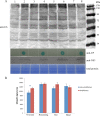
Ubiquitin conjugates accumulate in tissues with high levels of RSV. (A) Total proteins were extracted from different tissues, and an equal amount of protein was used for Western blot analysis. Antibodies against ubiquitin, NS3, and RSV were used to detect ubiquitinated substrates, NS3, and coat protein (CP), respectively. The lower panel represents a Coomassie blue-stained SDS-polyacrylamide gel for loading comparisons. Lanes 1, 5, and 7 represent protein extracts from the ovarium, tharm, and head, respectively, of nonviruliferous SBPH. Lane 3 represents the remaining tissues after removal of the ovarium, tharm, and head. Lanes 2, 4, 6, and 8 represent proteins extracted from the equivalent tissues of RSV-viruliferous SBPH. (B) Measurement of the ubiquitin signal was calculated using the Image Quant TL analysis tool (GE Company). We carried out three replicates for this assay. Asterisks indicate significant differences in ubiquitin levels (P < 0.05 by one-way ANOVA).
FIG 9
FIG 9
Silencing of SBPH RPN3 leads to increased accumulation of RSV. (A) Northern blots of total RNA isolated from viruliferous SBPH at 0, 2, 4, and 6 days after injection of dsRNA homologous to GFP or SBPH RPN3. After transfer to nylon membranes, blots were hybridized to 32P-labled in vitro transcriptional probes specific for the RSV genome. RNA 1, 2, 3, and 4 represent four RSV segments. (B) RSV titers detected using ELISA with antibody raised against RSV virions at 0, 2, 4, and 6 days after injection of viruliferous SBPH with dsRNA homologous to GFP or SBPH RPN3. (C) qRT-PCR analysis of RPN3 mRNA in total RNA isolated from viruliferous SBPH after treatment with dsRNA homologous to GFP or SBPH RPN3.

Similar articles

Cited by

References

    1. Wei TY, Yang JG, Liao FR, Gao FL, Lu LM, Zhang XT, Li F, Wu ZJ, Lin Q Y, Xie LH, Lin HX. 2009. Genetic diversity and population structure of Rice stripe virus in China. J Gen Virol 90:1025–1034. (Erratum, 90:1548.) doi:10.1099/vir.0.006858-0.. - DOI - PubMed
    1. Park HM, Choi MS, Kwak DY, Lee BC, Lee J H, Kim MK, Kim YG, Shin DB, Park SK, Kim YH. 2012. Suppression of NS3 and MP is important for the stable inheritance of RNAi-mediated Rice stripe virus (RSV) resistance obtained by targeting the fully complementary RSV-CP gene. Mol Cells 33:43–51. doi:10.1007/s10059-012-2185-5. - DOI - PMC - PubMed
    1. Kisimoto R. 1965. On the transovarial passage of the Rice stripe virus through the small brown planthopper, Laodelphax striatelllus Fallen, p 73–90. In Conference on relationships between arthropods and plantpathogenic viruses, Tokyo, Japan.
    1. Hibino H. 1996. Biology and epidemiology of rice viruses. Annu Rev Phytopathol 34:249–274. doi:10.1146/annurev.phyto.34.1.249. - DOI - PubMed
    1. Falk BW, Tsai JH. 1998. Biology and molecular biology of viruses in the genus Tenuivirus. Annu Rev Phytopathol 36:139–163. doi:10.1146/annurev.phyto.36.1.139. - DOI - PubMed

Publication types

Substances

LinkOut - more resources