. 2015 Mar 1;194(5):2369-79.
doi: 10.4049/jimmunol.1402412. Epub 2015 Feb 2.

IFN regulatory factor 8 represses GM-CSF expression in T cells to affect myeloid cell lineage differentiation

Affiliations

IFN regulatory factor 8 represses GM-CSF expression in T cells to affect myeloid cell lineage differentiation

Amy V Paschall et al. J Immunol. .

Abstract

During hematopoiesis, hematopoietic stem cells constantly differentiate into granulocytes and macrophages via a distinct differentiation program that is tightly controlled by myeloid lineage-specific transcription factors. Mice with a null mutation of IFN regulatory factor 8 (IRF8) accumulate CD11b(+)Gr1(+) myeloid cells that phenotypically and functionally resemble tumor-induced myeloid-derived suppressor cells (MDSCs), indicating an essential role of IRF8 in myeloid cell lineage differentiation. However, IRF8 is expressed in various types of immune cells, and whether IRF8 functions intrinsically or extrinsically in regulation of myeloid cell lineage differentiation is not fully understood. In this study, we report an intriguing finding that, although IRF8-deficient mice exhibit deregulated myeloid cell differentiation and resultant accumulation of CD11b(+)Gr1(+) MDSCs, surprisingly, mice with IRF8 deficiency only in myeloid cells exhibit no abnormal myeloid cell lineage differentiation. Instead, mice with IRF8 deficiency only in T cells exhibited deregulated myeloid cell differentiation and MDSC accumulation. We further demonstrated that IRF8-deficient T cells exhibit elevated GM-CSF expression and secretion. Treatment of mice with GM-CSF increased MDSC accumulation, and adoptive transfer of IRF8-deficient T cells, but not GM-CSF-deficient T cells, increased MDSC accumulation in the recipient chimeric mice. Moreover, overexpression of IRF8 decreased GM-CSF expression in T cells. Our data determine that, in addition to its intrinsic function as an apoptosis regulator in myeloid cells, IRF8 also acts extrinsically to repress GM-CSF expression in T cells to control myeloid cell lineage differentiation, revealing a novel mechanism that the adaptive immune component of the immune system regulates the innate immune cell myelopoiesis in vivo.


Conflict of interest statement

Conflict of interests: None

Figures
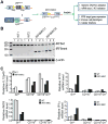
Figure 1
Figure 1. Creation of mice with IRF8 deficiency only in myeloid cells
A. Diagram of creation and analysis of mice with IRF8 deficiency only in the myeloid cells (IRF8 MKO mice). Mice with loxp-flanked Irf8 gene [B6(Cg)-Irf8tm1.1Hm/J] (irf8loxp+/+, top left) were crossed to the Lyz2-cre mice [B6.129P2-Lyz2tm1(cre)Ifo/J](Lyz2cre+/+, left bottom). irf8loxp+/+ mice: exon 2 (E2) was flanked with loxp. E1 and E3 indicate exon 1 and exon 3 of the irf8 gene. Lyz2cre+/+ mice: cre coding sequence was inserted in Lyz2 gene exon 1. E2: Lyz2 exon 2. Lyz2 P: Lyz2 promoter. B. Myeloid cells of IRF8 MKO mice express mutant IRF8 mRNA. Gr1+ (lane 1), CD11b+ (lane 2) and CD11b+Gr1+ (lane 3) cells were sorted from WT and IRF8 MKO mice, stimulated with IFN-γ (100 U/ml) and LPS (1 μg/ml) overnight and analyzed by RT-PCR for IRF8 mRNA levels. The WT IRF8 (IRF8WT) and truncated mutant IRF8 mRNA (IRF8mmt) are indicated at the right. C. IRF8 is functionally deficient in myeloid cells of IRF8 MKO mice. Cells as shown in B were analyzed for Il12b (IL12p40), Nos2 (iNOS), Cxcr3 (IP10), and Ccl3 (MIP1a) transcripts by real-time RT-PCR using β-actin as internal controls.
Figure 2
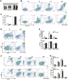
Figure 2. Mice with IRF8 deficiency only in myeloid cells exhibit normal myelopoiesis
A. Spleen morphology of three month old WT, IRF8 MKO and IRF8 KO mice. B. Percentages of CD11b+Gr1+ MDSCs in thymus (Thy), spleen (SP), lymph node (LN) and bone marrow (BM) of WT and IRF8 MKO mice. Shown are representative results from one mouse of three mice. C. Quantitation of CD11b+Gr1+ in SP and BM of WT (n=3) and IRF8 MKO (n=3) mice as shown in B. D. Subsets of Gr1+ (Ly6C+ and Ly6G+) myeloid cells SP and BM. Shown are representative results from one mouse of three mice. Column: mean, bar: SD. E. Quantification of subsets of Gr1+ myeloid cells in SP and BM of WT (n=3) and IRF8MKO (n=3) mice. F. The indicated tissues were collected from WT and IRF8 MKO mice, stained with the CD4- and CD8-specific mAbs. Shown are representative images CD4+ and CD8+ T cell profiles. G. Quantification of CD4+ and CD8+ T cells in the indicated tissues from WT (n=3) and IRF8 MKO (n=3) mice. Column: mean, bar: SD.
Figure 3
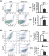
Figure 3. MDSCs from IRF8 MKO mice exhibit decrease spontaneous apoptosis
A. MDSCs from IRF8 KO mice exhibit decreased spontaneous apoptosis. Spleen and bone marrow were collected from three month-old WT (n=3) and IRF8 KO (n=3) mice. Cells were stained immediately under cold conditions with CD11b- and Gr1-specific mAbs plus Annexin V and DAPI. CD11b+Gr1+ cells were gated for Annexin V+DAPI+ cells. Shown are representative results of one of 3 pairs of mice. Apoptosis was quantified as % CD11b+Gr1+ cells that are also Annexin V+DAPI+ cells and presented at the right. Column: mean; bar: SD. ** p<0.01. B. MDSCs from IRF8 MKO mice also exhibit decreased spontaneous apoptosis. Spleen and BM cells were collected from WT (n=5) and IRF8 MKO (n=5) mice. Spontaneous apoptosis were determined as in A.
Figure 4
Figure 4. IRF8 deficiency in T cells results in accumulation of MDSCs
A. Spleen morphology of three month-old WT and IRF8 TKO mice. B. Percentages of CD11b+Gr1+ MDSCs in thymus (Thy), spleen (SP), lymph node (LN), bone marrow (BM) and blood of WT and IRF8 TKO mice. Shown are representative results from one mouse of three mice. C. Quantitation of CD11b+Gr1+ MDSCs in the indicated tissues of WT (n=3) and IRF8 TKO (n=3) mice. Column: mean, bar: SD. ** p<0.01. D. Subsets of Gr1+ (Ly6C+ and Ly6G+) myeloid cells in SP and BM. Shown are representative results from one mouse of three mice. E. Quantification of subsets of Gr1+ myeloid cells in SP and BM of WT (n=3) and IRF8 TKO (n=3) mice as shown in D. Column: mean, bar: SD. **p<0.01.
Figure 5
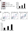
Figure 5. IRF8-deficient T cells induce MDSC accumulation
A. CD4+ T cells were purified from WT and IRF8 TKO mice and adoptively transferred to Rag1 KO mice. Mice were analyzed 14 days after cell transfer. Spleen morphology (left panel), and total spleen cells (right panel) are shown. B. MDSC profiles in the Rag1 KO mice, Rag1 KO mice that have received WT T cells, and Rag1 KO mice that have received IRF8 KO T cells as shown in A. Spleen cells were stained with H2Kb-, CD11b- and Gr1-specific mAbs. H-2Kb+ cells were gated and analyzed for CD11b+Gr1+ cells. Shown are representative results of one of three mice (top panel). MDSCs in each type of mice were quantified and presented at the bottom panel. Column: mean, bar: SD. ** p<0.01.
Figure 6
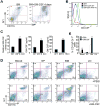
Figure 6. IRF8 represses GM-CSF expression in T cells to suppress MDSC accumulation
A. GM-CSF induces BM cell differentiation to CD11b+Gr1+ MDSCs. BM cells were prepared (left panel) and cultured in vitro in the presence of GM-CSF for 4 days (right panel) and analyzed for CD11b+ and Gr1+ cells. B. Purified CD4+ T cells from WT and IRF8 KO mice were stimulated with anti-CD3 and anti-CD28 mAbs for 3 days. Cells were stained intracellularly with GM-CSF-specific mAb and analyzed by flow cytometry. C. Left panel: CD4+ T cells were purified from spleens of WT and IRF8 KO mice and stimulated with anti-CD3 and anti-CD28 mAbs for 3 days. RNAs were prepared and analyzed by real-time RT-PCR for GM-CSF mRNA level using β-actin as internal control. Right panels: CD4+ T cells were purified from spleens of WT, IRF8 KO and IRF8 TKO mice and stimulated with anti-CD3 and anti-CD28 mAbs for 3 days. The culture supernatants were analyzed by ELISA for GM-CSF protein level. Column: mean, bar: SD. ** p<0.01. D. WT mice were injected intraperitoneally with recombinant GM-CSF protein (1 μg in 100 μl PBS per mouse) every two days for three times. Myeloid cells in the indicated tissues were analyzed by flow cytometry. Shown are representative results from one mouse of three mice. E. Quantification of CD11b+Gr1+ cells as shown in D. Column: mean, bar: SD. **p<0.01.
Figure 7
Figure 7. T cell produced GM-CSF induces MDSC differentiation
A. BALB/c mice (H-2d) were irradiated with 9 Gy from a 137Cs source, and were injected i.v. with 2×10⁶ T cell-depleted BM cells alone, or combined with 0.3×10⁶ CD4+ T cells isolated from C57BL/6 (H-2kb), C57BL/6-IRF8 KO, C57BL/6-GM-CSF KO mice. Mice were analyzed 14 days after cell transfer. Spleens of the indicated chimera mice are shown. Spleen cell numbers are presented at the right panel. B. The H-2kb+CD11b+ and H-2kb+Gr1+ myeloid cells in the indicated chimera mice were analyzed by flow cytometry in spleens 11 days after donor cell transfer. Shown are representative results from one mouse of three mice. C. Quantification of CD11b+ and Gr1+ cells in the indicated mice as shown in B. Column: mean, bar: SD. ** p<0.01.
Figure 8
Figure 8. Restoration of IRF8 expression decreases GM-CSF+ T cells
Naive CD4+ T cells from C57BL/6 WT and IRF8-/- mice were transduced with retrovirus encoding IRF8 or empty vector and the cells were activated with anti-CD3 and anti-CD28 antibodies for 3 days. The cells were re-stimulated with PMA/ionomycin for 5h and stained for intracellular GM-CSF and analyzed by flow cytometry. Representative FACS dot plots gated on CD4+ T cells and the percentage of GM-CSF-producing CD4+ T cells are shown. B. Quantification of CD4+GM-CSF+ cells in three independent experiments as shown in A. Column: mean, Bar: SD. Overexpression of IRF8 significantly decreased % GM-CSF+ cells in both WT and IRF8 KO CD4+ T cells (** p<0.01). C. Total RNA was prepared from cells as shown in A and analyzed for GM-CSF mRNA levels by real-time RT-PCR using β-actin as internal control. Overexpression of IRF8 significantly decreased GM-CSF mRNA levels in both WT and IRF8 KO CD4+ T cells (** p<0.01).
Figure 9
Figure 9. GM-CSF is not down-regulated in MDSCs from IRF8 KO mice
CD11b+Gr1+ MDSCs were purified from spleens of WT (n=4) and IRF8 KO (n=3) mice. Total RNA was isolated from these purified MDSCs and analyzed for IRF8 expression levels by real-time RT-PCR using β-actin as the internal control. The IRF8 expression level in MDSCs from the first WT mice was arbitrarily set at 1.0. The expression levels of IRF8 in MDSCs of the rest samples were the levels relative to the IRF8 expression level in MDSCs of first WT mice.
Figure 10
Figure 10. Model of IRF8 deficiency and MDSC accumulation
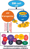
Under physiological conditions, IRF8 represses GM-CSF expression in T cells. Loss of IRF8 expression and function leads to elevated GM-CSF expression and secretion. T cell-produced GM-CSF binds to its receptor on myeloid progenitor cells to induce MDSC differentiation. On the other hand, loss of IRF8 expression or function in myeloid cells in IRF8 null mice leads to decreased spontaneous apoptosis of myeloid cells to reduce MDSC turnover. Thus, loss of IRF8 expression and function in T cells results in increased GM-CSF expression and resultant increased MDSC differentiation, which acts in concert with decreased myeloid cell spontaneous apoptosis and turnover to lead to MDSC accumulation.

Similar articles

Cited by

References

    1. Okabe Y, Medzhitov R. Tissue-specific signals control reversible program of localization and functional polarization of macrophages. Cell. 2014;157:832–844. - PMC - PubMed
    1. Wynn TA, Chawla A, Pollard JW. Macrophage biology in development, homeostasis and disease. Nature. 2013;496:445–455. - PMC - PubMed
    1. Gabrilovich DI, Nagaraj S. Myeloid-derived suppressor cells as regulators of the immune system. Nat Rev Immunol. 2009;9:162–174. - PMC - PubMed
    1. Gabrilovich DI, Ostrand-Rosenberg S, Bronte V. Coordinated regulation of myeloid cells by tumours. Nat Rev Immunol. 2012;12:253–268. - PMC - PubMed
    1. Metcalf D. The molecular control of cell division, differentiation commitment and maturation in haemopoietic cells. Nature. 1989;339:27–30. - PubMed

Publication types

MeSH terms

Substances