

Inhibition of prostate smooth muscle contraction and prostate stromal cell growth by the inhibitors of Rac, NSC23766 and EHT1864
- PMID: 25631101
- PMCID: PMC4439884
- DOI: 10.1111/bph.13099
Inhibition of prostate smooth muscle contraction and prostate stromal cell growth by the inhibitors of Rac, NSC23766 and EHT1864
Abstract
Background and purpose: Medical therapy of lower urinary tract symptoms (LUTS) suggestive of benign prostatic hyperplasia (BPH) targets smooth muscle contraction in the prostate, or prostate growth. However, current therapeutic options are insufficient. Here, we investigated the role of Rac in the control of smooth muscle tone in human prostates and growth of prostate stromal cells.
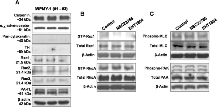
Experimental approach: Experiments were performed using human prostate tissues from radical prostatectomy and cultured stromal cells (WPMY-1). Expression of Rac was examined by Western blot and fluorescence staining. Effects of Rac inhibitors (NSC23766 and EHT1864) on contractility were assessed in the organ bath. The effects of Rac inhibitors were assessed by pull-down, cytotoxicity using a cell counting kit, cytoskeletal organization by phalloidin staining and cell growth using an 5-ethynyl-2'-deoxyuridine assay.
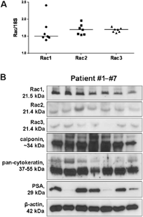
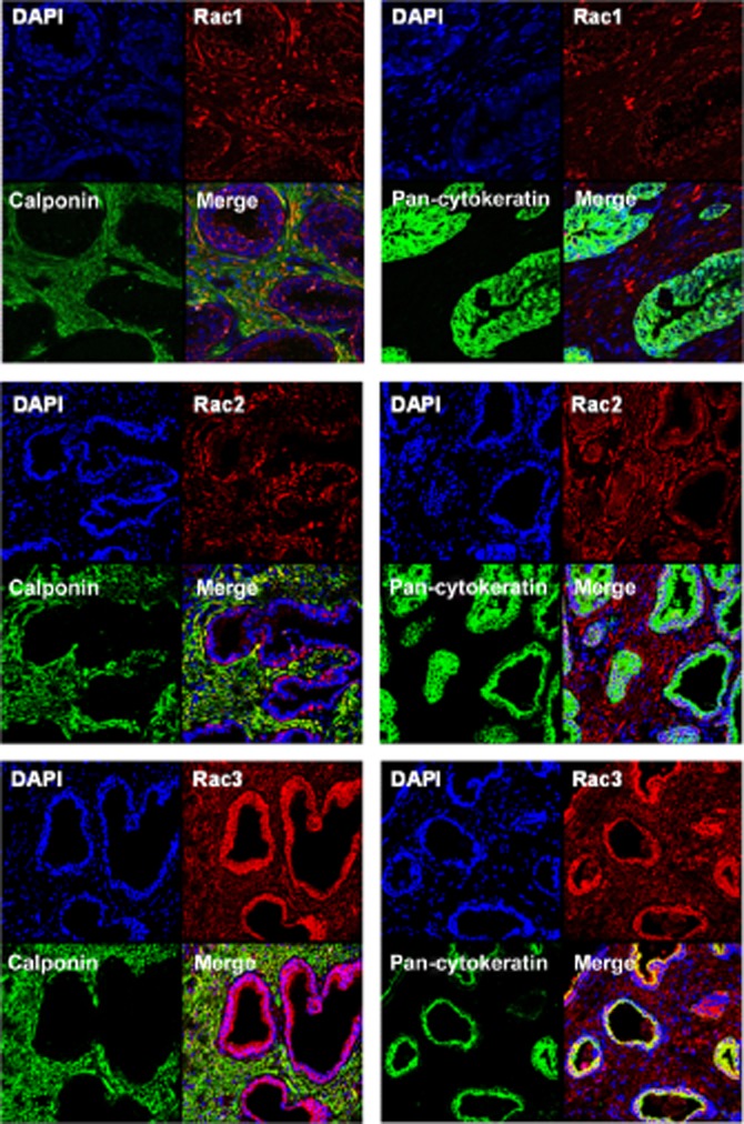
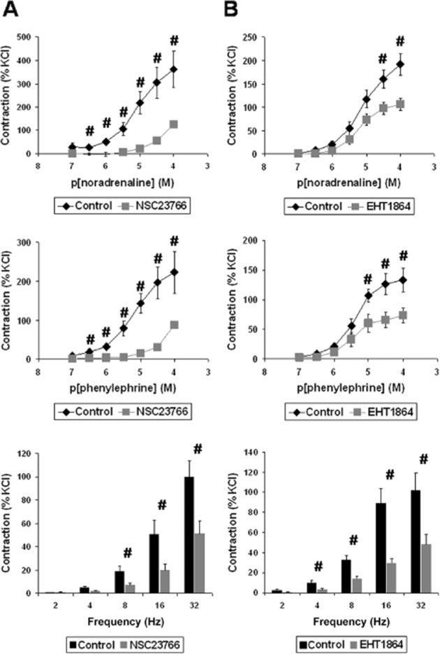
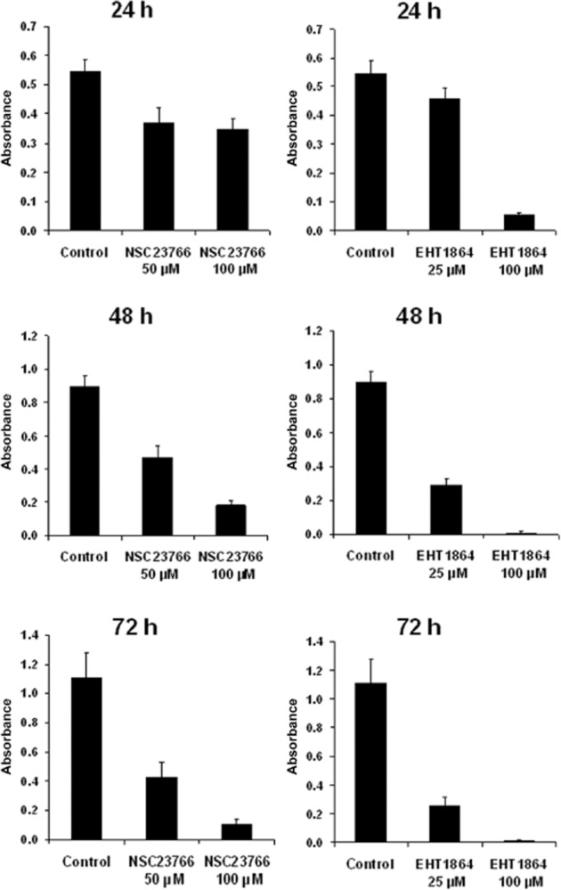
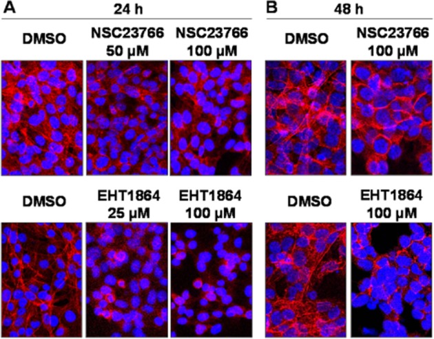
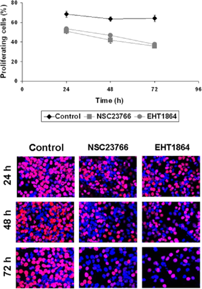
Key results: Expression of Rac1-3 was observed in prostate samples from each patient. Immunoreactivity for Rac1-3 was observed in the stroma, where it colocalized with the smooth muscle marker, calponin. NSC23766 and EHT1864 significantly reduced contractions of prostate strips induced by noradrenaline, phenylephrine or electrical field stimulation. NSC23766 and EHT1864 inhibited Rac activity in WPMY-1 cells. Survival of WPMY-1 cells ranged between 64 and 81% after incubation with NSC23766 (50 or 100 μM) or EHT1864 (25 μM) for 24 h. NSC23766 and EHT1864 induced cytoskeletal disorganization in WPMY-1 cells. Both inhibitors impaired the growth of WPMY-1 cells.
Conclusions and implications: Rac may be a link connecting the control of prostate smooth muscle tone with proliferation of smooth muscle cells. Improvements in LUTS suggestive of BPH by Rac inhibitors appears possible.
© 2015 The British Pharmacological Society.
Figures
Similar articles
-
Rac1 silencing, NSC23766 and EHT1864 reduce growth and actin organization of bladder smooth muscle cells.Life Sci. 2020 Nov 15;261:118468. doi: 10.1016/j.lfs.2020.118468. Epub 2020 Sep 19. Life Sci. 2020. PMID: 32961232
-
New strategies for inhibition of non-adrenergic prostate smooth muscle contraction by pharmacologic intervention.Prostate. 2019 May;79(7):746-756. doi: 10.1002/pros.23780. Epub 2019 Feb 27. Prostate. 2019. PMID: 30811062
-
P21-Activated Kinase Inhibitors FRAX486 and IPA3: Inhibition of Prostate Stromal Cell Growth and Effects on Smooth Muscle Contraction in the Human Prostate.PLoS One. 2016 Apr 12;11(4):e0153312. doi: 10.1371/journal.pone.0153312. eCollection 2016. PLoS One. 2016. PMID: 27071060 Free PMC article.
-
The autonomic and sensory innervation of the smooth muscle of the prostate gland: a review of pharmacological and histological studies.J Auton Pharmacol. 2000 Aug;20(4):193-206. doi: 10.1046/j.1365-2680.2000.00195.x. J Auton Pharmacol. 2000. PMID: 11260358 Review.
-
Development of EHop-016: a small molecule inhibitor of Rac.Enzymes. 2013;33 Pt A(Pt A):117-46. doi: 10.1016/B978-0-12-416749-0.00006-3. Epub 2013 Aug 8. Enzymes. 2013. PMID: 25033803 Free PMC article. Review.
Cited by
-
Inhibition of α1-Adrenergic, Non-Adrenergic and Neurogenic Human Prostate Smooth Muscle Contraction and of Stromal Cell Growth by the Isoflavones Genistein and Daidzein.Nutrients. 2022 Nov 22;14(23):4943. doi: 10.3390/nu14234943. Nutrients. 2022. PMID: 36500973 Free PMC article.
-
Effects of carvedilol on human prostate tissue contractility and stromal cell growth pointing to potential clinical implications.Pharmacol Rep. 2024 Aug;76(4):807-822. doi: 10.1007/s43440-024-00605-5. Epub 2024 Jun 11. Pharmacol Rep. 2024. PMID: 38858312 Free PMC article.
-
Inhibition of growth and contraction in human prostate stromal cells by silencing of NUAK1 and -2, and by the presumed NUAK inhibitors HTH01-015 and WZ4003.Front Pharmacol. 2023 Apr 28;14:1105427. doi: 10.3389/fphar.2023.1105427. eCollection 2023. Front Pharmacol. 2023. PMID: 37188272 Free PMC article.
-
A NAV2729-sensitive mechanism promotes adrenergic smooth muscle contraction and growth of stromal cells in the human prostate.J Biol Chem. 2019 Aug 9;294(32):12231-12249. doi: 10.1074/jbc.RA119.007958. Epub 2019 Jun 26. J Biol Chem. 2019. PMID: 31243101 Free PMC article.
-
Inhibition of Rac family protein impairs colitis and colitis-associated cancer in mice.Am J Cancer Res. 2018 Jan 1;8(1):70-80. eCollection 2018. Am J Cancer Res. 2018. PMID: 29416921 Free PMC article.
References
-
- Akbar H, Cancelas J, Williams DA, Zheng J, Zheng Y. Rational design and applications of a Rac GTPase-specific small molecule inhibitor. Methods Enzymol. 2006;406:554–565. - PubMed
-
- Alcaraz A, Hammerer P, Tubaro A, Schroder FH, Castro R. Is there evidence of a relationship between benign prostatic hyperplasia and prostate cancer? Findings of a literature review. Eur Urol. 2009;55:864–873. - PubMed
-
- Andersson KE, Lepor H, Wyllie MG. Prostatic alpha 1-adrenoceptors and uroselectivity. Prostate. 1997;30:202–215. - PubMed
Publication types
MeSH terms
Substances
LinkOut - more resources
Full Text Sources
Other Literature Sources
Research Materials
Miscellaneous
