. 2015 Apr;89(8):4058-68.
doi: 10.1128/JVI.03574-14. Epub 2015 Jan 28.

The endoplasmic reticulum membrane J protein C18 executes a distinct role in promoting simian virus 40 membrane penetration

Affiliations

The endoplasmic reticulum membrane J protein C18 executes a distinct role in promoting simian virus 40 membrane penetration

Parikshit Bagchi et al. J Virol. 2015 Apr.

Abstract

The nonenveloped simian virus 40 (SV40) hijacks the three endoplasmic reticulum (ER) membrane-bound J proteins B12, B14, and C18 to escape from the ER into the cytosol en route to successful infection. How C18 controls SV40 ER-to-cytosol membrane penetration is the least understood of these processes. We previously found that SV40 triggers B12 and B14 to reorganize into discrete puncta in the ER membrane called foci, structures postulated to represent the cytosol entry site (C. P. Walczak, M. S. Ravindran, T. Inoue, and B. Tsai, PLoS Pathog 10: e1004007, 2014). We now find that SV40 also recruits C18 to the virus-induced B12/B14 foci. Importantly, the C18 foci harbor membrane penetration-competent SV40, further implicating this structure as the membrane penetration site. Consistent with this, a mutant SV40 that cannot penetrate the ER membrane and promote infection fails to induce C18 foci. C18 also regulates the recruitment of B12/B14 into the foci. In contrast to B14, C18's cytosolic Hsc70-binding J domain, but not the lumenal domain, is essential for its targeting to the foci; this J domain likewise is necessary to support SV40 infection. Knockdown-rescue experiments reveal that C18 executes a role that is not redundant with those of B12/B14 during SV40 infection. Collectively, our data illuminate C18's contribution to SV40 ER membrane penetration, strengthening the idea that SV40-triggered foci are critical for cytosol entry.

Importance: Polyomaviruses (PyVs) cause devastating human diseases, particularly in immunocompromised patients. As this virus family continues to be a significant human pathogen, clarifying the molecular basis of their cellular entry pathway remains a high priority. To infect cells, PyV traffics from the cell surface to the ER, where it penetrates the ER membrane to reach the cytosol. In the cytosol, the virus moves to the nucleus to cause infection. ER-to-cytosol membrane penetration is a critical yet mysterious infection step. In this study, we clarify the role of an ER membrane protein called C18 in mobilizing the simian PyV SV40, a PyV archetype, from the ER into the cytosol. Our findings also support the hypothesis that SV40 induces the formation of punctate structures in the ER membrane, called foci, that serve as the portal for cytosol entry of the virus.


Figures

FIG 1
FIG 1
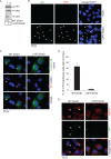
SV40 recruits C18 to the virus-induced B12/B14 foci which also harbor membrane penetration-competent virus. (A) CV-1 cells were transfected with S-tagged C18, and at 12 h after transfection, cells mock infected or infected with SV40 (MOI, 50) for 2 h and 14 h were fixed, stained with anti-S antibody and anti-BAP31 antibodies, and analyzed by immunofluorescence microscopy. (B and C) CV-1 cells were transfected with FLAG-tagged C18. At 12 h after transfection, cells infected with SV40 (MOI, 50) for 14 h were fixed, stained with anti-FLAG and anti-B12 (B) or anti-FLAG and anti-B14 antibodies (C), and analyzed by immunofluorescence microscopy. DAPI, 4′,6-diamidino-2-phenylindole (DAPI). (D and E) CV-1 cells were transfected with FLAG-tagged C18 and infected with SV40 (MOI, 50) for 14 h, fixed, stained with anti-FLAG and anti-VP1 (D) or anti-FLAG and anti-VP2/3 antibodies (E), and analyzed by immunofluorescence microscopy. (F) Quantification of the percentage of FLAG-C18 foci colocalizing with VP1 or VP2/3 foci. Values represent means ± SD from three independent experiments.
FIG 2
FIG 2
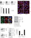
SV40 lacking VP3 does not induce C18 foci. (A) Purified WT SV40 and ΔVP3 SV40 were immunoblotted using the indicated antibodies. (B) CV-1 cells were mock infected or infected with ΔVP3 SV40 for 8 h, fixed, stained with anti-VP1 or VP2/3 antibodies, and analyzed by immunofluorescence microscopy. (C) FLAG-C18-transfected CV-1 cells were infected with the same amount (20 μg) of either purified WT or ΔVP3 SV40 for 8 h, fixed, stained with anti-FLAG and anti-VP1 antibodies, and analyzed by immunofluorescence microscopy. (D) Quantification data showed the percentage of FLAG-C18-expressing cells with C18 foci after WT SV40 or ΔVP3 SV40 infection. Cells were scored positive if at least one focus was present in the cell. Values represent means ± SD from three independent experiments. (E) CV-1 cells were infected with the same amount (20 μg) of either purified WT or ΔVP3 SV40 for 8 h, fixed, stained with anti-BAP31 and anti-VP1 antibodies, and analyzed by immunofluorescence microscopy.
FIG 3
FIG 3
C18 regulates B12, B14, and BAP31 focus formation. (A) RT-PCR results showing the knockdown efficiency of C18 (lanes 1 and 2)-, B12 (lanes 3 and 4)-, and B14 (lanes 5 and 6)-specific siRNAs. The expression of GAPDH mRNA was used as a loading control. (B) CV-1 cells were reverse transfected with scrambled or C18 siRNA. After 48 h of transfection, cells were infected with SV40 (MOI, 15) for 14 h, fixed, stained with the indicated antibodies, and analyzed by immunofluorescence microscopy. (C) Quantification data from panel B, where cells were scored positive if at least one focus was present in the cell. Values represent means ± SD from three independent experiments. (D) S/His-tagged Sel1L-transfected CV-1 cells were infected with SV40 (MOI, 50), fixed at 14 hpi, stained with anti-S and anti-BAP31 antibodies, and analyzed by immunofluorescence microscopy. (E) BAP31 interaction with C18, B14, and B12 were analyzed by using lysates derived from HEK 293T cells transfected with the indicated construct. The S-tagged proteins were affinity purified (AP) using S-agarose beads, and the precipitated samples were immunoblotted with the indicated antibodies. (F, G, and H) Interaction between C18-S/B14-S with endogenous B12 (F), C18-S/B12-S with endogenous B14 (G), and C18-S with endogenous Hsc70 and SGTA (H) were assessed by affinity purification using HEK 293T lysates harboring the indicated construct using S-agarose beads, followed by immunoblotting the precipitated material with the indicated antibodies. (I) S-tagged C18-expressing vector can rescue the C18 knockdown effect. CV-1 cells were reverse transfected with either scrambled or C18 siRNA (50 nM) for 24 h. Cells then were transfected with the indicated construct for 24 h, infected with SV40 (MOI, 0.5) for 20 h, fixed, and stained using anti-large T antigen, anti-FLAG, or anti-S antibodies. The percentages of large T antigen-positive cells were determined in cells expressing either GFP-FLAG or C18-S* by immunofluorescence microscopy. C18-S* is a construct designed to be resistant to the C18 siRNA. Values represent means ± SD from three independent experiments.
FIG 4
FIG 4
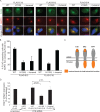
C18, B12, and B14 display various domain requirements for recruitment into foci. (A) FLAG-tagged WT or the indicated mutant forms of C18, B12, or B14 transfected in CV-1 cells were infected with SV40 (MOI, 50) for 14 h. Cells then were fixed, stained with the indicated antibodies, and analyzed by immunofluorescence microscopy. (B) Quantification of data from panel A showing the focus-forming ability of WT and mutant C18 and B12. Values represent means ± SD from three independent experiments. (C) Schematic diagram depicting functional domains (orange) of C18, B12, and B14 essential for SV40-induced focus formation. (D) CV-1 cells were reverse transfected with scrambled or C18 siRNA for 24 h prior to transfection with the indicated constructs for 24 h. Cells then were infected with SV40 (MOI, 0.5) for 20 h, fixed, and stained with anti-FLAG and anti-large T antigen antibodies. The percentages of large T antigen-positive cells were determined in GFP-expressing or WT or mutant C18-expressing cells by using immunofluorescence microscopy. Values represent means ± SD from three independent experiments.
FIG 5
FIG 5
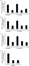
C18, B12, and B14 play nonredundant roles during SV40 infection. (A, B, and C) CV-1 cells were reverse transfected with scrambled or the indicated siRNAs for 24 h prior to transfection with the indicated FLAG-tagged constructs for 24 h. Cells then were infected with SV40 (MOI, 0.5) for 20 h, fixed, and stained with anti-FLAG and anti-large T antigen antibodies. The percentages of T antigen-positive cells were determined in GFP-, C18-, B12-, or B14-expressing cells by using immunofluorescence microscopy. Values represent means ± SD from three independent experiments. (D) CV-1 cells were transfected with scrambled or the indicated siRNAs for 48 h prior to infection with SV40 (MOI, 0.5) for 20 h. Cells were fixed and stained with anti-large T antigen antibodies, and the percentages of large T antigen-positive cells were scored by using immunofluorescence microscopy. Values represent means ± SD from three independent experiments.
FIG 6
FIG 6
Working model describing C18's role during SV40 infection. (A) At steady state, C18 binds to BAP31, which also interacts with the B12-B14 complex. (B) During ER membrane penetration of SV40, the VP2/3-exposed viral particle promotes C18, along with the B12-B14 complex and BAP31, to rearrange into foci in the bilayer of the ER membrane. Formation of foci requires both VP2 and VP3. While the precise mechanism by which C18 controls the recruitment of the B12-B14 complex and BAP31 to the foci remains unclear, it may be due to interaction of C18 with BAP31, which also binds to the B12-B14 complex. Regardless, focus formation likely serves to concentrate cytosolic Hsc70 machinery used to eject the virus into the cytosol, as suggested by our earlier study (31). Functional domains of C18, B12, and B14 essential for SV40-induced focus formation are colored in orange.

Similar articles

Cited by

References

    1. DeCaprio JA, Garcea RL. 2013. A cornucopia of human polyomaviruses. Nat Rev Microbiol 11:264–276. doi:10.1038/nrmicro2992. - DOI - PMC - PubMed
    1. Jiang M, Abend JR, Johnson SF, Imperiale MJ. 2009. The role of polyomaviruses in human disease. Virology 384:266–273. doi:10.1016/j.virol.2008.09.027. - DOI - PMC - PubMed
    1. Chen XS, Stehle T, Harrison SC. 1998. Interaction of polyomavirus internal protein VP2 with the major capsid protein VP1 and implications for participation of VP2 in viral entry. EMBO J 17:3233–3240. doi:10.1093/emboj/17.12.3233. - DOI - PMC - PubMed
    1. Stehle T, Gamblin SJ, Yan Y, Harrison SC. 1996. The structure of simian virus 40 refined at 3.1 A resolution. Structure 4:165–182. doi:10.1016/S0969-2126(96)00020-2. - DOI - PubMed
    1. Liddington RC, Yan Y, Moulai J, Sahli R, Benjamin TL, Harrison SC. 1991. Structure of simian virus 40 at 3.8-A resolution. Nature 354:278–284. doi:10.1038/354278a0. - DOI - PubMed

Publication types

MeSH terms