

Localization and local translation of Arc/Arg3.1 mRNA at synapses: some observations and paradoxes
- PMID: 25628532
- PMCID: PMC4290588
- DOI: 10.3389/fnmol.2014.00101
Localization and local translation of Arc/Arg3.1 mRNA at synapses: some observations and paradoxes
Abstract
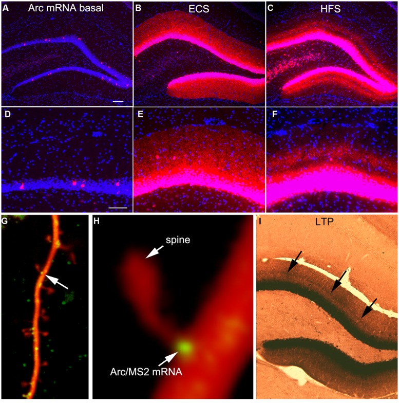
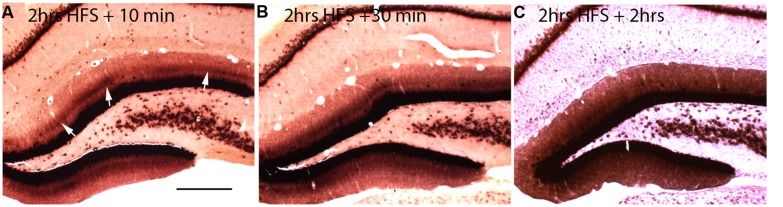
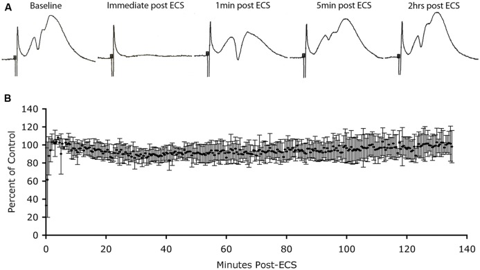
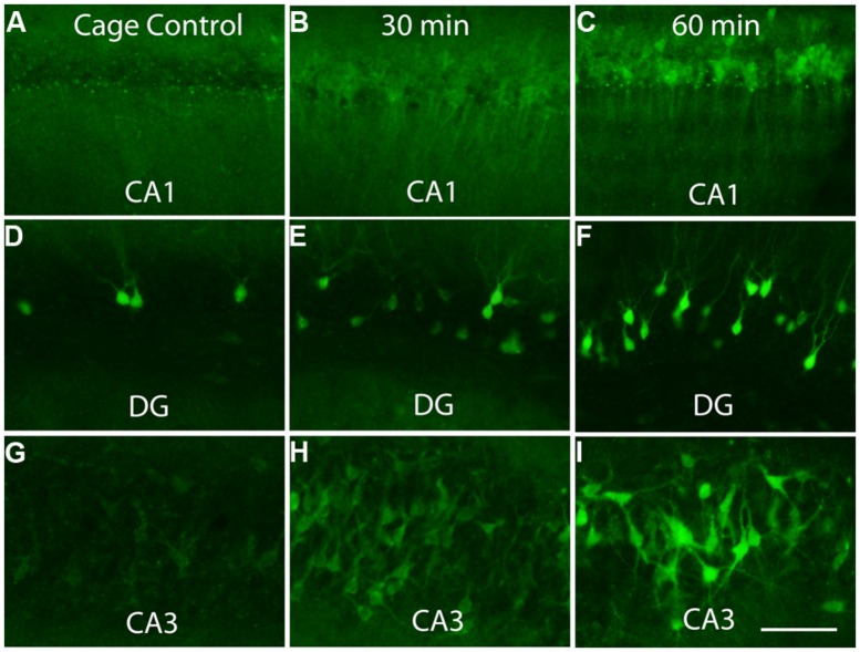
Arc is a unique immediate early gene whose expression is induced as synapses are being modified during learning. The uniqueness comes from the fact that newly synthesized Arc mRNA is rapidly transported throughout dendrites where it localizes near synapses that were recently activated. Here, we summarize aspects of Arc mRNA translation in dendrites in vivo, focusing especially on features of its expression that are paradoxical or that donot fit in with current models of how Arc protein operates. Findings from in vivo studies that donot quite fit include: (1) Following induction of LTP in vivo, Arc mRNA and protein localize near active synapses, but are also distributed throughout dendrites. In contrast, Arc mRNA localizes selectively near active synapses when stimulation is continued as Arc mRNA is transported into dendrites; (2) Strong induction of Arc expression as a result of a seizure does not lead to a rundown of synaptic efficacy in vivo as would be predicted by the hypothesis that high levels of Arc cause glutamate receptor endocytosis and LTD. (3) Arc protein is synthesized in the perinuclear cytoplasm rapidly after transcriptional activation, indicating that at least a pool of Arc mRNA is not translationally repressed to allow for dendritic delivery; (4) Increases in Arc mRNA in dendrites are not paralleled by increases in levels of exon junction complex (EJC) proteins. These results of studies of mRNA trafficking in neurons in vivo provide a new perspective on the possible roles of Arc in activity-dependent synaptic modifications.
Keywords: Arc/Arg3.1; LTP; dendrite; dendritic mRNA; dendritic spines; immediate early genes; protein synthesis; synaptic plasticity.
Figures

Similar articles
-
Delayed Degradation and Impaired Dendritic Delivery of Intron-Lacking EGFP-Arc/Arg3.1 mRNA in EGFP-Arc Transgenic Mice.Front Mol Neurosci. 2018 Jan 31;10:435. doi: 10.3389/fnmol.2017.00435. eCollection 2017. Front Mol Neurosci. 2018. PMID: 29445324 Free PMC article.
-
Actin polymerization and ERK phosphorylation are required for Arc/Arg3.1 mRNA targeting to activated synaptic sites on dendrites.J Neurosci. 2007 Aug 22;27(34):9054-67. doi: 10.1523/JNEUROSCI.2410-07.2007. J Neurosci. 2007. PMID: 17715342 Free PMC article.
-
Selective localization of arc mRNA in dendrites involves activity- and translation-dependent mRNA degradation.J Neurosci. 2014 Mar 26;34(13):4481-93. doi: 10.1523/JNEUROSCI.4944-13.2014. J Neurosci. 2014. PMID: 24671994 Free PMC article.
-
A cellular mechanism for targeting newly synthesized mRNAs to synaptic sites on dendrites.Proc Natl Acad Sci U S A. 2001 Jun 19;98(13):7062-8. doi: 10.1073/pnas.131146398. Proc Natl Acad Sci U S A. 2001. PMID: 11416188 Free PMC article. Review.
-
Control of synaptic consolidation in the dentate gyrus: mechanisms, functions, and therapeutic implications.Prog Brain Res. 2007;163:453-71. doi: 10.1016/S0079-6123(07)63025-8. Prog Brain Res. 2007. PMID: 17765733 Review.
Cited by
-
Translational Control in the Brain in Health and Disease.Cold Spring Harb Perspect Biol. 2019 Aug 1;11(8):a032912. doi: 10.1101/cshperspect.a032912. Cold Spring Harb Perspect Biol. 2019. PMID: 30082469 Free PMC article. Review.
-
Delayed Degradation and Impaired Dendritic Delivery of Intron-Lacking EGFP-Arc/Arg3.1 mRNA in EGFP-Arc Transgenic Mice.Front Mol Neurosci. 2018 Jan 31;10:435. doi: 10.3389/fnmol.2017.00435. eCollection 2017. Front Mol Neurosci. 2018. PMID: 29445324 Free PMC article.
-
Dendritic trafficking faces physiologically critical speed-precision tradeoffs.Elife. 2016 Dec 30;5:e20556. doi: 10.7554/eLife.20556. Elife. 2016. PMID: 28034367 Free PMC article.
-
Differential Expression of Endogenous Retroviruses and Inflammatory Mediators in Female and Male Offspring in a Mouse Model of Maternal Immune Activation.Int J Mol Sci. 2022 Nov 11;23(22):13930. doi: 10.3390/ijms232213930. Int J Mol Sci. 2022. PMID: 36430402 Free PMC article.
-
Bridging Synaptic and Epigenetic Maintenance Mechanisms of the Engram.Front Mol Neurosci. 2018 Oct 5;11:369. doi: 10.3389/fnmol.2018.00369. eCollection 2018. Front Mol Neurosci. 2018. PMID: 30344478 Free PMC article.
References
Grants and funding
LinkOut - more resources
Full Text Sources
Other Literature Sources