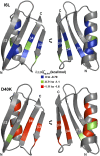
Quinary structure modulates protein stability in cells
- PMID: 25624496
- PMCID: PMC4330749
- DOI: 10.1073/pnas.1417415112
Quinary structure modulates protein stability in cells
Abstract
Protein quinary interactions organize the cellular interior and its metabolism. Although the interactions stabilizing secondary, tertiary, and quaternary protein structure are well defined, details about the protein-matrix contacts that comprise quinary structure remain elusive. This gap exists because proteins function in the crowded cellular environment, but are traditionally studied in simple buffered solutions. We use NMR-detected H/D exchange to quantify quinary interactions between the B1 domain of protein G and the cytosol of Escherichia coli. We demonstrate that a surface mutation in this protein is 10-fold more destabilizing in cells than in buffer, a surprising result that firmly establishes the significance of quinary interactions. Remarkably, the energy involved in these interactions can be as large as the energies that stabilize specific protein complexes. These results will drive the critical task of implementing quinary structure into models for understanding the proteome.
Keywords: H/D exchange; protein NMR; protein thermodynamics; quinary interactions.
Conflict of interest statement
The authors declare no conflict of interest.
Figures


Similar articles
-
Electrostatic Contributions to Protein Quinary Structure.J Am Chem Soc. 2016 Oct 12;138(40):13139-13142. doi: 10.1021/jacs.6b07323. Epub 2016 Oct 4. J Am Chem Soc. 2016. PMID: 27676610
-
Intracellular pH modulates quinary structure.Protein Sci. 2015 Nov;24(11):1748-55. doi: 10.1002/pro.2765. Epub 2015 Aug 30. Protein Sci. 2015. PMID: 26257390 Free PMC article.
-
Intact ribosomes drive the formation of protein quinary structure.PLoS One. 2020 Apr 24;15(4):e0232015. doi: 10.1371/journal.pone.0232015. eCollection 2020. PLoS One. 2020. PMID: 32330166 Free PMC article.
-
Quinary protein structure and the consequences of crowding in living cells: leaving the test-tube behind.Bioessays. 2013 Nov;35(11):984-93. doi: 10.1002/bies.201300080. Epub 2013 Aug 14. Bioessays. 2013. PMID: 23943406 Review.
-
NMR studies of protein folding and binding in cells and cell-like environments.Curr Opin Struct Biol. 2015 Feb;30:7-16. doi: 10.1016/j.sbi.2014.10.004. Epub 2014 Dec 3. Curr Opin Struct Biol. 2015. PMID: 25479354 Review.
Cited by
-
General method to stabilize mesophilic proteins in hyperthermal water.iScience. 2021 May 2;24(5):102503. doi: 10.1016/j.isci.2021.102503. eCollection 2021 May 21. iScience. 2021. PMID: 34113834 Free PMC article.
-
Extant fold-switching proteins are widespread.Proc Natl Acad Sci U S A. 2018 Jun 5;115(23):5968-5973. doi: 10.1073/pnas.1800168115. Epub 2018 May 21. Proc Natl Acad Sci U S A. 2018. PMID: 29784778 Free PMC article.
-
Cosolute and Crowding Effects on a Side-By-Side Protein Dimer.Biochemistry. 2017 Feb 21;56(7):971-976. doi: 10.1021/acs.biochem.6b01251. Epub 2017 Feb 9. Biochemistry. 2017. PMID: 28102665 Free PMC article.
-
Radio Signals from Live Cells: The Coming of Age of In-Cell Solution NMR.Chem Rev. 2022 May 25;122(10):9267-9306. doi: 10.1021/acs.chemrev.1c00790. Epub 2022 Jan 21. Chem Rev. 2022. PMID: 35061391 Free PMC article. Review.
-
Amyloid Oligomers: A Joint Experimental/Computational Perspective on Alzheimer's Disease, Parkinson's Disease, Type II Diabetes, and Amyotrophic Lateral Sclerosis.Chem Rev. 2021 Feb 24;121(4):2545-2647. doi: 10.1021/acs.chemrev.0c01122. Epub 2021 Feb 5. Chem Rev. 2021. PMID: 33543942 Free PMC article. Review.
References
-
- Zimmerman SB, Trach SO. Estimation of macromolecule concentrations and excluded volume effects for the cytoplasm of Escherichia coli. J Mol Biol. 1991;222(3):599–620. - PubMed
-
- Ebbinghaus S, Dhar A, McDonald JD, Gruebele M. Protein folding stability and dynamics imaged in a living cell. Nat Methods. 2010;7(4):319–323. - PubMed
Publication types
MeSH terms
Substances
LinkOut - more resources
Full Text Sources
Other Literature Sources
Miscellaneous

