. 2015 Apr;35(7):1281-98.
doi: 10.1128/MCB.01156-14. Epub 2015 Jan 26.

Estrogen-related receptor α (ERRα) and ERRγ are essential coordinators of cardiac metabolism and function

Affiliations

Estrogen-related receptor α (ERRα) and ERRγ are essential coordinators of cardiac metabolism and function

Ting Wang et al. Mol Cell Biol. 2015 Apr.

Abstract

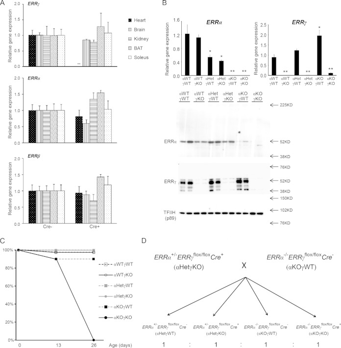
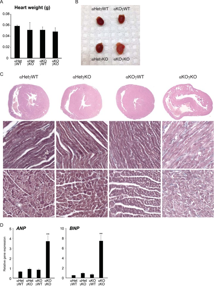
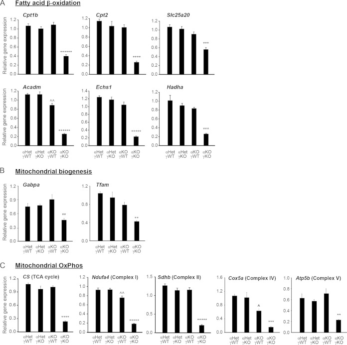
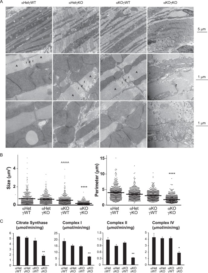
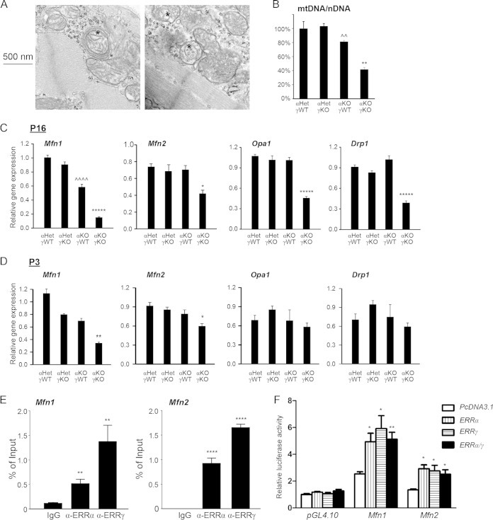
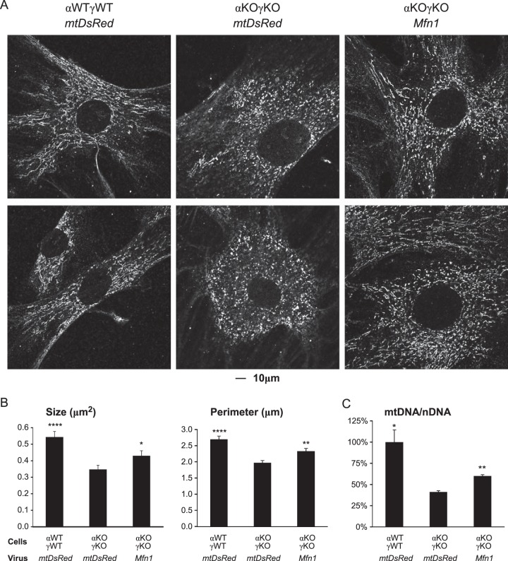
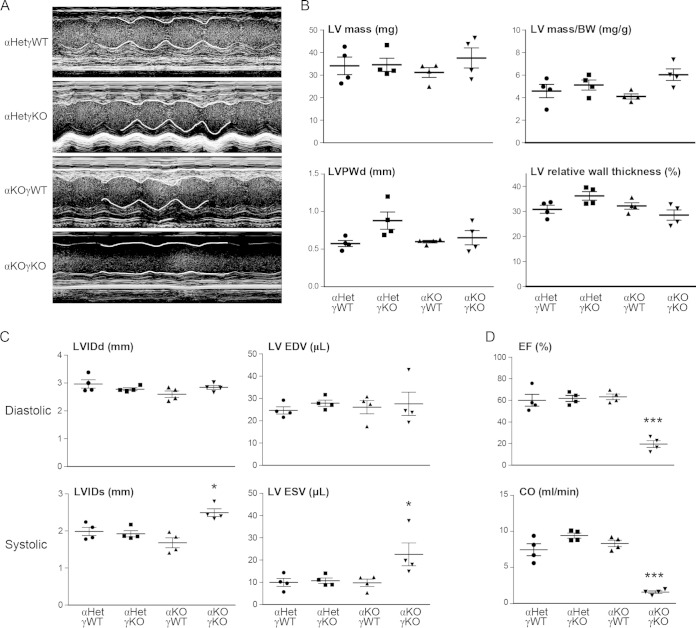
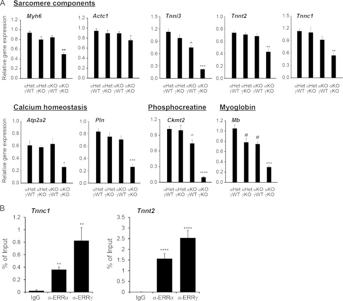
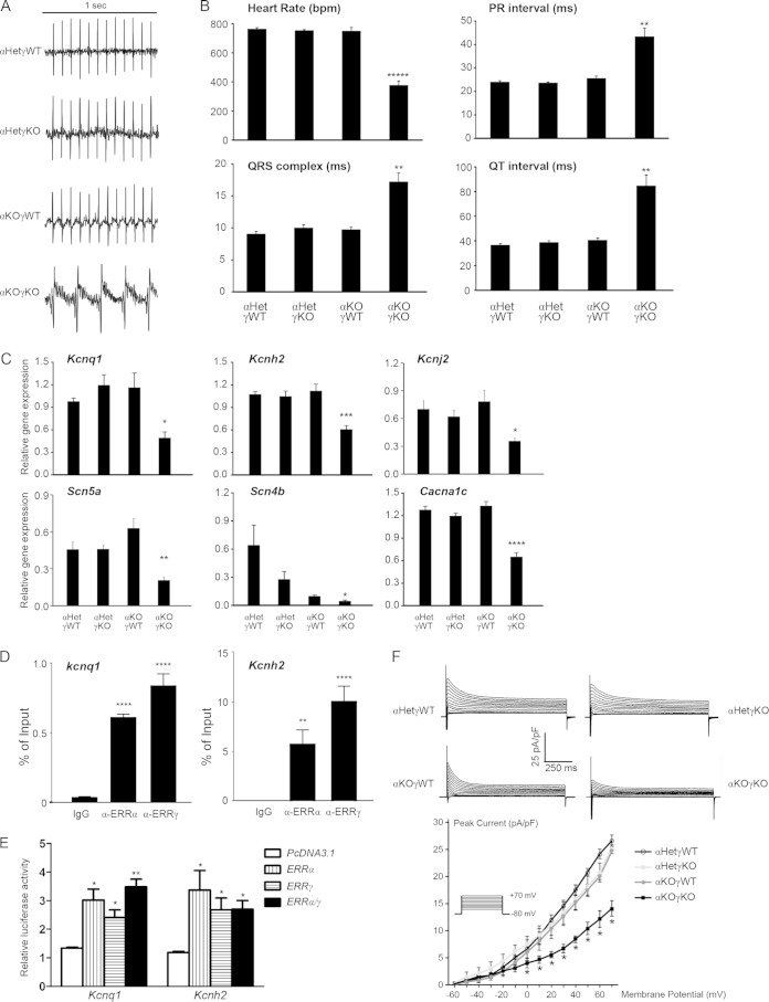
Almost all cellular functions are powered by a continuous energy supply derived from cellular metabolism. However, it is little understood how cellular energy production is coordinated with diverse energy-consuming cellular functions. Here, using the cardiac muscle system, we demonstrate that nuclear receptors estrogen-related receptor α (ERRα) and ERRγ are essential transcriptional coordinators of cardiac energy production and consumption. On the one hand, ERRα and ERRγ together are vital for intact cardiomyocyte metabolism by directly controlling expression of genes important for mitochondrial functions and dynamics. On the other hand, ERRα and ERRγ influence major cardiomyocyte energy consumption functions through direct transcriptional regulation of key contraction, calcium homeostasis, and conduction genes. Mice lacking both ERRα and cardiac ERRγ develop severe bradycardia, lethal cardiomyopathy, and heart failure featuring metabolic, contractile, and conduction dysfunctions. These results illustrate that the ERR transcriptional pathway is essential to couple cellular energy metabolism with energy consumption processes in order to maintain normal cardiac function.


Figures

FIG 1
FIG 1
Mice lacking cardiac ERRα and ERRγ die postnatally. (A) Myh6-Cre-mediated cardiac tissue-specific loss of ERRγ. ERRγ, ERRα, and ERRβ RNAs in different tissues from 2-month-old control (Cre) and cardiac ERRγ KO (Cre+) mice (n = 4 or 5) were determined by qRT-PCR. **, P < 0.01, between Cre and Cre+ mice. (B) ERRα and ERRγ RNA (top; n = 4 to 8) and nuclear protein (bottom; n = 2) levels in 3-day-old mouse hearts was determined by qRT-PCR and Western blotting, respectively. *, P < 0.05; **, P < 0.01, between indicated genotype and αWTγWT mice. (C) Survival rate of pups at 0, 13, and 26 days of age (n = 10 to 20). (D) Breeding strategy to generate experimental cohorts. All values are means plus standard errors of the means.
FIG 2
FIG 2
Mice lacking cardiac ERRα and ERRγ die postnatally with cardiomyopathy. (A) Heart weight of 16-day-old mice (n = 7 to 11). (B) Representative picture of hearts of 16-day-old mice. (C) Representative pictures of H&E-stained heart sections of 16-day-old mice, as follows: cross-section of the heart (top row), longitudinal view of the muscle fibers (middle row), and transverse view of the muscle fibers (bottom row). (D) Expression of ANP and BNP in 16-day-old mouse hearts (n = 6 to 8). **, P < 0.01, between αKOγKO mice and the other three genotypes. All values are means plus standard errors of the means.
FIG 3
FIG 3
ERRα and ERRγ are essential for expression of genes important in cardiomyocyte metabolism, especially mitochondrial functions. Expression levels of genes important in mitochondrial fatty acid oxidation (A), biogenesis (B), and OxPhos (C) were determined in 16-day-old mouse hearts (n = 6 to 8). **, P < 0.01; ***, P < 0.001; ****, P < 0.0001; *****, P < 0.00001; ******, P < 0.000001, between αKOγKO mice and the other three genotypes; ^, P < 0.05; ^, P < 0.01, between αKOγWT and αHetγWT/αHetγKO. All values are means plus standard errors of the means.
FIG 4
FIG 4
ERRα and ERRγ are essential for normal mitochondrial functions. (A) Representative EM pictures of hearts of 16-day-old mice, as follows: magnification of ×10,000 to show the overall cardiac myofibril structure including the sarcomeres and the mitochondria (top row); magnification of ×60,000 to show the mitochondrial ultrastructure and the sarcomere Z line (Z), I band (I), and A band (A) (middle row); magnification of ×60,000 to focus on the mitochondrial ultrastructure (bottom row). (B) Mitochondrial size (two-dimensional area) and perimeter were quantified from five EM fields (magnification of ×10,000 with at least 100 mitochondria per field) using ImageJ. (C) Activity of TCA cycle enzyme citrate synthase and different mitochondrial ETC complexes in 16-day-old mouse hearts was measured by enzymatic assays (n = 3). *, P < 0.05; **, P < 0.01; ****, P < 0.0001, between αKOγKO and the other three genotypes; ^^^^, P < 0.0001, between αKOγWT and αHetγWT/αHetγKO mice. All values are means plus standard errors of the means.
FIG 5
FIG 5
ERRα and ERRγ are important for integral mitochondrial dynamics. (A) Representative EM pictures of 16-day-old αKOγKO hearts. Magnification of ×75,000 to show some mitochondria surrounded by multiple double membranes (*). (B) mtDNA/nDNA content in 16-day-old mouse hearts (n = 4). **, P < 0.01, between αKOγKO and the other three genotypes; ^^, P < 0.01, between αKOγWT and αHetγKO only. (C and D) Expression of Mfn1, Mfn2, Opa1, and Drp1 in 16-day-old (C) and 3-day-old (D) mouse hearts (n = 6 to 8). *, P < 0.05; **, P < 0.01; *****, P < 0.00001, between αKOγKO and the other three genotypes; ^^^^, P < 0.0001 between αKOγWT and αHetγWT/αHetγKO. (E) ERRα and ERRγ bind to ERRE within the first intron of the mouse Mfn1 gene and in the promoter region of the mouse Mfn2 gene. ChIP was performed in mouse HL-1 cardiomyocytes. **, P < 0.01; ****, P < 0.0001, compared to IgG control. (F) ERRα and ERRγ directly activate the ERRE of the mouse Mfn1 and Mfn2 genes. Transient transfection was performed in 293 cells. The value was normalized to the pcDNA3.1/PGL4.10 group. *, P < 0.05; **, P < 0.01, compared to pcDNA3.1 empty plasmid control. All values are means plus standard errors of the means.
FIG 6
FIG 6
Overexpression of Mfn1 partially rescues the mitochondrial morphology and mtDNA defects in αKOγKO MEFs. (A) Representative confocal microscopy images show the mitochondrial morphology (revealed via ATP5b protein staining) in WT (αWTγWT) and ERRα−/− ERRγ−/− (αKOγKO) MEFs infected with a control mtDsRed or Mfn1 retrovirus expression vector. (B) Mitochondrion (ATP5b-positive staining) size (two-dimensional area) and perimeter in αWTγWT and αKOγKO MEFs were quantified (20 to 25 images per group). (C) mtDNA/nDNA content in αWTγWT and αKOγKO MEFs (n = 3). *, P < 0.05; **, P < 0.01; ****, P < 0.0001, between indicated genotype/treatment and the αKOγKO MEFs with mtDsRed. All values are means plus standard errors of the means.
FIG 7
FIG 7
ERRα and ERRγ are vital for cardiac contractile function. (A) Representative echocardiography images (M-mode) of 15- to 16-day-old mice (n = 4). Part of the contraction track was marked by white lines for easy visualization. Please note that since αKOγKO mice have lower heart rates (Fig. 9A and B), the x axis time scale of the αKOγKO echocardiography images was different from scales of other genotypes. (B) LV mass and wall thickness in 15- to 16-day-old mice measured by echocardiography. LVPWd, diastolic LV posterior wall thickness. (C) Cardiac dimensions and volumes in 15- to 16-day-old mice (n = 4) measured by echocardiography. LVIDd, left ventricle internal dimension, diastolic; LV EDV, left ventricle volume, end of diastolic; LVIDs, left ventricle internal dimension, systolic; LV ESV, left ventricle volume, end of systolic. (D) Ejection fraction (EF) and cardiac output (CO) in 15- to 16-day-old mice (n = 4) measured by echocardiography. *, P < 0.05; ***, P < 0.001, between αKOγKO and the other three genotypes. Values are means plus standard errors of the means.
FIG 8
FIG 8
ERRα and ERRγ regulate cardiac contraction through transcriptional modulation of muscle contractile genes. (A) Expression of genes important in cardiac contraction in 16-day-old mouse hearts (n = 6 to 8). *, P < 0.05; **, P < 0.01; ***, P < 0.001; ****, P < 0.0001, between αKOγKO and the other three genotypes; ^, P < 0.05, between αKOγWT and αHetγWT/αHetγKO mice; #, P < 0.05, between αKOγWT/αHetγKO and αHetγWT mice. (B) ERRα and ERRγ bound to ERREs within the first introns of the mouse Tnnc1 and Tnnt2 genes. ChIP was performed in HL-1 cardiomyocytes. **, P < 0.01; ****, P < 0.0001, compared to the IgG control. All the values are means plus standard errors of the means.
FIG 9
FIG 9
ERRα and ERRγ are essential for normal myocardial conduction through transcriptional regulation of key potassium, sodium, and calcium channels. (A) Representative ECG of 16-day-old mice. (B) Heart rate, PR interval, QRS complex, and QT interval in 16-day-old mice (n = 7 to 9) measured by ECG. **, P < 0.01; *****, P < 0.00001, between αKOγKO and the other three genotypes. (C) Expression of ion channel genes implicated in human conduction disorders in 16-day-old mouse hearts (n = 6 to 8). *, P < 0.05; **, P < 0.01; ***, P < 0.001; ****, P < 0.0001, between αKOγKO and the other three genotypes. (D) ERRα and ERRγ bound to ERRE within the first intron of the mouse Kcnq1 and Kcnh2 genes. ChIP was performed in HL-1 cardiomyocytes. **, P < 0.01; ****, P < 0.0001, compared to the IgG control. (E) ERRα and ERRγ directly activate the ERRE of the mouse Kcnq1 and Kcnh2 genes. Transient transfection was performed in 293 cells. The value was normalized to the pcDNA3.1/PGL4.10 group. *, P < 0.05; **, P < 0.01, compared to pcDNA3.1 empty plasmid control. In panels B to E, all values are means + standard errors of the means. (F) Global potassium currents recorded from a single ventricular myocyte isolated from 12- to 16-day-old hearts (n = 5). Shown on the top are representative single-cell potassium currents recorded from each of the four different genotypes. The plot on the bottom shows the normalized peak current density versus membrane potential, and the inset depicts the pulse protocol applied to elicit the family of currents. Values are means ± standard errors of the means. *, P < 0.05, between αKOγKO and the other three genotypes.

Similar articles

Cited by

References

    1. Spiegelman BM, Flier JS. 2001. Obesity and the regulation of energy balance. Cell 104:531–543. doi:10.1016/S0092-8674(01)00240-9. - DOI - PubMed
    1. Huss JM, Kelly DP. 2005. Mitochondrial energy metabolism in heart failure: a question of balance. J Clin Invest 115:547–555. doi:10.1172/JCI24405. - DOI - PMC - PubMed
    1. Cortassa S, Aon MA, O'Rourke B, Jacques R, Tseng HJ, Marban E, Winslow RL. 2006. A computational model integrating electrophysiology, contraction, and mitochondrial bioenergetics in the ventricular myocyte. Biophys J 91:1564–1589. doi:10.1529/biophysj.105.076174. - DOI - PMC - PubMed
    1. Kane LA, Youle RJ. 2010. Mitochondrial fission and fusion and their roles in the heart. J Mol Med (Berl) 88:971–979. doi:10.1007/s00109-010-0674-6. - DOI - PubMed
    1. Jafri MS, Dudycha SJ, O'Rourke B. 2001. Cardiac energy metabolism: models of cellular respiration. Annu Rev Biomed Eng 3:57–81. doi:10.1146/annurev.bioeng.3.1.57. - DOI - PubMed

Publication types

MeSH terms

LinkOut - more resources