. 2015 Jan 22;11(1):e1004616.
doi: 10.1371/journal.ppat.1004616. eCollection 2015 Jan.

Chronic filarial infection provides protection against bacterial sepsis by functionally reprogramming macrophages

Affiliations

Chronic filarial infection provides protection against bacterial sepsis by functionally reprogramming macrophages

Fabian Gondorf et al. PLoS Pathog. .

Abstract

Helminths immunomodulate their hosts and induce a regulatory, anti-inflammatory milieu that prevents allergies and autoimmune diseases. Helminth immunomodulation may benefit sepsis outcome by preventing exacerbated inflammation and severe pathology, but the influence on bacterial clearance remains unclear. To address this, mice were chronically infected with the filarial nematode Litomosoides sigmodontis (L.s.) and the outcome of acute systemic inflammation caused by i.p. Escherichia coli injection was determined. L.s. infection significantly improved E. coli-induced hypothermia, bacterial clearance and sepsis survival and correlated with reduced concentrations of associated pro-inflammatory cytokines/chemokines and a less pronounced pro-inflammatory macrophage gene expression profile. Improved sepsis outcome in L.s.-infected animals was mediated by macrophages, but independent of the alternatively activated macrophage subset. Endosymbiotic Wolbachia bacteria that are present in most human pathogenic filariae, as well as L.s., signal via TLR2 and modulate macrophage function. Here, gene expression profiles of peritoneal macrophages from L.s.-infected mice revealed a downregulation of genes involved in TLR signaling, and pulsing of macrophages in vitro with L.s. extract reduced LPS-triggered activation. Subsequent transfer improved sepsis outcome in naïve mice in a Wolbachia- and TLR2-dependent manner. In vivo, phagocytosis was increased in macrophages from L.s.-infected wild type, but not TLR2-deficient animals. In association, L.s. infection neither improved bacterial clearance in TLR2-deficient animals nor ameliorated E. coli-induced hypothermia and sepsis survival. These results indicate that chronic L.s. infection has a dual beneficial effect on bacterial sepsis, reducing pro-inflammatory immune responses and improving bacterial control. Thus, helminths and their antigens may not only improve the outcome of autoimmune and allergic diseases, but may also present new therapeutic approaches for acute inflammatory diseases that do not impair bacterial control.


Conflict of interest statement

The authors have declared that no competing interests exist.

Figures
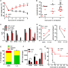
Figure 1
Figure 1. Chronic L. sigmodontis infection improves E. coli-induced sepsis.
(A) Kinetic of body temperature in response to i.p. E. coli injection of uninfected (n = 17) and chronically L. sigmodontis (L.s.)-infected mice (n = 19). (B) Peritoneal bacterial load and (C) serum cytokine/chemokine concentrations (n = 5–6/group) of uninfected and L. sigmodontis-infected mice at indicated time points and six hours post E. coli injection or mock injection, respectively. (D) Total number of peritoneal Gr1+ neutrophils and F4/80hiCD11bhi macrophages six hours post injection of E. coli or sterile LB broth (indicated as mock; n = 4–6/group). (E) Frequencies of propidium iodide-positive, Annexin V-positive and double negative (live) F4/80+ macrophages three hours post E. coli challenge. (F) CD80, CD86 and RELMα expression (mean fluorescence intensity) on F4/80hi macrophages (n = 4/group) six hours post E. coli injection; (G) survival of naïve and L. sigmodontis-infected mice after i.p. injection with E. coli (n = 8/group). (A) Shows pooled data from two independent experiments and (B-D, F) show one representative dataset of three independent experiments. Data in (A) is displayed as mean +/- SEM and was tested for statistical significance by 2-way ANOVA and Bonferroni post hoc test; data in (B-F) was tested for statistical significance by Mann-Whitney-U-test. Indicated statistical significant differences in (C) are based on comparisons of E. coli-treated groups and, in the case of IL-5, non-E. coli-treated groups (*p<0.05, **p<0.01, ***p<0.001); (G) was tested for statistical significance by Mantel-Cox Log-rank test (p = 0.0016).
Figure 2
Figure 2. Macrophage depletion renders L. sigmodontis-infected mice susceptible to E. coli-induced sepsis.
Chronic L. sigmodontis (L.s.)-infected BALB/c mice and uninfected controls (U) were i.p. injected with Clodronate- or PBS-containing liposomes before E. coli injection. (A) Kinetic of E. coli-induced hypothermia. (B) Peritoneal bacterial load and (C) serum TNFα and MIP-2β levels six hours after E. coli injection or mock treatment. (A) and (B) show pooled data from three independent experiments with at least 4 mice per group. (C) Pooled data from two independent experiments (mock treatment single experiment). Data in (A) is displayed as mean +/- SEM and was tested for statistical significance by 2-way ANOVA and Bonferroni post-hoc test (asterisks indicate significant differences between L.s. + PBS (n = 17) and L.s. + Clod. (n = 16), paragraphs indicate significant differences between L.s. + PBS and U + PBS (n = 15) treated mice (U + Clod. n = 13); in (B) and (C) data is presented as median and was tested for statistical significance by 1-way ANOVA followed by Dunn’s post-hoc test comparing only E. coli-treated groups. *p<0.05, **p<0.01, ***p<0.001.
Figure 3
Figure 3. Macrophage transcriptional analysis reveals a less inflammatory phenotype in L. sigmodontis-infected mice during E. coli challenge.
Gene expression profiles of peritoneal macrophages from L. sigmodontis (L.s.)-infected (left), L.s.-infected and E. coli challenged (middle) as well as E. coli challenged control mice (right) following PCR array analysis. Shared and exclusively regulated genes among the experimental groups were analyzed in comparison to gene expression of naïve control macrophages (cut-off: p<0.05 and >2-fold change). Statistical significances were tested by student’s t-test of the replicate 2^ (-Delta Ct) values for each gene in the control group and treatment groups.
Figure 4
Figure 4. L. sigmodontis-mediated protection against E. coli-induced sepsis is not compromised in AAM-deficient IL-4Rα-/-/IL-5-/- mice.
(A) Kinetic of body temperature in response to i.p. E. coli injection of uninfected (U) and chronic L. sigmodontis (L.s.)-infected wild type and IL-4Rα/IL-5-deficient mice. (B) Peritoneal bacterial load, (C) serum concentrations of TNFα, IL-6 and MIP-2β and (D) frequency of F4/80-positive peritoneal macrophages that are RELMα positive in those animals six hours post E. coli injection. (E) Representative histogram of RELMα fluorescence intensity of F4/80-positive peritoneal macrophages (isotype control (shaded), L.s.-infected IL-4Rα/IL-5-deficient mice (green), L.s.-infected wild type mice (red)). (A-D) Representative dataset from one of two independent experiments with at least 5 mice per group. Data shown in (A) is displayed as mean +/- SEM and was tested for statistical significance by 2-way ANOVA and Bonferroni post-hoc test (asterisks indicate significant differences between L.s.-infected and uninfected IL-4R/IL-5ko mice and paragraphs between L.s.-infected and uninfected wild type mice). (B, C) and (D) data was tested for statistical significance by 1-way ANOVA followed by Dunn’s post-hoc test. *p<0.05, **p<0.01.
Figure 5
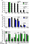
Figure 5. Macrophage stimulation with Wolbachia-containing preparations induces TLR2-dependent TNFα secretion and tolerance to subsequent LPS stimulation.
Thioglycollate-elicited peritoneal macrophages from wild type and either TLR2- (A), or TLR4-deficient (B) mice were stimulated with TLR4- (E. coli-LPS) and TLR2- (P3C, FSL1) specific ligands, L. sigmodontis adult worm extract (LsAg), LsAg from Wolbachia-depleted worms (Ls-tet), Wolbachia-infected (Wolb.) and sterile preparations of C6/36 insect cells. TNFα concentrations relative to LPS (A) and P3C (B) responses are shown. (C) Thioglycollate-induced macrophages from wild type and TLR2-deficient mice were primarily stimulated (prime) for 18 hours as described above before re-stimulation with LPS for an additional 18 hours. TNFα concentrations in supernatants of F4/80hi macrophages are plotted relative to the acute LPS response (medium prime/LPS stimulation). Shown are data representative of three independent experiments (n = 5/group for (A, B) and n = 3/group for (C)). Data shown is expressed as mean + SEM and was tested for statistical significance by student’s t-test. Asterisks above the bars indicate significant differences compared to med/LPS condition. *p< 0.05; **p< 0.01; ***p<0.001.
Figure 6
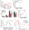
Figure 6. TLR2 is required for mediating protection against E. coli-induced sepsis in filarial-infected mice.
(A) Kinetic of body temperature in response to i.p. E. coli injection of uninfected (U) and chronic L. sigmodontis (L.s.)-infected wild type and TLR2-deficient mice. (B) Peritoneal bacterial load, (C) serum concentrations of IL-6, IL-10, and MIP-2β, and TNFα, (D) total peritoneal F4/80hi macrophage numbers and (E) their mean fluorescence intensity (MFI) of CD86 six hours post E. coli injection. (F) Survival after i.p. injection of E. coli into uninfected and L.s.-infected wild type (U: n = 15; L.s.: n = 18) and TLR2-deficient mice (U: n = 16; L.s.: n = 13). (A-D) shows a representative dataset from one of two independent experiments with at least 4 mice per group. Data shown in (A) is displayed as mean +/- SEM and was tested for statistical significance by 2-way ANOVA and Bonferroni post-hoc test (paragraphs indicate significant differences between L.s.-infected and uninfected wild type mice and asterisks in comparison to L.s.-infected TLR2-/- mice). Data shown in (B-E) was tested for statistical significance by 1-way ANOVA followed by Dunn’s post-hoc test. (F) Pooled data from two independent experiments. Differences between L.s.-infected BALB/c and TLR2-/- mice were tested for statistical significance by Mantel-Cox Log-rank test (p = 0.0035). *p<0.05; **p<0.01; ***p<0.001.
Figure 7
Figure 7. Anti-bacterial effector mechanisms are enhanced by L. sigmodontis infection in a TLR2 dependent manner.
(A) Colony forming units (cfu) obtained by a gentamycin assay using peritoneal macrophages derived from chronic L. sigmodontis (L.s.)-infected wild type and TLR2-/- mice and respective uninfected controls three hours after i.p. E. coli injection. (B) Nitrite concentrations of the same macrophages as in (A) after ex vivo cultivation for 48 hours. Frequency of peritoneal F4/80-positive macrophages from L.s.-infected and uninfected wild type and TLR2-/- mice (n = 5 per group) that phagocytosed pHrodo E. coli-BioParticles 90 minutes post injection (C) or from L.s.-infected and uninfected wild type mice (n = 5 per group) that phagocytosed pHrodo S. aureus-BioParticles three hours post injection (D). (A) Pooled data from two independent experiments with at least four mice per group. Data shown in (A-C) is illustrated as mean + SEM and was tested for statistical significance by 1-way ANOVA followed by Dunn’s multiple comparisons test; Data in (D) is also shown as mean + SEM and was tested for statistical significance by Mann-Whitney U test. *p< 0.05; **p< 0.01; ***p<0.001.
Figure 8
Figure 8. Repeated injections of Wolbachia-containing preparations and TLR2 ligands improve subsequent E. coli-induced sepsis in vivo.
Mice were injected three times with 40µg of L. sigmodontis extract (LsAg), L. sigmodontis extract obtained from tetracycline treated worms (Ls-tet), Wolbachia (WOLB) or Pam3Cys (P3C) every fourth day. Two days after the last injection sepsis was induced by i.p. E. coli injection. Body temperature (A), bacterial loads (B) and serum IL-6, IL-1β, and TNFα levels (C) were monitored six hours after E. coli challenge. Data in (A-C) is depicted as median and was tested for statistical significances using Kruskal-Wallis followed by Dunn’s multiple comparisons test.*p< 0.05;.**p< 0.01; ***p<0.001.
Figure 9
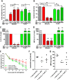
Figure 9. Transfer of primed macrophages improves E. coli-induced sepsis.
Macrophages derived from Thioglycollate-treated BALB/c or TLR2-/- mice were pre-treated in vitro with LsAg, Wolbachia or medium for 24 hours and subsequently transferred into naïve recipient wild type mice (n = 6/group). 12 hours after transfer sepsis was induced by i.p. E. coli injection. Body temperature (A), peritoneal bacterial loads (B) and serum IL-6 (C) and MIP-2β (D) were measured six hours after E. coli challenge. In a separate experiment wild type (WT) mice received peritoneal macrophages from either uninfected (U) wild type or chronic L. sigmodontis (L.s.)-infected WT or TLR2-deficient mice 12h before sepsis. Kinetic of body temperature (E), peritoneal bacterial loads (F) and serum MIP-2β (G) were measured six hours after E. coli challenge. Bar graphs in (A-D) show mean + SEM from recipient mice. Statistical significances were determined by 1-way ANOVA followed by Dunn’s multiple comparisons test. Data in (E) is displayed as mean +/- SEM and was tested for statistical significance by 2-way ANOVA and Bonferroni post-hoc test. Symbols in (F, G) show individual values of recipient mice that were tested for statistical significances by 1-way ANOVA followed by Dunn’s multiple comparisons test. *p< 0.05; **p< 0.01; ***p<0.001.

Similar articles

Cited by

References

    1. Maizels RM, Balic A, Gomez-Escobar N, Nair M, Taylor MD, et al. (2004) Helminth parasites--masters of regulation. Immunol Rev 201: 89–116. 10.1111/j.0105-2896.2004.00191.x - DOI - PubMed
    1. Allen JE, Maizels RM (2011) Diversity and dialogue in immunity to helminths. Nat Rev Immunol 11: 375–388. 10.1038/nri2992 - DOI - PubMed
    1. Anthony RM, Rutitzky LI, Urban JF, Stadecker MJ, Gause WC (2007) Protective immune mechanisms in helminth infection. Nat Rev Immunol 7: 975–987. 10.1038/nri2199 - DOI - PMC - PubMed
    1. Hoerauf a, Satoguina J, Saeftel M, Specht S (2005) Immunomodulation by filarial nematodes. Parasite Immunol 27: 417–429. 10.1111/j.1365-3024.2005.00792.x - DOI - PubMed
    1. Hübner MP, Shi Y, Torrero MN, Mueller E, Larson D, et al. (2012) Helminth protection against autoimmune diabetes in nonobese diabetic mice is independent of a type 2 immune shift and requires TGF-β. J Immunol 188: 559–568. 10.4049/jimmunol.1100335 - DOI - PMC - PubMed

Publication types

MeSH terms

Grants and funding

This work was funded by the German Research Foundation (HU 2144/1–1); intramural funding by the University Hospital of Bonn (BONFOR, 2010–1–16 and 2011–1–10); and the People Programme (Marie Curie Actions) of the European Union’s Seventh Framework Programme FP7/2007–2013 under Research Executive Agency Grant GA 276704. AB was supported by the German Academic Exchange Service (DAAD) and BCB was supported by the Jürgen Manchot Stiftung, Düsseldorf. AH is a member of the German Centre for Infection Research (DZIF), and of the Excellence Cluster Immunosensation (DFG, EXC 1023).