. 2015 Mar 6;290(10):6203-14.
doi: 10.1074/jbc.M114.635284. Epub 2015 Jan 21.

Tyrosyl-DNA phosphodiesterase I catalytic mutants reveal an alternative nucleophile that can catalyze substrate cleavage

Affiliations

Tyrosyl-DNA phosphodiesterase I catalytic mutants reveal an alternative nucleophile that can catalyze substrate cleavage

Evan Q Comeaux et al. J Biol Chem. .

Abstract

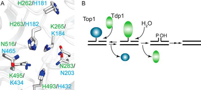
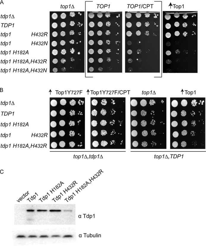
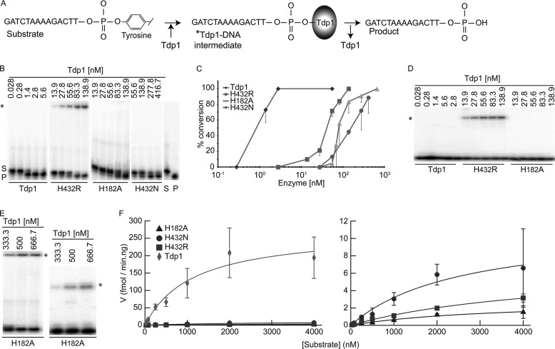
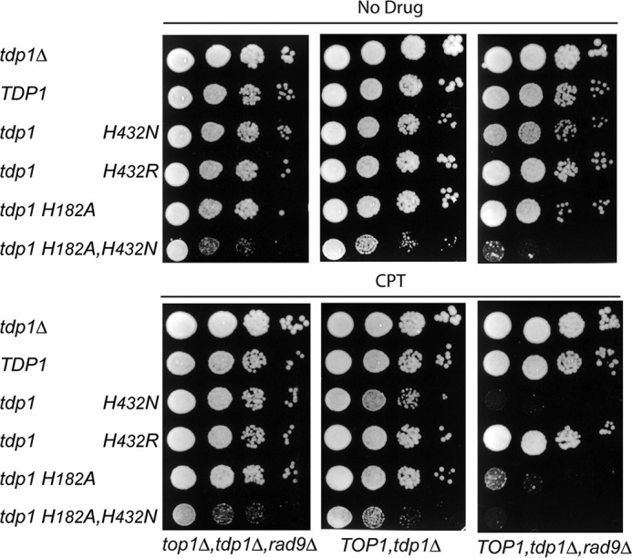
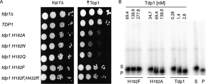
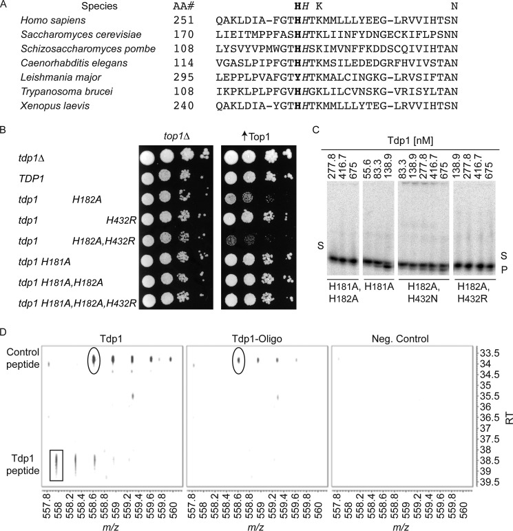
Tyrosyl-DNA phosphodiesterase I (Tdp1) catalyzes the repair of 3'-DNA adducts, such as the 3'-phosphotyrosyl linkage of DNA topoisomerase I to DNA. Tdp1 contains two conserved catalytic histidines: a nucleophilic His (His(nuc)) that attacks DNA adducts to form a covalent 3'-phosphohistidyl intermediate and a general acid/base His (His(gab)), which resolves the Tdp1-DNA linkage. A His(nuc) to Ala mutant protein is reportedly inactive, whereas the autosomal recessive neurodegenerative disease SCAN1 has been attributed to the enhanced stability of the Tdp1-DNA intermediate induced by mutation of His(gab) to Arg. However, here we report that expression of the yeast His(nuc)Ala (H182A) mutant actually induced topoisomerase I-dependent cytotoxicity and further enhanced the cytotoxicity of Tdp1 His(gab) mutants, including H432N and the SCAN1-related H432R. Moreover, the His(nuc)Ala mutant was catalytically active in vitro, albeit at levels 85-fold less than that observed with wild type Tdp1. In contrast, the His(nuc)Phe mutant was catalytically inactive and suppressed His(gab) mutant-induced toxicity. These data suggest that the activity of another nucleophile when His(nuc) is replaced with residues containing a small side chain (Ala, Asn, and Gln), but not with a bulky side chain. Indeed, genetic, biochemical, and mass spectrometry analyses show that a highly conserved His, immediately N-terminal to His(nuc), can act as a nucleophile to catalyze the formation of a covalent Tdp1-DNA intermediate. These findings suggest that the flexibility of Tdp1 active site residues may impair the resolution of mutant Tdp1 covalent phosphohistidyl intermediates and provide the rationale for developing chemotherapeutics that stabilize the covalent Tdp1-DNA intermediate.

Keywords: DNA Damage; DNA Repair; DNA Topoisomerase; Enzyme Mechanism; Enzyme Mutation; Protein-DNA Covalent Complexes; Spinocerebellar Ataxia with Axonal Neuropathy 1; Tyrosyl-DNA Phosphodiesterase I.


Figures

FIGURE 1.
FIGURE 1.
Tdp1 conserved catalytic pocket architecture and mechanism. A, overlay of the crystal structure of yeast and human Tdp1 active site residues that comprise the HXKXnN motifs. Human Tdp1 residues are labeled in green (Protein Data Bank code 1NOP) (22), and yeast Tdp1 is labeled in cyan (Protein Data Bank code 1Q32) (7). His263/182 functions as a nucleophile (Hisnuc), whereas His493/432 acts as a general acid-base (Hisgab). B, the two-step Tdp1 catalytic cycle. In step 1, Tdp1 hydrolysis of the Top1-DNA covalent complex by nucleophilic attack of Hisnuc on the 3′-phosphotyrosyl linkage, forming a 3′-phosphohistidyl bond (Tdp1Hisnuc-DNA covalent complex). In step 2, water is activated by the general acid/base histidine, Hisgab, to hydrolyze the Tdp1-DNA linkage, dissociating Tdp1 from the nicked DNA strand. The single-strand break ends need subsequent processing by polynucleotide kinase/phosphatase and DNA ligase to produce intact DNA.
FIGURE 2.
FIGURE 2.
Tdp1H182A induces a substrate-dependent toxicity. A, top1Δ,tdp1Δ cells were co-transformed with vector control (top1Δ) or YCpGPD-Top1·U (↑Top1) and control vector (tdp1Δ) or the indicated YCpGAL1-Tdp1·L, whereas tdp1Δ cells were co-transformed with vector control (TOP1) and control vector (tdp1Δ) or the indicated YCpGAL1-Tdp1·L. Exponential growing cultures were diluted to A595 = 0.3, then 10-fold serially diluted, and spotted onto 2% galactose selective media plates supplemented with or without 1 μg/ml CPT (only for tdp1Δ-transformants, shown between brackets). B, top1Δ,tdp1Δ cells were transformed with YCpGAL1-Top1Y727F·U (↑Top1Y727F) and indicated YCpGAL1-Tdp1·L or control vector (tdp1Δ), diluted, and spotted (as in A) onto selective media plates containing 2% galactose, with or without 5 μg/ml CPT. In addition, top1Δ,TDP1 cells were transformed with vector control (top1Δ) or YCpGPD-Top1·U (↑Top1) and the indicated YCpGAL1-Tdp1·L or vector control (tdp1Δ), diluted, and spotted (as in A) onto selective media plates containing 2% galactose. All plates were incubated for 4 days at 30 °C. C, total cell extracts collected from 6-h galactose-induced top1Δ,tdp1Δ cells co-transformed with vector control (top1Δ), and the indicated YCpGAL1-Tdp1vL or control vector (tdp1Δ) used in A were resolved on 12% SDS-PAGE and immunoblotted with anti-yeast Tdp1 and anti-α-tubulin.
FIGURE 3.
FIGURE 3.
Tdp1H182A is catalytically active. A, schematic of in vitro Tdp1 catalytic activity assay. 5′-32P-labeled 14-mer oligonucleotide substrate containing 3′-phosphotyrosine (Substrate) is covalently bound by Tdp1 (*Tdp1-DNA intermediate) and subsequently released as 3′-phosphate (Product). B, D, E, and F, various concentrations of indicated full-length Tdp1 enzyme were incubated with 16.7 nm (B, D, and E) or 25–4000 nm (F) of substrate for 10 min at 30 °C. For B, D, and E, reaction samples were split in two and analyzed as described in B and D by phosphorimaging. B, conversion of 3′-phosphotyrosine (S) to 3′-phosphate (P) resolved in 20% 8 m urea denaturing polyacrylamide gel. *, covalent Tdp1-DNA intermediates. C, the mean and S.D. of the substrate to product (enzyme-DNA intermediate + 3′-phosphoryl product) conversion (product/[product + substrate + intermediate]) from B were quantified by phosphorimaging analysis of at least three independent experiments. D, detection of covalent protein-DNA reaction intermediates via 12% SDS-PAGE. E, reactions with higher concentrations of Tdp1H182A protein analyzed as in B (left panel) and D (right panel), respectively. F, same reaction conditions as in B but with increasing substrate concentrations and constant enzyme concentration. The mean and standard deviation of four independent experiments are shown in a Michaelis-Menten plot (specific velocity (fmol product/min·ng of protein) versus [substrate] (nm)) generated with IGOR PRO 6 program 2010. The right panel shows an adjusted y axis scale from the left panel to visualize mutant enzyme results. In C and F, Tdp1 (diamond), H432R (square), H182A (triangle), and H432N (circle).
FIGURE 4.
FIGURE 4.
Rad9 DNA damage checkpoint suppresses the Top1-dependent toxicity of mutant Tdp1. top1Δ,tdp1Δ,rad9Δ, TOP1,tdp1Δ, and TOP1,tdp1Δ,rad9Δ cells were transformed with the indicated YCpGAL1-Tdp1·L or control vector (tdp1Δ) and then 10-fold serially diluted and spotted (as in Fig. 2A) onto 2% galactose selective media plates supplemented with or without 0.5 μg/ml CPT. The plates were incubated for 4 days at 30 °C.
FIGURE 5.
FIGURE 5.
Tdp1H182F protein is catalytically inactive. A, top1Δ,tdp1Δ cells were transformed with vector control (top1Δ) or YCpGPD-Top1·U (↑Top1) and the indicated YCpGAL1-Tdp1·L or control vector (tdp1Δ), 10-fold serially diluted and spotted (as in Fig. 2A) onto selective media plates with 2% galactose and incubated for 4 days at 30 °C. B, in vitro Tdp1 catalytic activity assay (as described in Fig. 3A), with various amounts (fmol) of indicated full-length Tdp1 enzymes resolved in 20% 8 m urea denaturing polyacrylamide gel to detect conversion of 3′-phosphotyrosine (S) to 3′-phosphate (P).
FIGURE 6.
FIGURE 6.
Tdp1H182A toxicity depends on a conserved adjacent histidine residue. A, alignment of a selection of Tdp1 orthologs N-terminal HHXKXnN motif amino acid sequence. The Hisnuc is italicized, and the adjacent histidine is in bold. AA#, amino acid number. UniProt KD/Swiss-Prot protein numbers were as follows: Homo sapiens (Q9NUW8), S. cerevisiae (P38319), Schizosaccharomyces pombe (Q9USG9), Caenorhabditis elegans (Q9TXV7), Leishmania major (Q4Q3N1), Trypanosoma brucei (Q586D1), and Xenopus laevis (Q6DFE8). B, top1Δ,tdp1Δ cells were transformed with vector control (top1Δ) or YCpGPD-Top1·U (↑Top1) and the indicated YCpGAL1-Tdp1·L or control vector (tdp1Δ), diluted, and spotted (as in Fig. 2A) onto selective media plates containing 2% galactose and incubated for 4 days at 30 °C. C, conversion of 3′-phosphotyrosine (S) to 3′-phosphate (P) resolved in 20% 8 m urea denaturing polyacrylamide gel as described for Fig. 3 (A and B) using 16.7 nm substrate. The mean and standard deviation were determined (graph not shown) as described for Fig. 3C, revealing a reduction of relative activity (versus wild type enzyme; Fig. 3, B and C) of ∼20-fold for H181A and ∼210-fold for H182A,H432N. Relative activity of H182A,H432R could not be determined because it did not convert 50% of substrate. D, two-dimensional plot of mass and time tags (557–560 m/z, 33–40 min.). The non-cross-linked His181-Ala182-containing tryptic fragment 170LIEITMPPFASHATK184 (Tdp1 peptide, rectangle) is illustrated with a mass and time tag of 557.96 m/z at 38.3 min (3+ charge state), with good signal intensity in the oligonucleotide-free sample (Tdp1) and yet not observed in the oligonucleotide reacted sample (Tdp1-Oligo). Serving as a control peptide 65IIDLTNQEQDLSER78 (control peptide, circle) presented with a similar mass and time tag of 558.62 m/z @ 33.7 min (3+ charge state) and was observed at nearly equal intensities in both samples ± oligonucleotide. Neither fragment was observed in the negative control sample (Neg. Control).
FIGURE 7.
FIGURE 7.
Position of Tdp1 181–182 residues within the catalytic pocket environment. A, overlay of the wild type Tdp1 catalytic histidine residues His181, His182, His432 in yellow (Protein Data Bank code 1Q32 (7)) and His181, Ala182, and His432 residues within the Tdp1H182A crystal structure in blue (Protein Data Bank code 3SQ3 (23)). In gray is shown the α-carbon backbone trace of Tdp1 protein and surrounding residues that might influence path of rotation. B, H182A catalytic pocket focusing on the proposed rotation (orange arrow, with the rectangle symbolizing end point after rotation) of His181 upon docking of the substrate to face the substrate within the active site, which would allow His181 to perform a nucleophilic attack. Note the increase distance from His432 to His182 in A and to potential location of His181 (after rotation, orange rectangle) in B.

Similar articles

Cited by

References

    1. Yang S. W., Burgin A. B., Jr., Huizenga B. N., Robertson C. A., Yao K. C., Nash H. A. (1996) A eukaryotic enzyme that can disjoin dead-end covalent complexes between DNA and type I topoisomerases. Proc. Natl. Acad. Sci. U.S.A. 93, 11534–11539 - PMC - PubMed
    1. Hsiang Y. H., Hertzberg R., Hecht S., Liu L. F. (1985) Camptothecin induces protein-linked DNA breaks via mammalian DNA topoisomerase I. J. Biol. Chem. 260, 14873–14878 - PubMed
    1. Hsiang Y. H., Lihou M. G., Liu L. F. (1989) Arrest of replication forks by drug-stabilized topoisomerase I-DNA cleavable complexes as a mechanism of cell killing by camptothecin. Cancer Res. 49, 5077–5082 - PubMed
    1. Pommier Y., Leo E., Zhang H., Marchand C. (2010) DNA topoisomerases and their poisoning by anticancer and antibacterial drugs. Chem. Biol. 17, 421–433 - PMC - PubMed
    1. Schoeffler A. J., Berger J. M. (2008) DNA topoisomerases: harnessing and constraining energy to govern chromosome topology. Q. Rev. Biophys. 41, 41–101 - PubMed

Publication types

MeSH terms