. 2015 Jan 20;10(1):e0117098.
doi: 10.1371/journal.pone.0117098. eCollection 2015.

Inhibition of bone morphogenetic protein signal transduction prevents the medial vascular calcification associated with matrix Gla protein deficiency

Affiliations

Inhibition of bone morphogenetic protein signal transduction prevents the medial vascular calcification associated with matrix Gla protein deficiency

Rajeev Malhotra et al. PLoS One. .

Abstract

Objective: Matrix Gla protein (MGP) is reported to inhibit bone morphogenetic protein (BMP) signal transduction. MGP deficiency is associated with medial calcification of the arterial wall, in a process that involves both osteogenic transdifferentiation of vascular smooth muscle cells (VSMCs) and mesenchymal transition of endothelial cells (EndMT). In this study, we investigated the contribution of BMP signal transduction to the medial calcification that develops in MGP-deficient mice.

Approach and results: MGP-deficient mice (MGP(-/-)) were treated with one of two BMP signaling inhibitors, LDN-193189 or ALK3-Fc, beginning one day after birth. Aortic calcification was assessed in 28-day-old mice by measuring the uptake of a fluorescent bisphosphonate probe and by staining tissue sections with Alizarin red. Aortic calcification was 80% less in MGP(-/-) mice treated with LDN-193189 or ALK3-Fc compared with vehicle-treated control animals (P<0.001 for both). LDN-193189-treated MGP(-/-) mice survived longer than vehicle-treated MGP(-/-) mice. Levels of phosphorylated Smad1/5 and Id1 mRNA (markers of BMP signaling) did not differ in the aortas from MGP(-/-) and wild-type mice. Markers of EndMT and osteogenesis were increased in MGP(-/-) aortas, an effect that was prevented by LDN-193189. Calcification of isolated VSMCs was also inhibited by LDN-193189.

Conclusions: Inhibition of BMP signaling leads to reduced vascular calcification and improved survival in MGP(-/-) mice. The EndMT and osteogenic transdifferentiation associated with MGP deficiency is dependent upon BMP signaling. These results suggest that BMP signal transduction has critical roles in the development of vascular calcification in MGP-deficient mice.


Conflict of interest statement

Competing Interests: Dr. Ravindra Kumar is an employee of Acceleron Pharma Inc. and has stock ownership in the company. This does not alter the authors’ adherence to PLOS ONE policies on sharing data and materials. The Massachusetts General Hospital has applied for patents related to pharmacologic BMP signaling inhibitors. These patents include: 1. Inhibitors of the bmp signaling pathway (Application # CA 2718403, PCT/US2009/001606). Drs. Yu and K. Bloch may be entitled to royalties. 2. Compositions and methods for cardiovascular disease (Application # PCT/US2012/022119). Drs. Malhotra, Derwall, Yu, and K. Bloch may be entitled to royalties. 3. Bmp inhibitors and methods of use thereof (Application # PCT/US2014/020360). Drs. Yu and K. Bloch may be entitled to royalties. These patents do not alter the authors’ adherence to PLOS ONE policies on sharing data and materials.

Figures

Figure 1
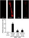
Figure 1. Pharmacologic inhibition of BMP signaling reduces osteogenic activity in the aortas of MGP-deficient mice.
MGP-/- mice were treated with vehicle, LDN-193189 (LDN), or ALK3-Fc starting at day 1 of life for 28 days. Wild-type mice (WT) were treated with vehicle. On day 27, OsteoSense-680 was injected via the tail vein. Aortas were harvested 24 hours later and imaged with near-infrared fluorescence (upper panel). Fluorescence intensities of the entire aorta normalized to vehicle-treated MGP-/- mouse aortas were quantified (lower panel). Inhibition of BMP signaling reduced vascular calcification by 80% in MGP-/- mice. * P<0.001 compared to vehicle-treated WT mice. # P<0.001 compared to vehicle-treated MGP-/- mice.
Figure 2
Figure 2. Vascular calcification associated with MGP deficiency is dependent on bone morphogenetic protein signaling.
Aortas were harvested from mice at 28 days of age, sectioned, and stained for tissue calcium with Alizarin Red. Sections from wild-type (A) and MGP-/- (B-D) mice were photographed at 100x magnification. Starting at day 1 of age, MGP-/- mice were treated with i.p. injections of vehicle (B), LDN-193189 (LDN, 2.5 mg/kg daily, C), or ALK3-Fc (2 mg/kg every other day, D). Inhibition of BMP signaling reduced aortic calcification in MGP-/- mice.
Figure 3
Figure 3. Inhibition of BMP signaling improves survival of MGP-/- mice.
MGP-/- mice were treated once daily with LDN-193189 (LDN) or vehicle starting at day 1 of life. Kaplan-Meier survival was compared using the log rank test. MGP-/- mice treated with LDN-193189 survived longer than vehicle-treated mice.
Figure 4
Figure 4. Vascular calcification associated with MGP deficiency occurs in the absence of vascular inflammation.
(A) At 27 days of age, OsteoSense-680 and Prosense-750 were injected via the tail vein of wild-type (WT) and MGP-/- mice. Aortas were harvested 24 hours later and imaged. Although aortas from MGP-/- mice exhibited extensive vascular calcification, this calcification was not associated with increased macrophage activity. (B) Aortas were harvested from WT and MGP-/- mice at 28 days of age, sectioned, and stained for macrophages with an antibody directed towards MAC-2. Aortas from LDLR-/- mice on a high fat diet were used as a positive control. Nuclei were stained with DAPI. Similar to WT mice, macrophages were not detected by immunohistochemistry in the aortas of MGP-/- mice.
Figure 5
Figure 5. BMP signaling is not increased in aortas of MGP-/- mice.
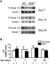
(A) Protein lysates were isolated from the aortas of 7-, 14-, and 28-day-old WT and MGP-/- mice. Each lane represents protein isolated from four pooled aortas. PVDF membranes were incubated with antibodies directed against phosphorylated Smad 1/5 (P-Smad 1/5) and total Smad 1. The ratio of P-Smad 1/5 to total Smad 1 was the same in aortas derived from WT and MGP-/- mice. (B) RNA was isolated from aortas of WT and MGP-/- mice at 1, 7, 14, and 28 days of (n = 6–8 in each group, as indicated). No difference in aortic Id1 mRNA levels was observed between MGP-/- and WT mice.
Figure 6
Figure 6. MGP deficiency does not alter basal BMP signaling or responsiveness to BMP-2 in VSMCs.
(A) VSMCs were isolated from the aortas of wild-type and MGP-/- mice. VSMCs were treated without or with recombinant human BMP-2 (for 2 hours at the indicated doses). Groups were compared using a 2-way ANOVA. Both WT and MGP-/- VSMCs exhibited similar Id1 mRNA levels, both at baseline and in response to exogenous BMP-2. (B) Cultured aortic VSMCs from wild-type mice were transfected with either scrambled siRNA (siSC) or siRNA targeting MGP (siMGP) at 20 nM. RNA was isolated from cells after 4 days. siMGP decreased MGP mRNA levels in WT VSMCs by >95% compared with siSC-treated cells. However, depletion of MGP in WT VSMCs did not alter Id1 mRNA levels. **P<0.0001 compared to siSC-treated VSMCs. (C) VSMCs isolated from wild-type mice were treated with 20 nM of either scrambled siRNA (siSC) or siRNA specific for MGP (siMGP). Cells were incubated with or without BMP-2 (20 ng/mL) for 1 h prior to protein harvest. Western blots were probed with antibodies specific for phosphorylated Smad 1/5 (P-Smad 1/5) and total Smad 1. Depletion of MGP in WT VSMCs did not alter the ratio of P-Smad 1/5 levels to total Smad 1 levels, both at baseline and in response to exogenous BMP-2.
Figure 7
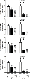
Figure 7. Aortic expression of VSMC markers in wild-type and MGP-/- mice.
RNA was isolated from aortas of WT and MGP-/- mice and from LDN-193189-treated MGP-/- mice at 7 and 14 days of age (n = 4–8 in each group). Levels of mRNAs encoding myocardin, α smooth muscle actin (SMA), transgelin, and calponin are depicted. The aortas of 14-day-old MGP-/- mice have decreased expression of VSMC markers compared to WT mice. Treatment with LDN-193189 did not restore the expression of VSMC markers to WT levels. # P<0.05 compared to 7-day-old MGP-/- mice.
Figure 8
Figure 8. BMP signaling is required for the increased aortic expression of osteogenic markers associated with MGP deficiency.
RNA was isolated from aortas of WT and MGP-/- mice at 1, 7, 14, and 28 days of age and from LDN-193189-treated MGP-/- mice at 7, 14, and 28 days of age (n = 4–11 in each group, as indicated). Expression of genes encoding Runx2 and osteopontin (OPN) was measured. MGP-/- mice had increased levels of aortic Runx2 and OPN mRNA compared to WT mice. Treatment of MGP-/- mice with LDN-193189 reduced aortic Runx2 and OPN mRNA levels. * P<0.001 compared to WT mice of same age. # P<0.05 compared to age-matched MGP-/- mice treated with vehicle.
Figure 9
Figure 9. Restoration of MGP levels decreases calcification of MGP-/- vascular smooth muscle cells while siRNA-mediated depletion of MGP increases calcification of wild-type vascular smooth muscle cells in a BMP-dependent manner.
Cultured aortic VSMCs isolated from MGP-/- mice were infected with either (A) a control adenovirus (Ad.GFP) or (B) an adenovirus expressing MGP (Ad.MGP) at a multiplicity of infection of 10 and placed in DMEM supplemented with 10% FBS and 2 mM sodium phosphate. Cultured aortic VSMCs isolated from wild-type mice were transfected with either (C) scrambled siRNA (siSC) or (D & E) siRNA targeting MGP (siMGP) at 20 nM and placed in DMEM supplemented with 10% FBS and 2 mM sodium phosphate. Cells were also treated without (C & D) or with (E) 100 nM LDN-193189 (LDN). Cells were stained after 7 days using the von Kossa method. Serial fields of view were photographed for each condition and von Kossa stain was quantified using image J software after background subtraction (F & G). In (F), *P = 0.03 compared to Ad.GFP. In (G), **P<0.0001 compared to siSC-treated cells. #P = 0.0003 compared to siMGP + control. Restoration of MGP expression reduced phosphate-induced calcification of MGP-/- VSMCs, while depletion of MGP increased calcification of WT VSMCs and this calcification was partially inhibited by treatment with LDN-193189.
Figure 10
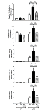
Figure 10. Aortic expression of endothelial and multipotency markers in wild-type and MGP-/- mice.
RNA was isolated from aortas of WT and MGP-/- mice and from LDN-193189-treated MGP-/- mice at 7 and 14 days of age (n = 4–8 in each group). Levels of mRNAs encoding endothelial markers (VE-Cadherin and CD31) and multipotency markers (nanog, Oct 3/4, and Sox2) are depicted. The aortas of 14-day-old MGP-/- mice have increased endothelial and multipotency markers compared to WT mice. Treatment with LDN-193189 normalized the endothelial and multipotency markers to WT levels in MGP-/- mice. * P ≤ 0.01 compared to 7-day-old WT mice. # P<0.05 compared to 7-day-old MGP-/- mice.

Similar articles

Cited by

References

    1. Abedin M, Tintut Y, Demer LL (2004) Vascular calcification: mechanisms and clinical ramifications. Arterioscler Thromb Vasc Biol 24: 1161–1170. 10.1161/01.ATV.0000133194.94939.42 - DOI - PubMed
    1. Huang H, Virmani R, Younis H, Burke AP, Kamm RD, et al. (2001) The impact of calcification on the biomechanical stability of atherosclerotic plaques. Circulation 103: 1051–1056. 10.1161/01.CIR.103.8.1051 - DOI - PubMed
    1. Vengrenyuk Y, Carlier S, Xanthos S, Cardoso L, Ganatos P, et al. (2006) A hypothesis for vulnerable plaque rupture due to stress-induced debonding around cellular microcalcifications in thin fibrous caps. Proc Natl Acad Sci U S A 103: 14678–14683. 10.1073/pnas.0606310103 - DOI - PMC - PubMed
    1. Virmani R, Burke AP, Farb A, Kolodgie FD (2006) Pathology of the vulnerable plaque. J Am Coll Cardiol 47: C13–18. 10.1016/j.jacc.2005.10.065 - DOI - PubMed
    1. Amann K (2008) Media calcification and intima calcification are distinct entities in chronic kidney disease. Clin J Am Soc Nephrol 3: 1599–1605. 10.2215/CJN.02120508 - DOI - PubMed

Publication types

MeSH terms

LinkOut - more resources