Distinctive expression patterns of glycoprotein non-metastatic B and folliculin in renal tumors in patients with Birt-Hogg-Dubé syndrome
- PMID: 25594584
- PMCID: PMC4376441
- DOI: 10.1111/cas.12601
Distinctive expression patterns of glycoprotein non-metastatic B and folliculin in renal tumors in patients with Birt-Hogg-Dubé syndrome
Abstract
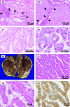
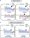
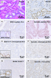
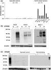
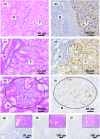
Birt-Hogg-Dubé syndrome (BHD) is an inherited disorder associated with a germline mutation of the folliculin gene (FLCN). The affected families have a high risk for developing multiple renal cell carcinomas (RCC). Diagnostic markers that distinguish between FLCN-related RCC and sporadic RCC have not been investigated, and many patients with undiagnosed BHD fail to receive proper medical care. We investigated the histopathology of 27 RCCs obtained from 18 BHD patients who were diagnosed by genetic testing. Possible somatic mutations of RCC lesions were investigated by DNA sequencing. Western blotting and immunohistochemical staining were used to compare the expression levels of FLCN and glycoprotein non-metastatic B (GPNMB) between FLCN-related RCCs and sporadic renal tumors (n = 62). The expression of GPNMB was also evaluated by quantitative RT-PCR. Histopathological analysis revealed that the most frequent histological type was chromophobe RCC (n = 12), followed by hybrid oncocytic/chromophobe tumor (n = 6). Somatic mutation analysis revealed small intragenic mutations in six cases and loss of heterozygosity in two cases. Western blot and immunostaining analyses revealed that FLCN-related RCCs showed overexpression of GPNMB and underexpression of FLCN, whereas sporadic tumors showed inverted patterns. GPNMB mRNA in FLCN-related RCCs was 23-fold more abundant than in sporadic tumors. The distinctive expression patterns of GPNMB and FLCN might identify patients with RCCs who need further work-up for BHD.
Keywords: Birt-Hogg-Dubé syndrome (BHD); familial cancer; folliculin (FLCN); glycoprotein non-metastatic B (GPNMB); renal tumor.
© 2015 The Authors. Cancer Science published by Wiley Publishing Asia Pty Ltd on behalf of Japanese Cancer Association.
Figures

Similar articles
-
Immunohistochemical characterization of renal tumors in patients with Birt-Hogg-Dubé syndrome.Pathol Int. 2015 Mar;65(3):126-32. doi: 10.1111/pin.12254. Epub 2015 Jan 19. Pathol Int. 2015. PMID: 25597876
-
Fluorescent and chromogenic in situ hybridization of CEN17q as a potent useful diagnostic marker for Birt-Hogg-Dubé syndrome-associated chromophobe renal cell carcinomas.Hum Pathol. 2016 Jun;52:74-82. doi: 10.1016/j.humpath.2016.01.004. Epub 2016 Feb 4. Hum Pathol. 2016. PMID: 26980015
-
Genome-Wide Uniparental Disomy and Copy Number Variations in Renal Cell Carcinomas Associated with Birt-Hogg-Dubé Syndrome.Am J Pathol. 2016 Feb;186(2):337-46. doi: 10.1016/j.ajpath.2015.10.013. Am J Pathol. 2016. PMID: 26776076
-
Birt-Hogg-Dubé syndrome: Clinical and molecular aspects of recently identified kidney cancer syndrome.Int J Urol. 2016 Mar;23(3):204-10. doi: 10.1111/iju.13015. Epub 2015 Nov 25. Int J Urol. 2016. PMID: 26608100 Review.
-
Birt-Hogg-Dubé syndrome in an overall view: Focus on the clinicopathological prospects in renal tumors.Semin Diagn Pathol. 2024 May;41(3):119-124. doi: 10.1053/j.semdp.2024.01.008. Epub 2024 Jan 6. Semin Diagn Pathol. 2024. PMID: 38242750 Review.
Cited by
-
FLCN alteration drives metabolic reprogramming towards nucleotide synthesis and cyst formation in salivary gland.Biochem Biophys Res Commun. 2020 Feb 19;522(4):931-938. doi: 10.1016/j.bbrc.2019.11.184. Epub 2019 Dec 2. Biochem Biophys Res Commun. 2020. PMID: 31806376 Free PMC article.
-
Loss of FLCN inhibits canonical WNT signaling via TFE3.Hum Mol Genet. 2019 Oct 1;28(19):3270-3281. doi: 10.1093/hmg/ddz158. Hum Mol Genet. 2019. PMID: 31272105 Free PMC article.
-
Splice-site mutation causing partial retention of intron in the FLCN gene in Birt-Hogg-Dubé syndrome: a case report.BMC Med Genomics. 2018 May 2;11(1):42. doi: 10.1186/s12920-018-0359-5. BMC Med Genomics. 2018. PMID: 29720200 Free PMC article.
-
Pulmonary Neoplasms in Patients with Birt-Hogg-Dubé Syndrome: Histopathological Features and Genetic and Somatic Events.PLoS One. 2016 Mar 14;11(3):e0151476. doi: 10.1371/journal.pone.0151476. eCollection 2016. PLoS One. 2016. PMID: 26974543 Free PMC article.
-
TFE3 Xp11.2 Translocation Renal Cell Carcinoma Mouse Model Reveals Novel Therapeutic Targets and Identifies GPNMB as a Diagnostic Marker for Human Disease.Mol Cancer Res. 2019 Aug;17(8):1613-1626. doi: 10.1158/1541-7786.MCR-18-1235. Epub 2019 May 1. Mol Cancer Res. 2019. PMID: 31043488 Free PMC article.
References
-
- Menko FH, van Steensel MA, Giraud S, et al. Birt–Hogg–Dube syndrome: diagnosis and management. Lancet Oncol. 2009;10:1199. - PubMed
-
- Nickerson ML, Warren MB, Toro JR, et al. Mutations in a novel gene lead to kidney tumors, lung wall defects, and benign tumors of the hair follicle in patients with the Birt–Hogg–Dube syndrome. Cancer Cell. 2002;2:157. - PubMed
-
- Zbar B, Alvord WG, Glenn G, et al. Risk of renal and colonic neoplasms and spontaneous pneumothorax in the Birt–Hogg–Dube syndrome. Cancer Epidemiol Biomarkers Prev. 2002;11:393. - PubMed
-
- Pavlovich CP, Walther MM, Eyler RA, et al. Renal tumors in the Birt–Hogg–Dube syndrome. Am J Surg Pathol. 2002;26:1542. - PubMed
Publication types
MeSH terms
Substances
LinkOut - more resources
Full Text Sources
Other Literature Sources
Medical

