. 2015 Mar 6;290(10):6558-73.
doi: 10.1074/jbc.M114.623181. Epub 2015 Jan 14.

Distinct requirements for HIV-cell fusion and HIV-mediated cell-cell fusion

Affiliations

Distinct requirements for HIV-cell fusion and HIV-mediated cell-cell fusion

Naoyuki Kondo et al. J Biol Chem. .

Abstract

Whether HIV-1 enters cells by fusing with the plasma membrane or with endosomes is a subject of active debate. The ability of HIV-1 to mediate fusion between adjacent cells, a process referred to as "fusion-from-without" (FFWO), shows that this virus can fuse with the plasma membrane. To compare FFWO occurring at the cell surface with HIV-cell fusion through a conventional entry route, we designed an experimental approach that enabled the measurements of both processes in the same sample. The following key differences were observed. First, a very small fraction of viruses fusing with target cells participated in FFWO. Second, whereas HIV-1 fusion with adherent cells was insensitive to actin inhibitors, post-CD4/coreceptor binding steps during FFWO were abrogated. A partial dependence of HIV-cell fusion on actin remodeling was observed in CD4(+) T cells, but this effect appeared to be due to the actin dependence of virus uptake. Third, deletion of the cytoplasmic tail of HIV-1 gp41 dramatically enhanced the ability of the virus to promote FFWO, while having a modest effect on virus-cell fusion. Distinct efficiencies and actin dependences of FFWO versus HIV-cell fusion are consistent with the notion that, except for a minor fraction of particles that mediate fusion between the plasma membranes of adjacent cells, HIV-1 enters through an endocytic pathway. We surmise, however, that cell-cell contacts enabling HIV-1 fusion with the plasma membrane could be favored at the sites of high density of target cells, such as lymph nodes.

Keywords: Cytoskeleton; Endocytosis; Fluorescence; Membrane Fusion; Plasma Membrane.


Figures

FIGURE 1.
FIGURE 1.
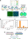
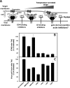
Parallel measurements of HIV-cell and HIV-mediated cell-cell fusion. A, dual readout assay to measure virus-cell fusion and virus-mediated cell-cell fusion (FFWO) based on the BlaM (scissors) and dual-split GFP-luciferase (DSP, yellow segments/circles) activities, respectively. Top inset, illustration of the DSP assay for cell-cell fusion. DSP-1 and DSP-2 are the N- and C-terminal fragments of DSP that recombine to form functional GFP and luciferase. Bottom inset, images of noninfected (left panel) and infected (right panel) cells loaded with the BlaM substrate (green). Cleavage of the BlaM substrate in the cytoplasm by viral β-lactamase results in a blue shift in fluorescence. B, kinetics of HXB2pp escape from the fusion inhibitory peptide, C52L, and from the low temperature block. A 1:1 mixture of confluent DSP-1 and DSP-2 cells was inoculated with HXB2 pseudovirus at m.o.i. = 1 by spinoculation (see “Experimental Procedures”);unbound viruses were washed away, and viral fusion was triggered by quickly bringing the temperature to 37 °C (designated as 0 min). At the indicated time, the fusion reaction was stopped by adding a fully inhibitory concentration (1 μm) of C52L or lowering the temperature by placing cells on ice. The resulting virus-cell fusion was measured by the BlaM assay. Data points are means ± S.E. from two experiments with triplicate samples. C, inhibition of FFWO by HIV-1 fusion inhibitors, as measured by the DSP assay (luciferase signal). DSP-1 and DSP-2 cells (1:1 ratio) were grown to confluency on microplates. Cells were inoculated with HXB2 pseudovirus (m.o.i. = 3) and incubated at 37 °C for 1.5 h, either in the absence or in the presence of fusion inhibitors BMS806 (10 μm), AMD3100 (30 μm), or C52L (1 μm). Fusion between CD4+/CXCR4+ DSP-1 cells and DSP-2 cells expressing only CD4 is shown as a negative control (last bar). Bars show the means ± S.E. from a representative experiment with triplicate samples. R.L.U., relative light units. D, illustration of the cell co-culture and overlay formats used in the FFWO experiments. DSP-1 and DSP-2 cells are shown in blue and green, respectively. The overlay format minimizes the adverse effects of actin inhibitors on cell-virus-cell contacts.
FIGURE 2.
FIGURE 2.
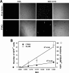
HIV-mediated cell-cell fusion occurs much less frequently than virus-cell fusion. A, comparison of virus-cell fusion (BlaM assay, top panel) and FFWO (DSP assay for GFP expression, bottom panel). The panels show images of DSP-1 and DSP-2 cells grown to confluency and infected with HXB2 pseudoviruses containing BlaM-Vpr at an m.o.i. 0.015. Differential interference contrast images of the same fields are also shown; the contrast appears to be improved on the top image panels, because DSP-1 and DSP-2 cells are less flat after an overnight incubation at 12 °C compared with cells incubated at 37 °C for a total of 6 h (bottom panel). B, dependence of the number of BlaM- and GFP-positive cells in parallel samples inoculated with low multiplicities of infection of HXB2 pseudoviruses. Data points are the average number of scored cells from six image fields after removing the background fluorescent signal obtained from the matching C52L control samples. Error bars are S.E. Solid lines are linear regressions. Note the ×10 different scales for y axis for the BlaM (right) and GFP (left) data.
FIGURE 3.
FIGURE 3.
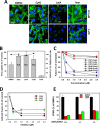
FFWO but not HIV-cell fusion requires intact actin. A, images of DSP-2 cells pretreated with the indicated concentrations of three actin inhibitors (CytD, LatA, or Jasp) or left untreated (DMSO). Cells were fixed, permeabilized, and stained with fluorescent phalloidin (green) and the nuclear stain, Hoechst-33342 (blue). Images were acquired on a Carl Zeiss LSM780 confocal microscope. Scale bar, 35 μm. B, effects of actin inhibitors on HXB2 pseudovirus uptake by DSP-2 cells. Cells were treated with 3 μm cytoskeleton inhibitors or left untreated prior to binding of HXB2pp (m.o.i. = 1) in the cold. Virus uptake was initiated by shifting to 37 °C for 90 min in the presence or absence of the inhibitors, and the amount of internalized virus was determined by p24 ELISA. Parallel samples incubated at 4 °C (Cold) to prevent endocytosis were used as a negative control. Filled circles show parallel measurements of DSP-1 and DSP-2 cell viability using a 3-(4,5-dimethylthiazol-2-yl)-5-(3-carboxymethoxyphenyl)-2-(4-sulfophenyl)-2H-tetrazolium assay. Data points (means ± S.E. from one representative triplicate experiment) are normalized to the amount of internalized virus in the vehicle control (DMSO). N.S., not significant. C, effects of actin inhibitors CytD, Jasp, and LatA on HXB2-cell fusion (red symbols) and on HXB2-mediated FFWO between DSP-1 and DSP-2 cells (blue symbols) in the overlay format; HXB2 pseudoviruses (m.o.i. = 3) were prebound to adherent DSP-2 cells in the cold, and the virus-cell complexes were overlaid with DSP-1 cells detached from a culture dish. Fusion was allowed to proceed for 2 h at 37 °C. D, effect of actin inhibitors on fusion between 293T/DSP-1 cells transiently expressing HXB2 Env and target DSP-2 cells, as measured by the DSP assay. Data points are means ± S.E. from one representative triplicate experiment. E, treatment of only one fusion partner with actin inhibitors blocks HIV-1-mediated FFWO. DSP-1 or DSP-2 cells were pretreated with 3 μm of LatA, Jasp, or mock-treated (DMSO) and washed to remove the drugs. DSP-1 cells were then overlaid onto DSP-2 cells decorated with HXB2 viruses (m.o.i. = 3) and incubated at 37 °C for 2 h. Data points in C and E are means ± S.E. from two independent triplicate experiments. At 10 μm, all drugs statistically significantly inhibited FFWO (p < 0.001).
FIGURE 4.
FIGURE 4.
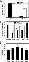
FFWO dependence on cytoskeleton. A, effects of LatA on FFWO (DSP assay) and HIV-cell fusion (BlaM assay) in control experiments in which HXB2 pseudoviruses were prebound to cells without spinoculation. DSP-2 cells were allowed to bind viruses for 1.5 h at 20 °C (to minimize virus uptake), overlaid with DSP-1 cells, further incubated at 20 °C, and shifted to 37 °C to allow fusion. Data are means ± S.E. from two experiments with triplicate samples. B, effects of 10 μm nocodazole (Nocod), paclitaxel (Pacl), blebbistatin (Blebb), and 1.5 μm nilotinib (NIL) on FFWO between DSP-1 and DSP-2 cells mediated by HXB2 pseudoviruses and on pseudovirus fusion with DSP-1/DSP-2 cells (measured by the DSP and BlaM assays, respectively). FFWO experiments were performed in the cell co-culture format, as illustrated in Fig. 1D. Data points are means ± S.E. from two independent triplicate experiments. ***, p < 0.001. C, effect of cytoskeleton inhibitors on the ability of DSP-1 and DSP-2 fragments to form a functional DSP protein in vitro. Lysates of DSP-1 and DSP-2 cells were prepared and mixed in the absence (DMSO) or in the presence of inhibitors. The luciferase activity was measured after incubation for 10 min at 25 °C and normalized to the DMSO control. Jasp, LatA, and CytD were used at 3 μm each and blebbistatin at 100 μm. Data points are means ± S.E. from a representative experiment performed in triplicate.
FIGURE 5.
FIGURE 5.
Post-CD4/coreceptor binding steps of HIV-1-mediated cell-cell fusion require intact actin. A, illustration of the TAS of HIV-1 fusion obtained by prolonged coincubation of viruses and cells at a subthreshold temperature, usually between 18 and 24 °C. B, FFWO induced by raising the temperature after establishing TAS is sensitive to actin inhibitors and gp41 refolding inhibitors but not to antagonists of CD4 or coreceptor binding. HXB2 pseudoviruses (m.o.i. = 3) were prebound to a mixture of co-cultured DSP-1 and DSP-2 cells in the cold by spinoculation and incubated at 22 °C for 2.5 h. The creation of TAS was verified based on partial or nearly full resistance of HXB2pp fusion to inhibitors of CD4 binding (10 μm BMS-806) and CXCR4 binding (30 μm AMD3100) added at this stage, just prior to shifting to 37 °C. A potent block of FFWO from TAS by the C52L peptide demonstrates that the fusion reaction did not progress beyond the CD4/coreceptor binding steps. CytD, LatA, and Jasp added at 3 μm each after establishing TAS potently blocked cell-cell fusion. C, after measuring FFWO (using the DSP activity, as described in B), the extent of HXB2-cell fusion was determined in parallel using the BlaM assay. Data points are means ± S.E. from two independent triplicate experiments.
FIGURE 6.
FIGURE 6.
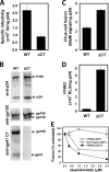
Deletion of the gp41 CT modulates HIV-1 fusion competence and the ability to promote FFWO. Pseudoviruses bearing NL4-3 Env were used in these experiments. A, infectious titers of WT and ΔCT viruses in TZM-bl cells were determined using the β-galactosidase assay (40) and normalized to the p24 content of the respective viral stocks. Data points are means ± S.E. from one triplicate experiment. B, Western blot analysis of Env cleavage/incorporation into pseudoviruses. Equal volumes of concentrated virus preparations were analyzed by SDS-PAGE and visualized using anti-gp120 and anti-gp41 antibodies. The HIV-1 p24 bands were detected using anti-HIV sera. The ΔCT preparation contained ∼2-fold more p24/ml than WT virus, as determined by ELISA (data not shown). C and D, analyses of virus-cell fusion (C) and FFWO (D) mediated by pseudoviruses. Co-cultures of DSP-1 and DSP-2 cells were spinoculated with viruses adjusted to the same amount of p24 (0.5 ng), washed, and incubated for 1.5 h at 37 °C to allow fusion. Data points are means ± S.E. from two experiments performed in triplicate. E, effect of Jasp pretreatment on FFWO mediated by WT and ΔCT viruses was measured in the overlay format. The BlaM activity of WT viruses is shown for comparison. Data points are means ± S.E. from a single triplicate experiment.
FIGURE 7.
FIGURE 7.
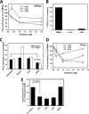
HIV-1 fusion with CEM.CCR5 cells is partially dependent on cytoskeleton. A, effects of cytoskeleton inhibitors on the HXB2 pseudovirus (HXB2pp, m.o.i. = 2) fusion with CEM.CCR5 cells. Data are normalized to the signal in DMSO-treated samples and are means ± S.E. from two experiments performed in triplicate. B, LatA abrogates FFWO between CEM.CCR5 cells mediated by HXB2 pseudoviruses. Two samples of CEM.CCR5 cells each labeled with a green or an orange cytoplasmic dye were mixed and treated with 3 μm LatA or left untreated prior to binding of HXB2pp (m.o.i. = 10) in the cold. Cells were washed to remove unbound viruses and allowed to fuse for 3 h at 37 °C in the presence or absence of LatA or 10 μm C52L (negative control). Data are the average numbers (and S.E.) of fused cells normalized to the total number of cells from 10 image fields. C, effects of cytoskeletal inhibitors, nocodazole (10 μm), paclitaxel (10 μm), and blebbistatin (50 μm) on HXB2pp fusion with CEM.CCR5 cells. Data are normalized to the signal in untreated samples and are means ± S.E. from two experiments performed in triplicate. To simplify the washing steps, cells were attached to poly-lysine-coated microplates prior to fusion experiments, as described under “Experimental Procedures.” In this format, a robust BlaM signal was detected after the virus binding in the cold and incubation at permissive temperature. Blebbistatin caused significant (p < 0.001) reduction in HIVpp and VSVpp fusion. D, VSVpp (m.o.i. = 2) fusion with CEM.CCR5 cells in the presence of actin inhibitors. For details, see Fig. 6A. E, actin inhibitors decrease HIV-1 internalization by CEM.CCR5 cells. Cells were treated with 3 μm of cytoskeleton inhibitors or left untreated prior to binding of HXB2pp (m.o.i. = 2) in the cold. Virus internalization was initiated by shifting the samples at 37 °C for 2 h in the presence or absence of the inhibitors. The amount of internalized virus was determined by p24 ELISA, as described under “Experimental Procedures.” Data are normalized to the total amount of cell-bound virus and are means ± S.E. from one triplicate experiment.

Similar articles

Cited by

References

    1. Gallo S. A., Finnegan C. M., Viard M., Raviv Y., Dimitrov A., Rawat S. S., Puri A., Durell S., Blumenthal R. (2003) The HIV Env-mediated fusion reaction. Biochim. Biophys. Acta 1614, 36–50 - PubMed
    1. Melikyan G. B. (2011) Membrane fusion mediated by human immunodeficiency virus envelope glycoprotein. Curr. Top. Membr. 68, 81–106 - PubMed
    1. Wilen C. B., Tilton J. C., Doms R. W. (2012) Molecular mechanisms of HIV entry. Adv. Exp. Med. Biol. 726, 223–242 - PubMed
    1. Melikyan G. B. (2014) HIV entry: a game of hide-and-fuse? Curr. Opin. Virol. 4, 1–7 - PMC - PubMed
    1. Permanyer M., Ballana E., Esté J. A. (2010) Endocytosis of HIV: anything goes. Trends Microbiol. 18, 543–551 - PubMed

Publication types

Substances